

Customized 3D-Printed Titanium Mesh Developed to Regenerate a Complex Bone Defect in the Aesthetic Zone: A Case Report Approached with a Fully Digital Workflow
- PMID: 32887390
- PMCID: PMC7503418
- DOI: 10.3390/ma13173874
Customized 3D-Printed Titanium Mesh Developed to Regenerate a Complex Bone Defect in the Aesthetic Zone: A Case Report Approached with a Fully Digital Workflow
Abstract
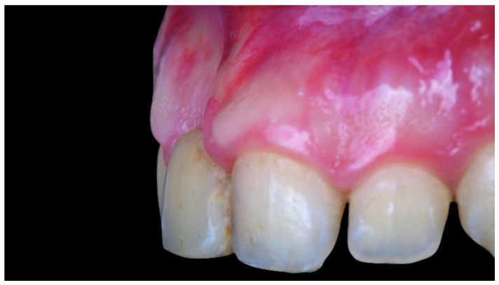
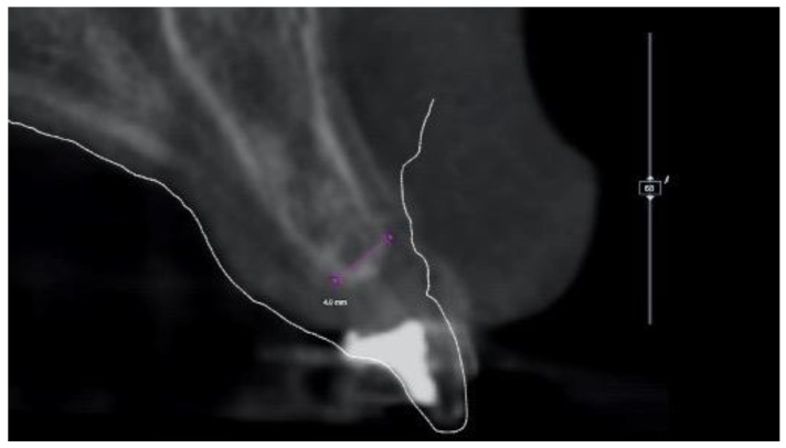
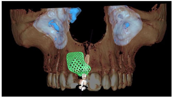
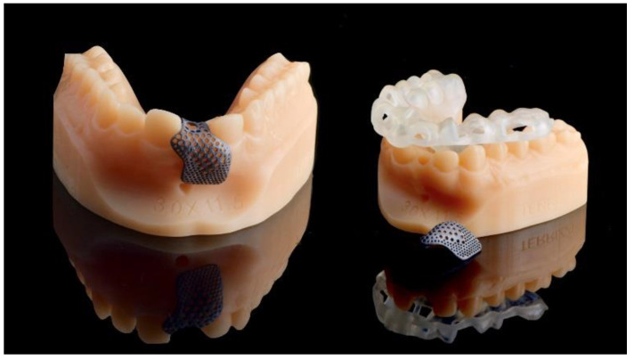
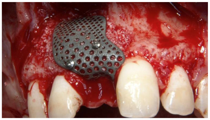
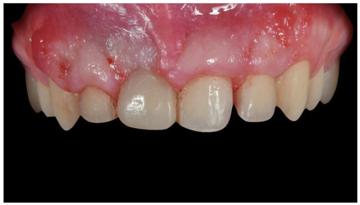
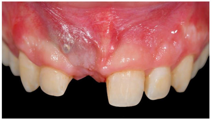
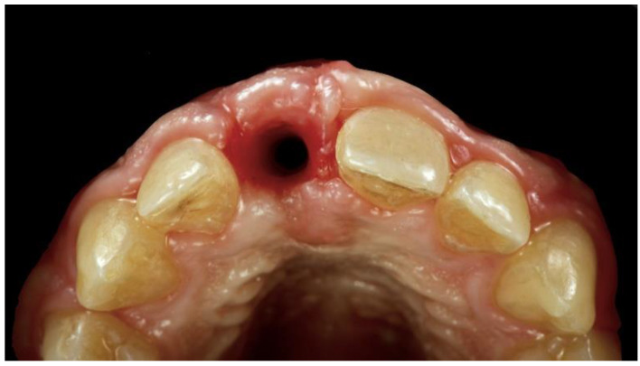
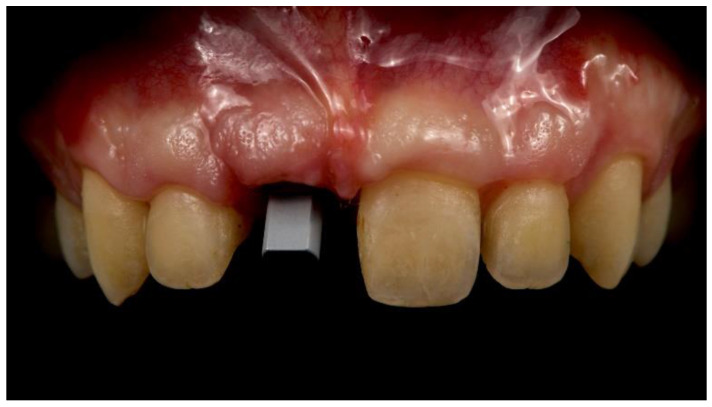
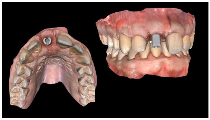
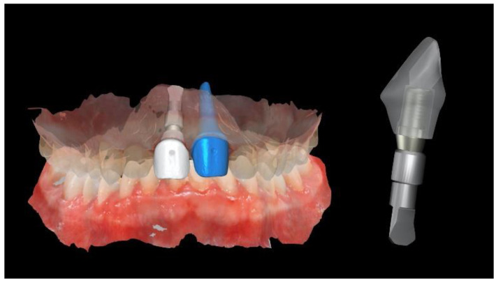
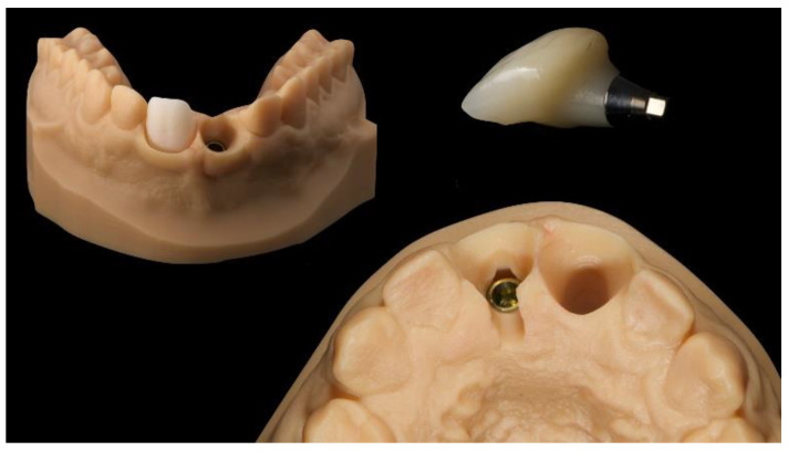
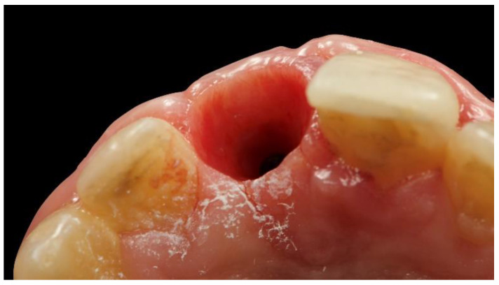
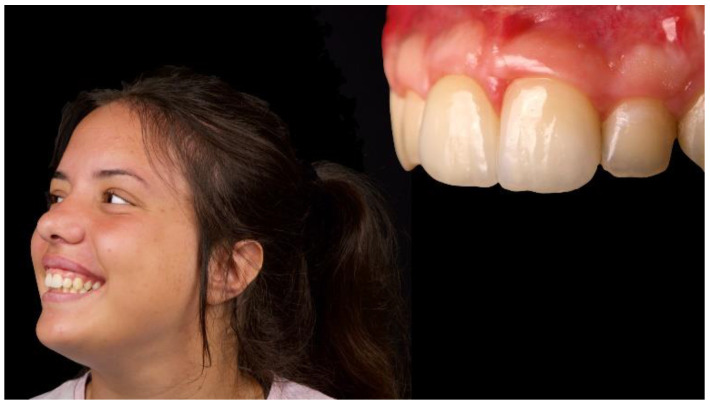
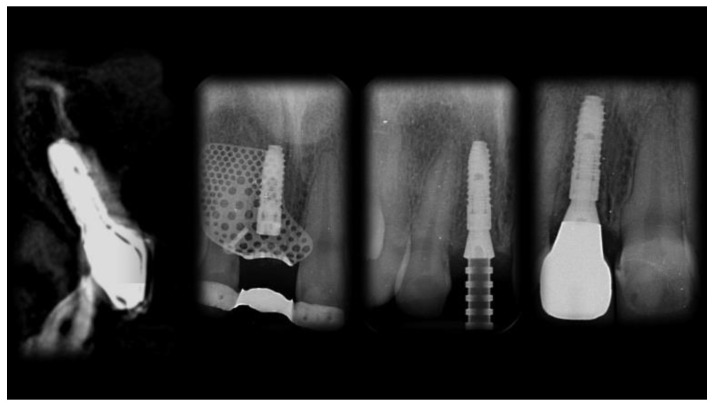
Alveolar-ridge augmentation, anterior aesthetics, and digital technologies are probably the most popular topics in the dental-implant field. The aim of this report is to present a clinical case of severe atrophy of the anterior maxilla in a younger female patient, treated with a titanium membrane customized with computer-aided design/computer-aided manufacturing (CAD/CAM), simultaneous guided implant placement, and a fully digital workflow. A young female patient with a history of maxillary trauma was treated and followed-up for 1 year after implant placement. A narrow implant was inserted in a prosthetically driven position with the aid of computer-guided surgery. In the same surgical section, a customized implantable titanium mesh was applied. The scaffold was designed according to the contralateral maxillary outline in order to recreate a favorable maxillary bone volume. Finally, highly aesthetic, CAD/CAM, metal-free restorations were delivered using novel digital technologies.
Keywords: anterior maxilla; dental implants; digital workflow; guided bone regeneration; titanium mesh scaffold.
Conflict of interest statement
The authors declare no conflict of interest.
Figures


References
-
- Esposito M., Trullenque-Eriksson A., Tallarico M. Endodontic retreatment versus dental implants of teeth with an uncertain endodontic prognosis: 3-year results from a randomised controlled trial. Eur. J. Oral Implantol. 2018;11:423–438. - PubMed
-
- Meloni S.M., Lumbau A., Baldoni E., Pisano M., Spano G., Massarelli O., Tallarico M. Platform switching versus regular platform single implants: 5-year post-loading results from a randomised controlled trial. Int. J. Oral Implantol. (New Malden) 2020;13:43–52. - PubMed
-
- Meloni S.M., Lumbau A., Spano G., Baldoni E., Pisano M., Tullio A., Tallarico M. Sinus augmentation grafting with anorganic bovine bone versus 50% autologous bone mixed with 50% anorganic bovine bone: 5 years after loading results from a randomised controlled trial. Int. J. Oral Implantol. (New Malden) 2019;12:483–492. - PubMed
-
- Meloni S.M., Spano G., Ceruso F.M., Gargari M., Lumbau A., Baldoni E., Massarelli O., Pisano M., Tallarico M. Upper jaw implant restoration on six implants with flapless guided template surgery and immediate loading: 5 years results of a prospective case series. Oral Implantol. 2020;12:151–160. doi: 10.1111/clr.45_13508. - DOI
-
- Tallarico M., Cervino G., Scrascia R., Uccioli U., Lumbau A.I., Meloni S.M. Minimally invasive treatment of edentulous maxillae with overdenture fully supported by a cad/cam Titanium Bar with a low-profile attachment screwed on four or six implants: A case series. Prosthesis. 2020;2:53–64. doi: 10.3390/prosthesis2020006. - DOI
Publication types
LinkOut - more resources
Full Text Sources
Miscellaneous
