. 2020 Sep 4;13(1):120.
doi: 10.1186/s13045-020-00954-7.

SARS-CoV-2 binds platelet ACE2 to enhance thrombosis in COVID-19

Affiliations

SARS-CoV-2 binds platelet ACE2 to enhance thrombosis in COVID-19

Si Zhang et al. J Hematol Oncol. .

Abstract

Background: Critically ill patients diagnosed with COVID-19 may develop a pro-thrombotic state that places them at a dramatically increased lethal risk. Although platelet activation is critical for thrombosis and is responsible for the thrombotic events and cardiovascular complications, the role of platelets in the pathogenesis of COVID-19 remains unclear.

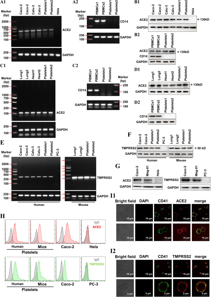
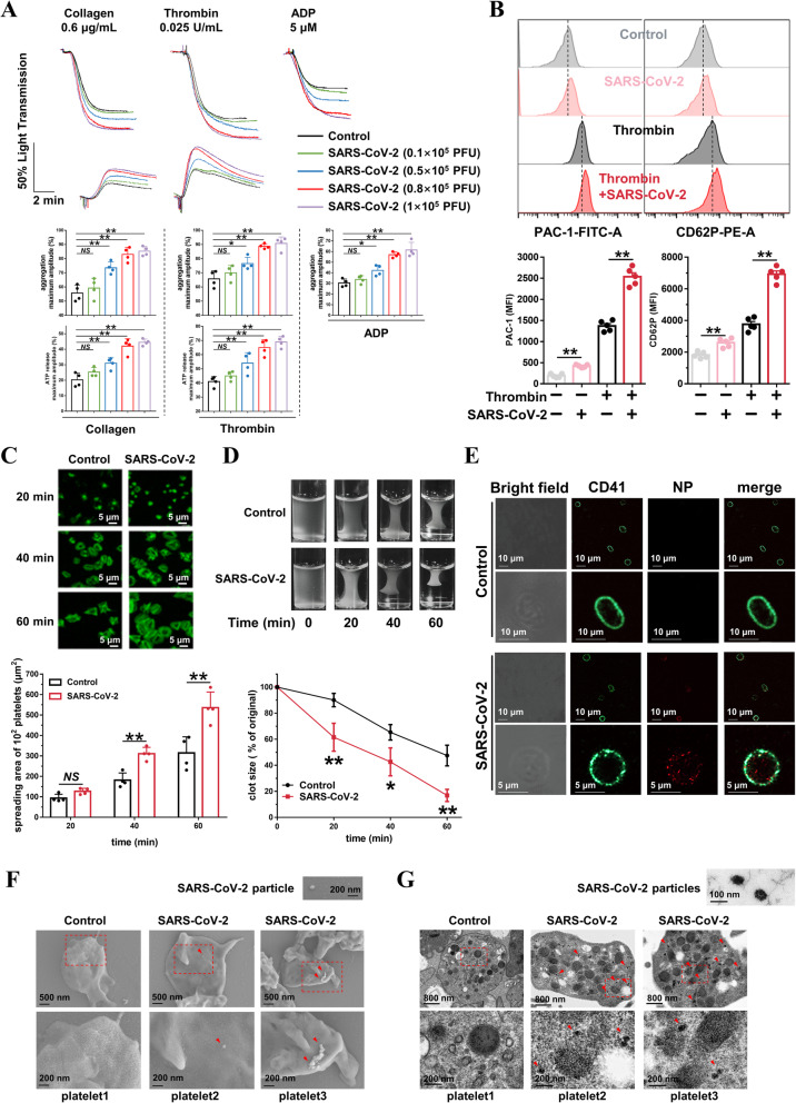
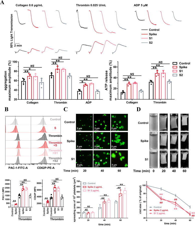
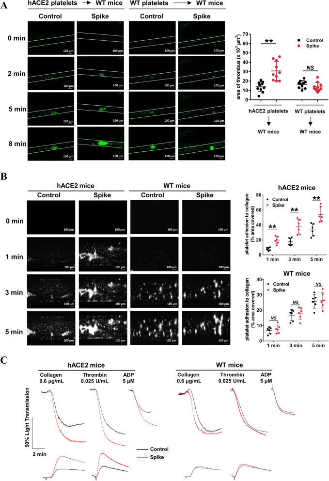
Methods: Using platelets from healthy volunteers, non-COVID-19 and COVID-19 patients, as well as wild-type and hACE2 transgenic mice, we evaluated the changes in platelet and coagulation parameters in COVID-19 patients. We investigated ACE2 expression and direct effect of SARS-CoV-2 virus on platelets by RT-PCR, flow cytometry, Western blot, immunofluorescence, and platelet functional studies in vitro, FeCl3-induced thrombus formation in vivo, and thrombus formation under flow conditions ex vivo.

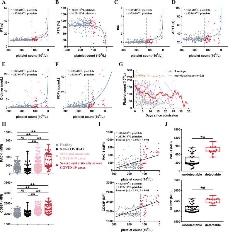
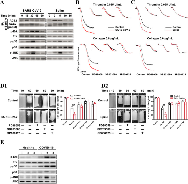
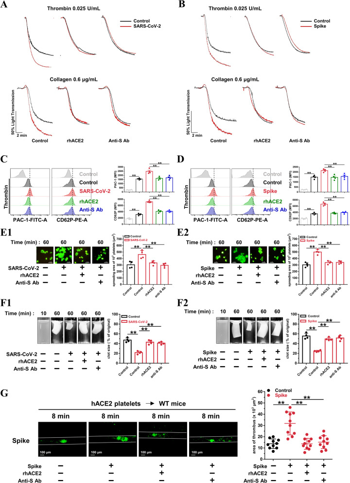
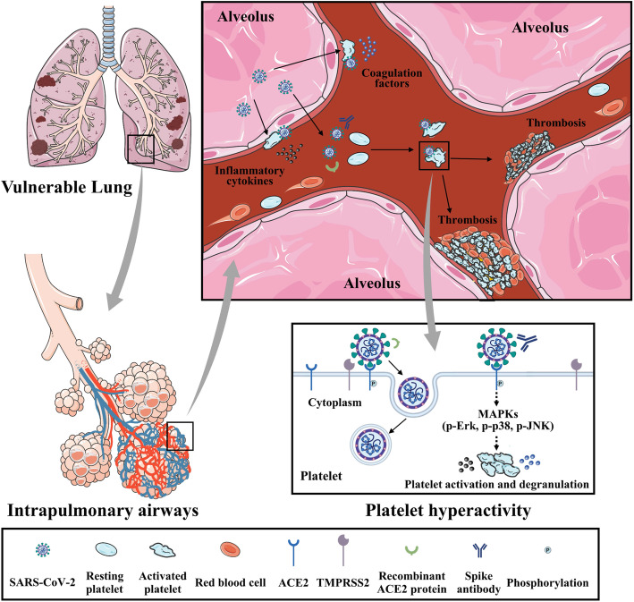
Results: We demonstrated that COVID-19 patients present with increased mean platelet volume (MPV) and platelet hyperactivity, which correlated with a decrease in overall platelet count. Detectable SARS-CoV-2 RNA in the blood stream was associated with platelet hyperactivity in critically ill patients. Platelets expressed ACE2, a host cell receptor for SARS-CoV-2, and TMPRSS2, a serine protease for Spike protein priming. SARS-CoV-2 and its Spike protein directly enhanced platelet activation such as platelet aggregation, PAC-1 binding, CD62P expression, α granule secretion, dense granule release, platelet spreading, and clot retraction in vitro, and thereby Spike protein enhanced thrombosis formation in wild-type mice transfused with hACE2 transgenic platelets, but this was not observed in animals transfused with wild-type platelets in vivo. Further, we provided evidence suggesting that the MAPK pathway, downstream of ACE2, mediates the potentiating role of SARS-CoV-2 on platelet activation, and that platelet ACE2 expression decreases following SARS-COV-2 stimulation. SARS-CoV-2 and its Spike protein directly stimulated platelets to facilitate the release of coagulation factors, the secretion of inflammatory factors, and the formation of leukocyte-platelet aggregates. Recombinant human ACE2 protein and anti-Spike monoclonal antibody could inhibit SARS-CoV-2 Spike protein-induced platelet activation.

Conclusions: Our findings uncovered a novel function of SARS-CoV-2 on platelet activation via binding of Spike to ACE2. SARS-CoV-2-induced platelet activation may participate in thrombus formation and inflammatory responses in COVID-19 patients.

Keywords: ACE2; COVID-19; Platelet activation; TMPRSS2; Thrombosis.


Conflict of interest statement

The authors declare that they have no competing interests.

Figures

Fig. 1
Fig. 1
Increased platelet activation in patients with SARS-CoV-2 infection. af Dot plot showing the correlation between platelet count and PT (a), platelet count and PTA (b), platelet count and INR (c), platelet count and APTT (d), platelet count and d-dimer (e), as well as platelet count and FDPs (F) in COVID-19 patients (n = 241). Each circle represents a different patient. g Dynamics of platelet count in COVID-19 patients with critically severe illness after hospital admission. The platelet counts values were obtained from 22 independent patients. Different colors were used for different patients. h Increased expression of platelet integrin αIIbβ3 activation (PAC-1 binding) and P-selectin (CD62P) expression in COVID-19 patients compared with healthy 11 donors and non-COVID-19 patients. Each circle represents a different individual from healthy donors (n = 166), non-COVID-19 cases (n = 60), the mild and moderate COVID-19 cases (n = 184) or the severe and critically severe COVID-19 cases (n = 57). I, PAC-1 binding and CD62P expression are correlated with platelet count in COVID- 19 patients (n = 241). Each solid circle represents a different individual. j PAC-1binding and CD62P expression in severe and critically severe type COVID-19 patients with detectable blood virus RNA (detectable, n = 12) and with undetectable blood virus RNA (undetectable, n = 45). Statistical analyses were performed using Kruskal-Wallis test with Bonferroni correction in (h), Pearson’s correlation analysis in (i) and nonparametric Mann-Whitney U test in (j). NS no significance; **P < 0.01. PT prothrombin time, PTA prothrombin time activity, INR international normalized ratio, APTT activated partial thromboplastin time, FDPs fibrinogen degradation products, undetectable: severe and critically severe type COVID-19 patients with undetectable blood virus RNA, detectable: severe and critically severe type COVID-19 patients with detectable blood virus RNA
Fig. 2
Fig. 2
Both human 1 and mouse platelets express ACE2 and TMPRSS2. A, RT2 PCR detection of ACE2 (A1) and monocyte-specific CD14 (A2) in healthy human platelets. B, Western blot detection of ACE2 and monocyte-specific CD14 (B2) in healthy human platelets. For A and B, the human colon cell line Caco-2 and the human lung cell line Calu-3 were used as positive controls of ACE2, and the human Hela cell line was used as a negative control of ACE2. The peripheral blood mononuclear cells (PBMCs) from healthy human were used as a positive control of CD14. C, RT-PCR detection of ACE2 (C1) in lungs, hearts, and platelets from wild-type mice. D, Western blot detection of ACE2 in lungs, hearts, and platelets from wild-type mice. For C and D, PBMCs from mice were used as a positive control of CD14. E, RT-PCR detection of TMPRSS2 in platelets from healthy human and wild-type mice. F, Western blot detection of TMPRSS2 in platelets from healthy human and wild-type mice. For E and F, the colon cell line Caco-2 and the human lung cell line Calu-3 from human and the lungs from mice were used as positive controls of TMPRSS2, and the human prostate cell line PC-3 was used as a negative control of TMPRSS2. For A to F, platelet-rich plasma prepared as previously described was filtered through a Sepharose 2B column equilibrated in Tyrode’s solution to isolate platelets. Platelets1 in A, B, E left panel and F left panel were platelets from 1 healthy blood sample and platelets2 in A, B, E left panel and F left panel were platelets mixture from 20 healthy donors. Platelets1 in C, D, E right panel and F right panel were platelets from 1 wild-type mouse and platelets2 in C, D, E right panel and F right panel were platelets mixture from 5 wild-type mice. PBMCs were isolated by centrifugation on a Ficoll-Paque from two different blood samples of healthy donors (PBMCs1 and PBMCs2 in A and B) and from two different blood samples of wild-type mice (PBMCs1 and PBMCs2 in C and D). The two different lung (lung1 and lung2 in C, D, E and F) and heart (heart1 and heart2 in C and D) tissues were dissected from different wild-type mice. G, Western blot detection of ACE2 and TMPRSS2 in megakaryocyte cell line (Meg-01). H, Detecting ACE2 and TMPRSS2 expression on healthy human and wild-type mice platelets by flow cytometry. I, Imaging of ACE2 (I1) and TMPRSS2 (I2) expression in healthy human platelets using confocal microscopy. ACE2, the angiotensin converting enzyme 2; TMPRSS2, transmembrane protease serine 2; and RT-PCR, reverse transcription polymerase chain reaction. Images were representative of three independent RT-PCR, Western blot or flow cytometry experiments
Fig. 3
Fig. 3
SARS-CoV-2 directly enhances 1 platelet activation in vitro. a SARS-CoV-2 dose-dependently potentiated platelets aggregation and ATP release in response to collagen, thrombin, and ADP in vitro. Washed platelets from healthy donors were preincubated with SARS-CoV-2 in the indicated concentration for 30 min, then stimulated with collagen (0.6 μg/mL), thrombin (0.025 U/mL), or ADP (5 μM). Aggregation and ATP release (with luciferase) were assessed under stirring at 1200 rpm. Representative results and summary data of 4 experiments are presented. b SARS8 CoV-2 induced PAC-1 binding and CD62P expression in the absence of agonist; and potentiated integrin PAC-1 binding and CD62P expression induced by thrombin in platelets. Platelets were preincubated with SARS-CoV-2 virus (1 × 10⁵ PFU, 60 min) or with SARS-CoV-2 virus (1×10⁵ PFU, 30 min), and treated with thrombin (0.025 U/mL, 10 min), and then analyzed using a flow cytometer. Representative flow cytometry histograms and summary data of 5 experiments are presented. c Representative confocal fluorescence images (phalloidin) showing that SARS-CoV-2 potentiated platelet spreading on immobilized fibrinogen (100 μg/mL). After preincubation with SARS-CoV-2 (1 × 10⁵ PFU) for 30 min, platelets were allowed to spread on the fibrinogen-coated surfaces at 37 °C for indicated times. Representative results and summary data of 4 experiments are presented. d SARS-CoV-2 potentiated clot retraction induced by thrombin. Platelets from healthy donors were normalized at a concentration of 4 × 10⁸/mL and preincubated with SARS-CoV-2 (1 × 10⁵ PFU) for 30 min, then stimulated with thrombin (1 U/mL). Representative results and summary data of 4 experiments are presented. e Immunofluorescent staining of Nucleocapsid protein (NP, red) and CD41 (green) in human platelets incubated with SARS-CoV-2 virus (1 × 10⁵ PFU) for 3 h. Representative images from 3 experiments using platelets from different healthy donors. f Scanning electron microscope (SEM) of SARS-CoV-2 particles on the surface of platelets. SEM of healthy human platelets (3 × 10⁸ platelets/mL) incubated with SARS-CoV-2 (1 × 10⁵ PFU) for 30 min. Platelets were washed for 3 times and fixed immediately after incubation and processed for SEM experiment. Representative images of single platelet from control group (platelet1) and SARS-CoV-2 treatment group (platelet2 and platelet3) are shown from three different experiments. Arrows point toward the SARS-CoV-2 virus. g. Transmission electron microscopy (TEM) of SARS-CoV-2 particles in platelets. TEM of healthy human platelets (3 × 10⁸ platelets/mL) incubated with SARS-CoV-2 (1 × 10⁵ PFU) for 3 h. Platelets were washed for 3 times and fixed immediately after incubation and processed for TEM experiment. Representative images from control group (platelet1) and SARS CoV-2 treatment group (platelet2 and platelet3) are shown from three different experiments. Arrows point toward the SARS-CoV-2 particles. Statistical analyses were performed using unpaired two-tailed Student’s t test in (a), (b) and (c). NS no significance; *P < 0.05; **P < 0.01. Two-way ANOVA and Tukey’s post hoc test was performed in (d); *P < 0.05 and **P < 0.01 compared with control group
Fig. 4
Fig. 4
SARS-CoV-2 Spike 1 protein directly enhances human platelet activation. a SARS-CoV-2 Spike protein and Spike subunit 1 (S1) potentiated platelet aggregation and ATP release in response to collagen, thrombin, and ADP in vitro, whereas Spike subunit 2 (S2) did not. Washed platelets from healthy donors were preincubated with Spike protein, S1 or S2 at 2 μg/mL for 5 min, then stimulated with collagen (0.6 μg/mL), thrombin (0.025 U/mL), or ADP (5 μM). Aggregation and ATP release (with luciferase) were assessed under stirring at 1200 rpm. Representative results and summary data of 4 experiments are presented. b Spike protein stimulated platelets for PAC-1 binding and CD62P expression in the absence of agonist. In addition, Spike protein and S1 increased PAC-1 binding and CD62P expression induced by thrombin in platelets. Platelets were incubated with Spike protein (2 μg/mL, 60 min) in the absence of agonist, or preincubated with Spike protein, S1 or S2 at 2 μg/mL for 5 min and stimulated with thrombin (0.025 U/mL) for 10 min, and then analyzed using a flow cytometer. Representative results and summary data of 4 experiments are presented. c Spike protein (2 μg/mL) and S1 (2 μg/mL) potentiated platelet spreading on immobilized fibrinogen. After preincubation with Spike protein (2 μg/mL) for 5 min, platelets were allowed to spread on the fibrinogen-coated surfaces at 37 °C for the indicated times. Representative results and summary data of 3 experiments are presented. d Spike protein and S1 potentiated clot retraction induced by thrombin. Platelets from healthy donors were normalized at a concentration of 4 × 10⁸/mL and preincubated with Spike protein (2 μg/mL) or S1 (2 μg/mL) for 5 min, then stimulated with thrombin (1 U/mL). Representative images and summary data are presented from 3 experiments using platelets from different donors. Statistical analyses were performed using one-way ANOVA, followed by Tukey’s post hoc analysis in (a), (b) and (c). NS no significance; *P < 0.05; **P < 0.01. Two-way ANOVA and Tukey’s post hoc test was performed in (d); ## significant difference (P < 0.01) between Spike and control group; ** significant difference (P < 0.01) between S1 and control group
Fig. 5
Fig. 5
ACE2/MAPK mediates the 1 potentiating effects of SARS-CoV-2 on platelet activation. A SARS-CoV-2 virus and its Spike protein phosphorylate ACE2, Erk, p38, and JNK in human platelets. Platelets from healthy donors were pretreated with SARS-CoV-2 (1 × 10⁵ PFU) or its Spike protein (2 μg/mL) for various times, as 5 indicated. For ACE2 phosphorylation detection, cell lysates were prepared and subjected to immunoprecipitation (IP) with anti-phospho-Ser/Thr antibody, followed by immunoblotting analysis with anti-ACE2 antibody. Cell lysates without the process of immunoprecipitation (10% of input) were analyzed in parallel as loading controls. For p-Erk, p-p38, and p-JNK detection, cell lysates were prepared and directly subjected to immunoblotting. Representative results are presented from 4 experiments using platelets from different healthy donors and summary data is presented in the Additional file 1: Online Figure 7. B Enhanced platelet aggregation by SARS-CoV- 2 is abolished by MAPK inhibitors. The healthy human platelets were pretreated with 10 μM PD98059 (ERK1/2 inhibitor), 10 μM SB203580 (p38 inhibitor), or 10 μM SP600125 (JNK inhibitor) for 10 min, then treated with SARS-CoV-2 (1 × 10⁵ PFU, 30 min) before stimulation by 0.025 U/mL thrombin or 0.6 μg/mL collagen. Representative results are presented from 3 experiments using platelets from different donors. C Enhanced platelet aggregation by Spike protein is abolished by MAPK inhibitors. Human platelets were pretreated with 10 μM PD98059, 10 μM SB203580, or 10 μM SP600125 for 10 min, then treated with Spike protein (2 μg/mL, 5 min) or vehicle as control before stimulation by 0.025 U/mL thrombin or 0.6 μg/mL collagen. Representative results are presented from 3 experiments using platelets from different healthy donors. D Accelerated clot retraction by SARS-CoV-2 (D1) or its Spike protein (D2) is abolished by MAPK inhibitors. Platelets were pretreated with 10 μM PD98059, 10 μM SB203580, or 10 μM SP600125 for 10 min, and then incubated with SARS26 CoV-2 (1 × 10⁵ PFU, 30 min) or Spike protein (2 μg/mL, 5 min) as in B and C. Clot retraction was initiated as in Fig. 3d. Representative images and summary data of 3 experiments are presented using platelets from different healthy donors. Representative images and summary data are presented from 3 experiments using platelets from different donors. E Increased phosphorylation of Erk, p38, and JNK in platelets from COVID-19 patients, compared with healthy donors. Representative results are presented using platelets from 6 individuals from different COVID-19 patients (n = 3) and healthy donors (n = 3). Statistical analyses were performed using unpaired two34 tailed Student’s t test in (D1) and (D2). *P < 0.05; **P < 0.01. MAPK indicates mitogen activated protein kinase; PD, PD98059; SB, SB203580; SP, SP600125
Fig. 6
Fig. 6
SARS-CoV-2 Spike 1 protein directly enhances thrombosis potential in vivo. a Washed platelets from wild-type or hACE2 transgenic mice were infused into WT mice. After intravenous injection 200 μg/kg Spike protein or control (saline), FeCl3-induced arterial thrombus formation was initiated, and the thrombus area was recorded. Representative image of thrombus formation and the relative fluorescence at different time points are shown. Statistically analysis of FeCl3-induced thrombosis by assess thrombus area at 8 min (n = 10). b Spike protein-treated whole blood from hACE2 mice showed accelerated thrombus formation over an immobilized collagen surface at a shear rate of 1000 s−1, whereas Spike protein-treated whole blood from wild type mice did not. The whole blood from mice was fluorescently labeled by mepacrine (100 μM, 30 min) and incubated with Spike protein (2 μg/mL) for 5 min, and then perfused through fibrillar collagen-coated bioflux plates for 5 min. Representative images and time courses of thrombus formation challenged with control or Spike protein at the indicated time points are presented. Dot plot showing thrombus formation area (n = 6). c Spike protein-treated platelets from hACE2 mice presented increased platelet aggregation and ATP release in response to collagen, thrombin, and ADP, whereas Spike protein-treated platelets from wild-type mice did not. Washed platelets from mice were pretreated with control or Spike protein (2 μg/mL) for 5 min, and then stimulated with collagen (0.6 μg/mL), thrombin (0.025 U/mL), or ADP (5 μM). Aggregation and ATP release (with luciferase) were assessed under stirring at 1200 rpm. Representative results are presented from 4 experiments using platelets from different mice and summary data are presented in Additional file 1: Online Figure 8. Statistical analyses were performed using unpaired two-tailed Student’s t test in (a) and (b). NS no significance; **P < 0.01. WT indicates wild-type; hACE2 indicates hACE2 transgenic
Fig. 7
Fig. 7
Recombinant human ACE2 protein and anti-Spike monoclonal antibody suppress SARS-CoV-2-induced platelet activation. A, Enhanced platelet aggregation by SARS-CoV-2 is abolished by recombinant human ACE2 protein and anti-Spike monoclonal antibody (targeting the receptor-binding domain [RBD] of SARS-CoV-2). Representative results are presented from 3 experiments using platelets from different donors. B, Enhanced platelet aggregation by Spike protein is suppressed by ACE2 protein and anti-Spike antibody. Representative results are presented from 3 experiments using platelets from different healthy donors. For A and B, the healthy human platelets were pretreated with ACE2 protein (10 μg/mL) or anti-Spike antibody (4 μg/mL) for 10 min, then treated with SARS-CoV-2 (1 × 10⁵ PFU, 30 min) or Spike protein (2 μg/mL, 5 min) before stimulation by 0.025 U/mL thrombin or 0.6 μg/mL collagen. C and D, The ACE2 protein and anti-Spike antibody reversed PAC-1 binding and CD62P expression induced by SARS-CoV-2 (C) or Spike protein (D). Representative images and summary data are presented from 4 experiments using platelets from different healthy donors. E, Enhanced platelet spreading induced by SARS-CoV-2 (E1) or its Spike protein (E2) are abolished by ACE2 protein and anti-Spike monoclonal antibody. Representative images and summary data of 4 experiments are presented using platelets from different healthy donors. F, Accelerated clot retraction induced by SARS-CoV-2 (F1) or its Spike protein (F2) are abolished by ACE2 protein and anti-Spike monoclonal antibody. Representative images and summary data of 4 experiments are presented using platelets from different healthy donors. For C, D, E and F, platelets were pretreated with ACE2 protein (10 μg/mL) or anti-Spike antibody (4 μg/mL) for 10 min, and incubated with SARS-CoV-2 (1 × 10⁵ PFU, 30 min) or Spike protein (2 μg/mL, 5 min), and then subjected to flow cytometry of thrombin-activated platelets, platelet spreading assay, and clot retraction assay. G, Increased thrombus area induced by Spike protein in wild-type mice transfused with platelets from hACE2 transgenic mice is suppressed by ACE2 protein and anti-Spike antibody. Representative photographs of FeCl3-induced thrombus formation at the indicated time points within 30 min after intravenous administration of Spike protein (200 μg/kg) with ACE2 protein (1 mg/kg) or anti-Spike monoclonal antibody (400 μg/kg). Dot plot showing thrombus area for control or Spike protein treated mice (n = 10). Statistical analyses were performed using one-way ANOVA, followed by Tukey’s post hoc analysis in (C), (D), (E), (F), and (G). *P < 0.05; **P < 0.01. RhACE2 indicates recombinant human ACE2 protein; anti-S Ab, anti-Spike antibody
Fig. 8
Fig. 8
Summary schemes illustrating SARS-CoV-2 activates platelets and enhances thrombosis in COVID-19. Global schema illustrating SARS-CoV-2 from alveolus binds and activates platelets, which enhances thrombosis formation and inflammatory reaction in capillaries, and subsequently contributes to development of disseminated intravascular coagulation and acute respiratory distress syndrome. SARS-CoV-2 Spike protein binds to ACE2 and phosphorylates ACE2, leading to MAPK signaling activation (phosphorylation of Erk, p-38, and JNK) and subsequent platelet activation, coagulation factors release, and inflammatory cytokines secretion. Interaction between SARS-CoV-2 Spike protein and platelet ACE2 confers the platelet activation, which is suppressed by the recombinant human ACE2 protein and anti-Spike monoclonal antibody (central illustration)

References

    1. Huang C, Wang Y, Li X, Ren L, Zhao J, Hu Y, et al. Clinical features of patients infected with 2019 novel coronavirus in Wuhan China. Lancet. 2020;395(10223):497–506. - PMC - PubMed
    1. Clerkin KJ, Fried JA, Raikhelkar J, Sayer G, Griffin JM, Masoumi A, et al. COVID-19 and Cardiovascular Disease. Circulation. 2020;141(20):1648–1655. - PubMed
    1. Madjid M, Safavi-Naeini P, Solomon SD, Vardeny O. Potential Effects of coronaviruses on the Cardiovascular System: A Review. JAMA Cardiol. 2020. - PubMed
    1. Driggin E, Madhavan MV, Bikdeli B, Chuich T, Laracy J, Biondi-Zoccai G, et al. Cardiovascular considerations for patients, health care workers, and health systems during the COVID-19 pandemic. J Am Coll Cardiol. 2020;75(18):2352–2371. - PMC - PubMed
    1. Chen N, Zhou M, Dong X, Qu J, Gong F, Han Y, et al. Epidemiological and clinical characteristics of 99 cases of 2019 novel coronavirus pneumonia in Wuhan, China: a descriptive study. Lancet. 2020;395(10223):507–513. - PMC - PubMed

Publication types

MeSH terms