. 2021 Feb;37(2):166-182.
doi: 10.1007/s12264-020-00565-9. Epub 2020 Sep 4.

Sex Differences in Electrophysiological Properties of Mouse Medial Preoptic Area Neurons Revealed by In Vitro Whole-cell Recordings

Affiliations

Sex Differences in Electrophysiological Properties of Mouse Medial Preoptic Area Neurons Revealed by In Vitro Whole-cell Recordings

Wen Zhang et al. Neurosci Bull. 2021 Feb.

Abstract

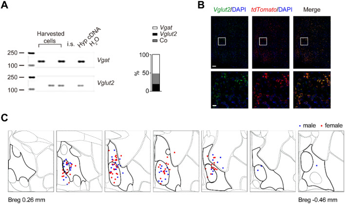
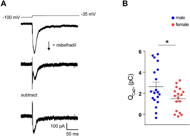
Despite extensive characterization of sex differences in the medial preoptic area (mPOA) of the hypothalamus, we know surprisingly little about whether or how male and female mPOA neurons differ electrophysiologically, especially in terms of neuronal firing and behavioral pattern generation. In this study, by performing whole-cell patch clamp recordings of the mPOA, we investigated the influences of sex, cell type, and gonadal hormones on the electrophysiological properties of mPOA neurons. Notably, we uncovered significant sex differences in input resistance (male > female) and in the percentage of neurons that displayed post-inhibitory rebound (male > female). Furthermore, we found that the current mediated by the T-type Ca2+ channel (IT), which is known to underlie post-inhibitory rebound, was indeed larger in male mPOA neurons. Thus, we have identified salient electrophysiological properties of mPOA neurons, namely IT and post-inhibitory rebound, that are male-biased and likely contribute to the sexually dimorphic display of behaviors.

Keywords: Electrophysiological properties; Post-inhibitory rebound; Sex differences; T-type calcium channels; mPOA.


Conflict of interest statement

The authors claim no conflict of interest.

Figures

Fig. 1
Fig. 1
Slice recordings from mPOA neurons. A Acute brain slices containing the mPOA (black outlines) were chosen. Images of neurons were captured under a 40 × objective just before the electrode reached the target neuron (scale bar, 10 μm) and were used to analyze the somatic morphology of the recorded neurons. After recordings, the tip of the electrode was visualized under a 5 × objective (scale bar, 100 μm) to document the coordinates of the recorded neuron. B Example voltage signals (upper traces) recorded from an mPOA neuron in response to stimulation currents (lower traces). The first spike elicited in this neuron (insert on right) was processed to calculate the electrophysiological parameters of the action potential: threshold, amplitude, half-height duration, and after-hyperpolarization (AHP). C Representative traces of spontaneous excitatory postsynaptic currents (EPSCs; upper) and inhibitory postsynaptic currents (IPSCs; lower). D Locations of all neurons recorded in the wild-type mice mapped onto the reference mouse brain atlas of the Allen Brain Institute (from bregma 0.26 mm to −0.46 mm). The mPOA is highlighted with thick black lines in each section.
Fig. 2
Fig. 2
Slice recording from mPOAVglut2+ neurons. A Constitution of Vgat+ and Vglut2+ neurons in unbiased patches of the mPOA. Left panel: representative gel images showing single-cell RT-PCR of mPOA neurons with the genes indicated on the right. H2O and internal solution (i.s.) are negative controls and diluted hypothalamic cDNA (HypcDNA) is the positive control. Size markers are shown on the left. Right panel: percentages of Vgat+ and Vglut2+ neurons and neurons that co-express Vgat+ and Vglut2+ (Co). Out of 33 neurons patched, 21 were successfully reverse transcribed, and of these11 were Vgat+, 4 were Vglut2+, and 6 were positive for both. B Example images showing co-localization of tdTomato and Vglut2 signals in Vglut2::Ai9 animals. Images below are magnifications of the corresponding white squares in the images above (scale bars, 100 μm, above; 20 μm below). All tdTomato+ cells are Vglut2+ and account for ~95.4% of all Vglut2+ cells [n = 3 mice (2 males and 1 female)]. C Locations of all neurons recorded in Vglut2::Ai9 mice mapped onto the reference mouse brain atlas from the Allen Brain Institute (from bregma 0.26 mm to −0.46 mm). The mPOA is highlighted by thick black lines in each section.
Fig. 3
Fig. 3
Slice recording from mPOA neurons in sham and castrated males. A Locations of all neurons recorded in sham (blue filled squares) and castrated (cyan open squares) mice mapped onto the reference mouse brain atlas from the Allen Brain Institute (from bregma 0.26 mm to −0.46 mm). The mPOA is highlighted by thick black lines in each section. B Serum testosterone levels in recorded sham and castrated mice (N = 4 sham and 5 castrated mice; ***P < 0.001).
Fig. 4
Fig. 4
Comparison of electrophysiological properties of mPOA neurons with adjusted P values. Benjamini-Hochberg-adjusted P values are listed for all three groups, including wild-type males and females, Vglut2+::Ai9 males and females, and sham and castrated males. Colored cells represent parameters that show statistically significant differences after adjustment for a false discovery rate of 0.05.
Fig. 5
Fig. 5
Currents mediated by T-type Ca2+ channels are larger in males. A Representative current traces from a male mPOA neuron during voltage activation from −100 mV to −35 mV (upper trace) before and after applying mibefradil. The subtracted trace below illustrates Ca2+ currents mediated by T-type Ca2+channels. B Quantity of Ca2+ entering through activated T-type Ca2+channels are larger in males [n = 19 cells from 7 males (blue circles) and 17 cells from 6 females (red circles); *P < 0.05].

References

    1. Newman SW. The medial extended amygdala in male reproductive behavior. A node in the mammalian social behavior network. Ann N Y Acad Sci 1999, 877: 242–257. - PubMed
    1. O’Connell LA, Hofmann HA. Evolution of a vertebrate social decision-making network. Science. 2012;336:1154–1157. - PubMed
    1. Zha X, Xu X. Dissecting the hypothalamic pathways that underlie innate behaviors. Neurosci Bull. 2015;31:629–648. - PMC - PubMed
    1. Campi KL, Jameson CE, Trainor BC. Sexual dimorphism in the brain of the monogamous california mouse (peromyscus californicus) Brain Behav Evol. 2013;81:236–249. - PMC - PubMed
    1. Gorski RA, Gordon JH, Shryne JE, Southam AM. Evidence for a morphological sex difference within the medial preoptic area of the rat brain. Brain Res. 1978;148:333–346. - PubMed

LinkOut - more resources