. 2020 Sep 1;143(9):2771-2787.
doi: 10.1093/brain/awaa217.

Dystonia genes functionally converge in specific neurons and share neurobiology with psychiatric disorders

Collaborators, Affiliations

Dystonia genes functionally converge in specific neurons and share neurobiology with psychiatric disorders

Niccolò E Mencacci et al. Brain. .

Erratum in

Abstract

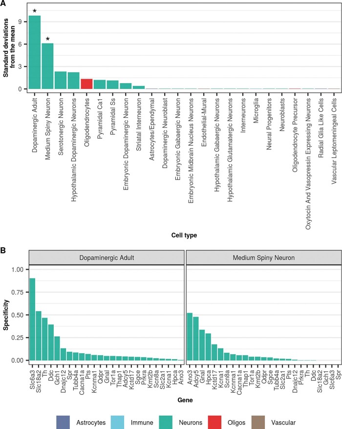
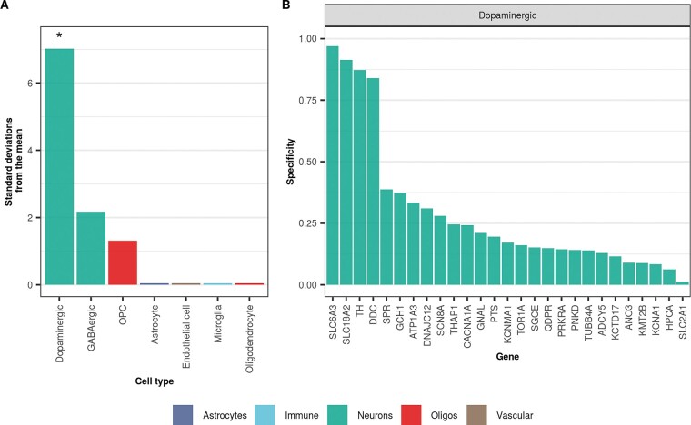
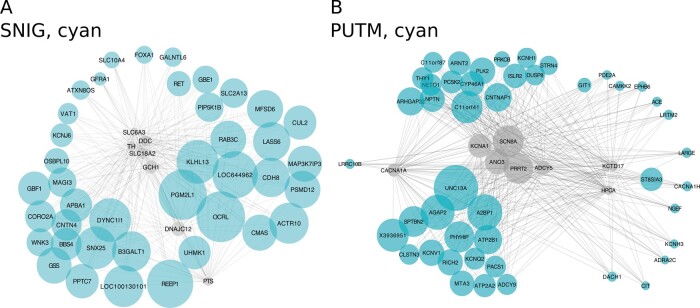
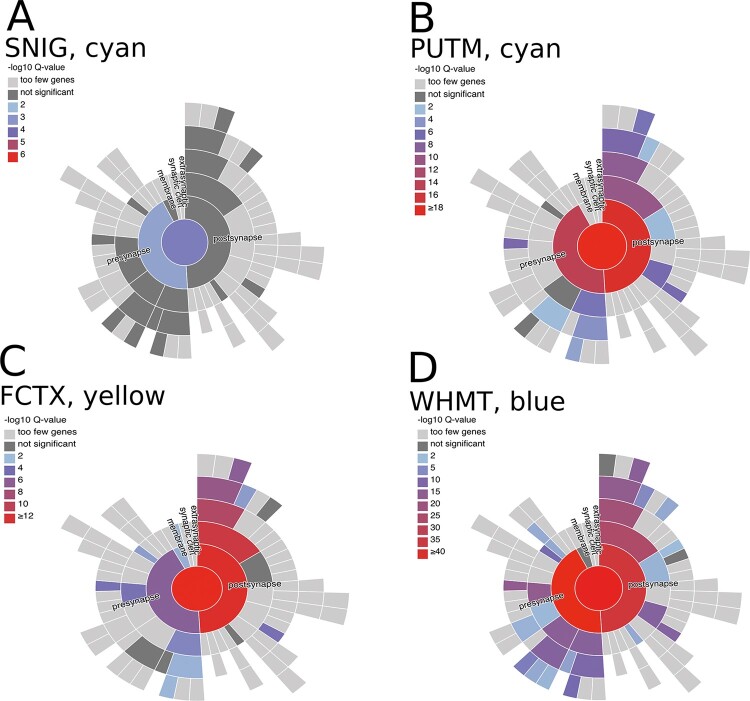
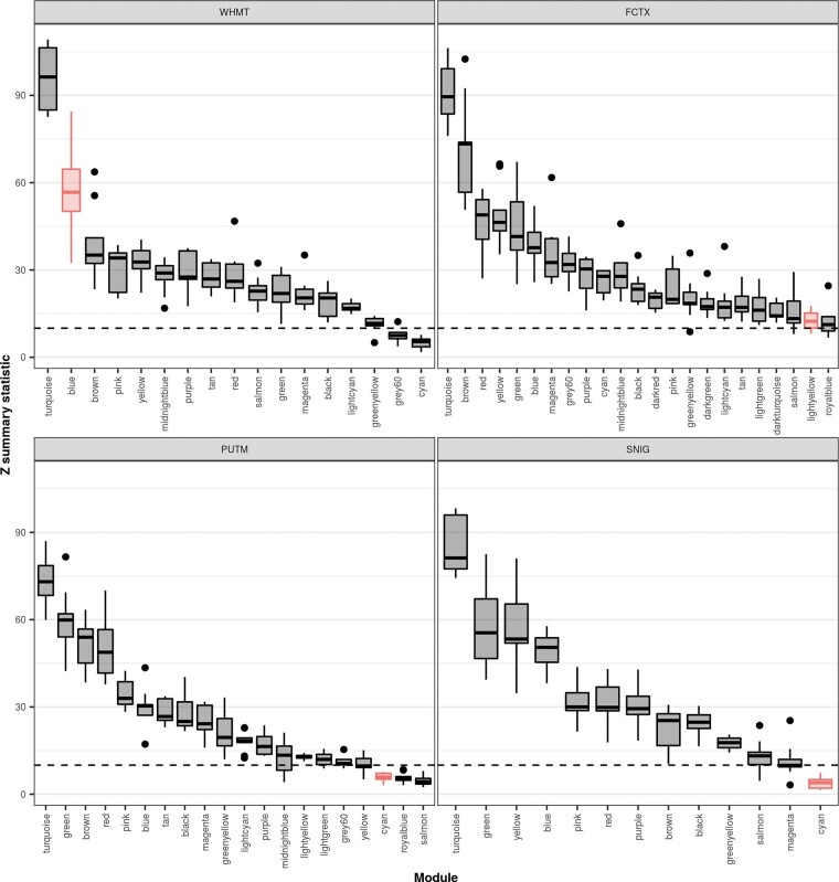
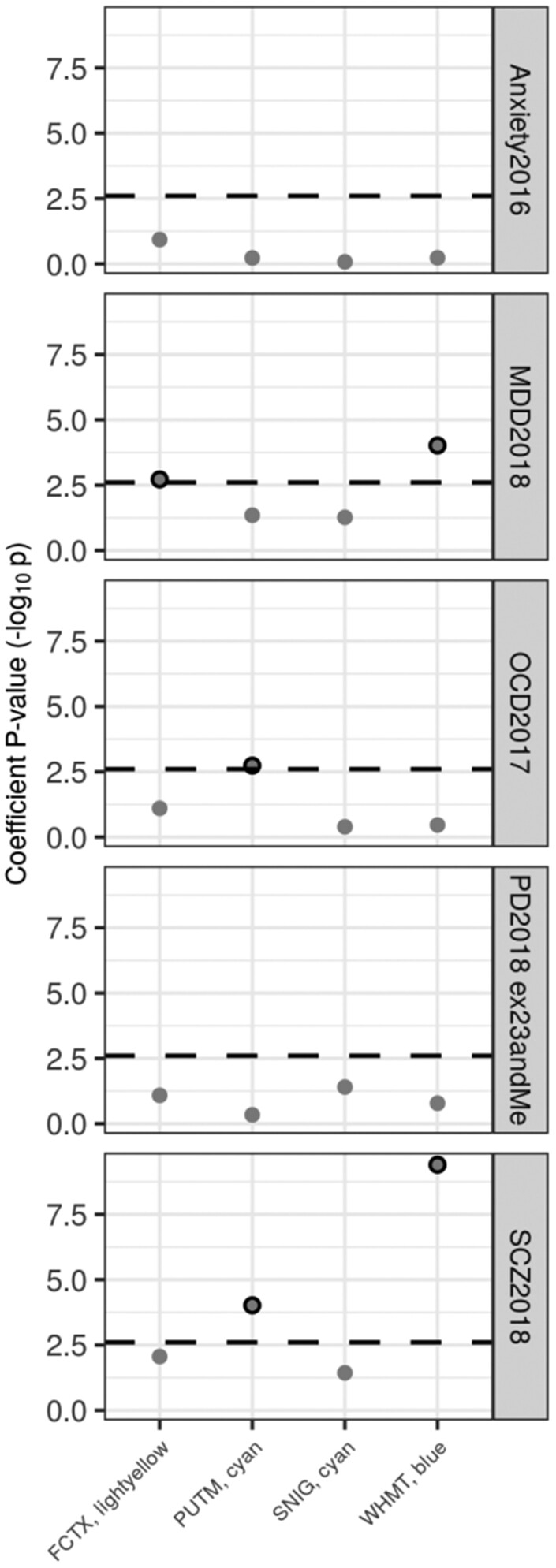
Dystonia is a neurological disorder characterized by sustained or intermittent muscle contractions causing abnormal movements and postures, often occurring in absence of any structural brain abnormality. Psychiatric comorbidities, including anxiety, depression, obsessive-compulsive disorder and schizophrenia, are frequent in patients with dystonia. While mutations in a fast-growing number of genes have been linked to Mendelian forms of dystonia, the cellular, anatomical, and molecular basis remains unknown for most genetic forms of dystonia, as does its genetic and biological relationship to neuropsychiatric disorders. Here we applied an unbiased systems-biology approach to explore the cellular specificity of all currently known dystonia-associated genes, predict their functional relationships, and test whether dystonia and neuropsychiatric disorders share a genetic relationship. To determine the cellular specificity of dystonia-associated genes in the brain, single-nuclear transcriptomic data derived from mouse brain was used together with expression-weighted cell-type enrichment. To identify functional relationships among dystonia-associated genes, we determined the enrichment of these genes in co-expression networks constructed from 10 human brain regions. Stratified linkage-disequilibrium score regression was used to test whether co-expression modules enriched for dystonia-associated genes significantly contribute to the heritability of anxiety, major depressive disorder, obsessive-compulsive disorder, schizophrenia, and Parkinson's disease. Dystonia-associated genes were significantly enriched in adult nigral dopaminergic neurons and striatal medium spiny neurons. Furthermore, 4 of 220 gene co-expression modules tested were significantly enriched for the dystonia-associated genes. The identified modules were derived from the substantia nigra, putamen, frontal cortex, and white matter, and were all significantly enriched for genes associated with synaptic function. Finally, we demonstrate significant enrichments of the heritability of major depressive disorder, obsessive-compulsive disorder and schizophrenia within the putamen and white matter modules, and a significant enrichment of the heritability of Parkinson's disease within the substantia nigra module. In conclusion, multiple dystonia-associated genes interact and contribute to pathogenesis likely through dysregulation of synaptic signalling in striatal medium spiny neurons, adult nigral dopaminergic neurons and frontal cortical neurons. Furthermore, the enrichment of the heritability of psychiatric disorders in the co-expression modules enriched for dystonia-associated genes indicates that psychiatric symptoms associated with dystonia are likely to be intrinsic to its pathophysiology.

Keywords: dystonia; medium-spiny neurons; network analysis; synaptic transmission; transcriptomic analysis.


Figures

Figure 1
Figure 1
Dystonia-associated genes are highly expressed in dopaminergic neurons and MSNs from mouse. (A) Enrichment of dystonia-associated genes in level 1 cell types from the Karolinska superset was determined using EWCE. Standard deviations from the mean indicate the distance of the mean expression of the target list from the mean expression of the bootstrap replicates. Asterisks denote significance at P < 0.05 after correcting for multiple testing with the Benjamini-Hochberg method over all level 1 cell types. Numerical results are reported in Supplementary Table 1. (B) Plot of specificity values for all dystonia-associated genes within adult nigral dopaminergic neurons and MSNs (level 1 cell types from the Karolinska single-cell RNA-sequencing superset). Specificity values were derived from Skene et al. (2018) who calculated specificity by dividing the mean expression of a gene in one cell type by the mean expression in all cell types. In other words, specificity is the proportion of a gene’s total expression attributable to one cell type, with a value of 0 meaning a gene is not expressed in that cell type and a value of 1 meaning that a gene is only expressed in that cell type. In both plots, cell types are coloured by the overall class they belong to (e.g. astrocyte, neuron, oligodendrocyte, etc.).
Figure 2
Figure 2
Dystonia-associated genes are highly expressed in human dopaminergic neurons. (A) Enrichment of dystonia-associated genes in level 1 cell types from human-derived substantia nigra single-nuclei RNA-sequencing data was determined using EWCE. Standard deviations from the mean indicate the distance of the mean expression of the target list from the mean expression of the bootstrap replicates. Asterisks denote significance at P < 0.05 after correcting for multiple testing with the Benjamini-Hochberg method over all level 1 cell types. Numerical results are reported in Supplementary Table 2. (B) Plot of specificity values for all dystonia-associated genes within dopaminergic neurons (level 1 cell type from human-derived substantia nigra single-nuclei RNA-sequencing data). Specificity values represent the proportion of a gene’s total expression attributable to one cell type, with a value of 0 meaning a gene is not expressed in that cell type and a value of 1 meaning that a gene is only expressed in that cell type. In both plots, cell types are coloured by the overall class they belong to (e.g. astrocyte, neuron, oligodendrocyte, etc.).
Figure 3
Figure 3
Gene co-expression modules enriched for dystonia genes in the substantia nigra and putamen. The substantia nigra (SNIG) ‘cyan’ (A) and putamen (PUTM) ‘cyan’ (B) dystonia-linked UKBEC modules visualized using ‘bottom-up’ plots. In each case, the dystonia genes within the modules are depicted as grey nodes with their most connected genes, as determined by the Topology Overlap Measure, depicted in cyan (to a maximum of seven genes per seed). Node size reflects connectivity. Plots were generated using Cytoscape 3.5.1 and the Edge-weighted Spring Embedded layout algorithm was used for rendering to a 2D canvas.
Figure 4
Figure 4
Variable enrichment of genes associated with presynaptic and postsynaptic structures within dystonia-linked UKBEC co-expression modules. Visualizations of the enrichment of SynGO ontology terms for synaptic location within the dystonia-linked UKBEC modules (the substantia nigra ‘cyan’, putamen ‘cyan’, frontal cortex ‘lightyellow’ and white matter ‘blue’ modules) provided by the SynGO web resource (https://www.syngoportal.org/index.html). The hierarchical structure of SynGO terms is represented by concentric rings with the most specific terms placed peripherally. The colour coding of the terms is based on the enrichment q-values. FCTX = frontal cortex; PUTM = putamen; SNIG = substantia nigra; WHMT = white matter.
Figure 5
Figure 5
Module preservation within UKBEC tissues containing dystonia-linked modules. Plot of module preservation for each module within UKBEC tissues containing dystonia-linked modules. Module preservation, as denoted by the Z summary statistic, was determined using a WGCNA-based preservation analysis applied across all 10 UKBEC co-expression networks. Thus, each box plot represents the preservation of the named module within the labelled tissue across the remaining nine tissues. The black dashed lines indicate the threshold for strong evidence of module preservation (Z summary statistic > 10), as defined by Langfelder et al. (2011). Modules enriched with dystonia genes are highlighted in red.
Figure 6
Figure 6
Enrichment of disease heritability within dystonia-linked UKBEC co-expression modules. Stratified LDSC using UKBEC co-expression modules. The black dashed lines indicate the cut-off for Bonferroni significance [P < 0.05 / (4 × 5)]. Bonferroni-significant results are marked with black borders. Numerical results are reported in Supplementary Table 6. FCTX = frontal cortex; MDD = major depressive disorder; OCD = obsessive compulsive disorder; PD = Parkinson’s disease; PUTM = putamen; SCZ = schizophrenia; SNIG = substantia nigra; WHMT = white matter.

Comment in

References

    1. Abecasis GR, Auton A, Brooks LD, DePristo MA, Durbin RM, Handsaker RE, et al. An integrated map of genetic variation from 1,092 human genomes. Nature 2012; 491: 56–65. - PMC - PubMed
    1. Albanese A, Bhatia K, Bressman SB, Delong MR, Fahn S, Fung VS, et al. Phenomenology and classification of dystonia: a consensus update. Mov Disord 2013; 28: 863–73. - PMC - PubMed
    1. Altshuler DM, Gibbs RA, Peltonen L, Altshuler DM, Gibbs RA, Peltonen L, et al. Integrating common and rare genetic variation in diverse human populations. Nature 2010; 467: 52–8. - PMC - PubMed
    1. Anikster Y, Haack TB, Vilboux T, Pode-Shakked B, Thony B, Shen N, et al. Biallelic mutations in DNAJC12 cause hyperphenylalaninemia, dystonia, and intellectual disability. Am J Hum Genet 2017; 100: 257–66. - PMC - PubMed
    1. Atasu B, Hanagasi H, Bilgic B, Pak M, Erginel-Unaltuna N, Hauser AK, et al. HPCA confirmed as a genetic cause of DYT2-like dystonia phenotype. Mov Disord 2018; 33: 1354–8. - PubMed

Publication types

MeSH terms