Review
. 2020 Sep 5;19(1):134.
doi: 10.1186/s12933-020-01109-1.

Type 2 diabetes and reduced exercise tolerance: a review of the literature through an integrated physiology approach

Affiliations
Review

Type 2 diabetes and reduced exercise tolerance: a review of the literature through an integrated physiology approach

Lorenzo Nesti et al. Cardiovasc Diabetol. .

Abstract

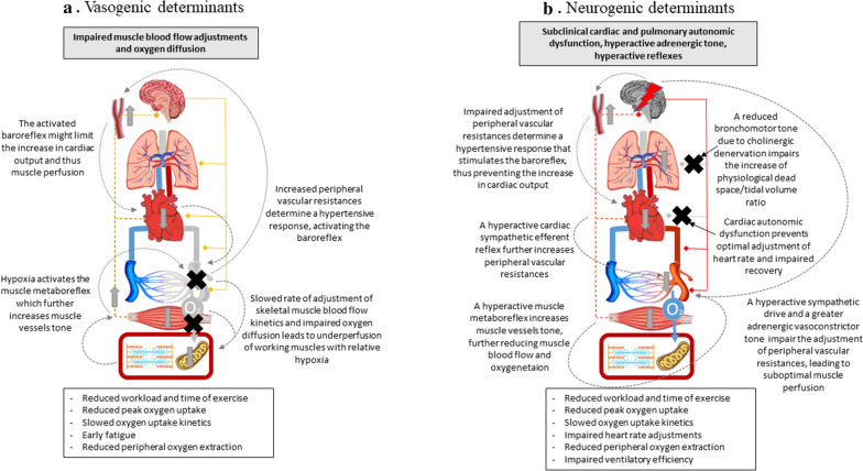
The association between type 2 diabetes mellitus (T2DM) and heart failure (HF) is well established. Early in the course of the diabetic disease, some degree of impaired exercise capacity (a powerful marker of health status with prognostic value) can be frequently highlighted in otherwise asymptomatic T2DM subjects. However, the literature is quite heterogeneous, and the underlying pathophysiologic mechanisms are far from clear. Imaging-cardiopulmonary exercise testing (CPET) is a non-invasive, provocative test providing a multi-variable assessment of pulmonary, cardiovascular, muscular, and cellular oxidative systems during exercise, capable of offering unique integrated pathophysiological information. With this review we aimed at defying the cardiorespiratory alterations revealed through imaging-CPET that appear specific of T2DM subjects without overt cardiovascular or pulmonary disease. In synthesis, there is compelling evidence indicating a reduction of peak workload, peak oxygen assumption, oxygen pulse, as well as ventilatory efficiency. On the contrary, evidence remains inconclusive about reduced peripheral oxygen extraction, impaired heart rate adjustment, and lower anaerobic threshold, compared to non-diabetic subjects. Based on the multiparametric evaluation provided by imaging-CPET, a dissection and a hierarchy of the underlying mechanisms can be obtained. Here we propose four possible integrated pathophysiological mechanisms, namely myocardiogenic, myogenic, vasculogenic and neurogenic. While each hypothesis alone can potentially explain the majority of the CPET alterations observed, seemingly different combinations exist in any given subject. Finally, a discussion on the effects -and on the physiological mechanisms-of physical activity and exercise training on oxygen uptake in T2DM subjects is also offered. The understanding of the early alterations in the cardiopulmonary response that are specific of T2DM would allow the early identification of those at a higher risk of developing HF and possibly help to understand the pathophysiological link between T2DM and HF.

Keywords: Aerobic capacity; Autonomic dysfunction; Cardiopulmonary exercise test; Diabetic cardiomyopathy; Diabetic complications; Diabetic lung; Diabetic myopathy; Exercise physiology; Exercise tolerance; Exercise training; Physical exercise; Type 2 diabetes mellitus.


Conflict of interest statement

The authors have no conflict of interest to declare.

Figures

Fig. 1
Fig. 1
Myocardiogenic and skeletal myogenic determinants of reduced exercise tolerance in type 2 diabetes. Schematic representation of two pathophysiological hypotheses explaining the alterations observed in type 2 diabetes mellitus through cardiopulmonary exercise testing. Central and peripheral (efferent fibers to lungs, heart, conduit vessels, muscle arterioles are represented on the right, while afferent fibers of muscle metaboreflex and cardiac sympathetic reflex are represented on the left of the picture) nervous system, the baroreceptor, lungs, heart, conduit vessels, muscular vasculature, oxygen diffusion from capillaries to muscle, skeletal muscle and muscle cells are schematically represented. In each hypothesis, the organ/system that is primarily impaired is colored in grey and is marked with a black cross, whilst the other organs maintain their colors. Secondary involvement of other organs/systems is indicated with vertical grey arrows indicating stimulation (arrow facing up) or inhibition (arrow facing down) through known physiological mechanisms, which are represented as grey dotted lines with captions. On top of the figure, the grey text panel explains the primary involvement of the organ/system identified with the black cross and grey shadowing; the text panel beneath the figure shows a list of the altered cardiopulmonary exercise test variables observed in type 2 diabetes mellitus that may be explained by the pathophysiological hypothesis here shown. a Myocardiogenic determinants: an insufficient cardiac output adjustment reduces muscle perfusion, force production, and thus exercise tolerance. b Skeletal myogenic determinants: reduced skeletal muscle aerobic capacity or early fatigue can account for the reduced oxygen uptake, peripheral oxygen extraction, whilst a reduced cardiac output may be secondary to an enhanced baroreflex activation (see main text for full description)
Fig. 2
Fig. 2
Vasogenic and neurogenic determinants of reduced exercise tolerance in type 2 diabetes. Schematic representation of two pathophysiological hypotheses explaining the alterations observed in type 2 diabetes mellitus through cardiopulmonary exercise testing. Central and peripheral (efferent fibers to lungs, heart, conduit vessels, muscle arterioles are represented on the right, while afferent fibers of muscle metaboreflex and cardiac sympathetic reflex are represented on the left of the picture) nervous system, the baroreceptor, lungs, heart, conduit vessels, muscular vasculature, oxygen diffusion from capillaries to muscle, skeletal muscle and muscle cells are schematically represented. In each hypothesis, the organ/system that is primarily impaired is colored in grey and is marked with a black cross, whilst the ones that are hyperactivated are marked with a red bolt and a red shadowing. Unaffected systems maintain their colors. Secondary involvement of other organs/systems is indicated with vertical grey arrows indicating stimulation (arrow facing up) or inhibition (arrow facing down) through known physiological mechanisms, which are represented as grey dotted lines with captions. On top of the figure, the grey text panel explains the primary involvement of the organ/system identified with the black cross and grey shadowing or the red bolt and red shadowing; the text panel beneath the figure shows a list of the altered cardiopulmonary exercise test variables observed in type 2 diabetes mellitus that may be explained by the pathophysiological hypothesis here shown. a Vasogenic determinants: impaired modulation of peripheral vascular resistances reduces muscle blood flow and oxygen diffusion, thus negatively affecting skeletal muscle performance, peripheral oxygen extraction and cardiac output through enhanced baroreflex and muscle metaboreflex activation. b Neurogenic determinants: altered autonomic tone and hyperactive muscle metaboreflex and cardiac sympathetic efferent reflex might account for impaired cardiac output, suboptimal vascular and pulmonary adjustments during exercise, eventually leading to reduced muscle perfusion and oxygen extraction (see main text for full description)
Fig. 3
Fig. 3
Proposed intervention trials. Four proposals for pharmacological interventions that could help address the relative importance of individual organ contributions to exercise intolerance in type 2 diabetes are here schematized. Grey vertical arrows indicate either enhancement (arrows facing up) or inhibition (arrows facing down) by the pharmacological treatment on the target organ. The red lines represent diabetic subjects and the blue ones the controls. The cardiopulmonary exercise test variables that investigate the organs implicated (schematized on top of each graphic) are qualitatively showed in the graphic on the left side of each subsection, while on the right there is the effect that we could expect if the organ schematized is the principal responsible for the impairment in the variable outlined (the gap between diabetic and non-diabetic patients is reduced or abolished). The Asterix (*) indicates the difference between these two populations, based on literature review. a The use of positive inotropes -like dobutamine- could confirm the weight of a reduced stroke work on oxygen uptake per heartbeat if a normalization is observed. b Muscle perfusion enhancers -like dipyridamole- could highlight the contribution of skeletal muscle vasculature adjustments by ameliorating oxygen uptake. c By reducing a hyperactive sympathetic restrain, sympathetic inhibitors might ameliorate cardiovascular adjustments and thus oxygen assumption, confirming the role of autonomic imbalance in exercise intolerance. d Anticholinergic bronchodilators could normalize “ventilatory efficiency” by facilitating the reduction of physiological dead space if the latter is due to bronchial cholinergic hyperactivation. VO2, oxygen uptake, VO2/HR, oxygen pulse; VE, ventilation

References

    1. Lehrke M, Marx N. Diabetes mellitus and heart failure. Am J Cardiol. 2017;120(1S):S37–S47. - PubMed
    1. Mancini DM, Eisen H, Kussmaul W, Mull R, Edmunds LH, Jr, Wilson JR. Value of peak exercise oxygen consumption for optimal timing of cardiac transplantation in ambulatory patients with heart failure. Circulation. 1991;83(3):778–786. - PubMed
    1. Blair SN, Kohl HW, Paffenbarger RS, Clark DG, Cooper KH, Gibbons LW. Physical fitness and all-cause mortality. A prospective study of healthy men and women. JAMA. 1989;262(17):2395–2401. - PubMed
    1. Nauman J, Nes BM, Lavie CJ, Jackson AS, Sui X, Coombes JS, Blair SN, Wisloff U. Prediction of cardiovascular mortality by estimated cardiorespiratory fitness independent of traditional risk factors: the HUNT study. Mayo Clin Proc. 2017;92(2):218–227. - PubMed
    1. Look ARG, Wing RR, Bolin P, Brancati FL, Bray GA, Clark JM, Coday M, Crow RS, Curtis JM, Egan CM, et al. Cardiovascular effects of intensive lifestyle intervention in type 2 diabetes. N Engl J Med. 2013;369(2):145–154. - PMC - PubMed

MeSH terms