. 2020 Sep 5;11(9):722.
doi: 10.1038/s41419-020-02920-0.

Membrane-tethering of cytochrome c accelerates regulated cell death in yeast

Affiliations

Membrane-tethering of cytochrome c accelerates regulated cell death in yeast

Alexandra Toth et al. Cell Death Dis. .

Abstract

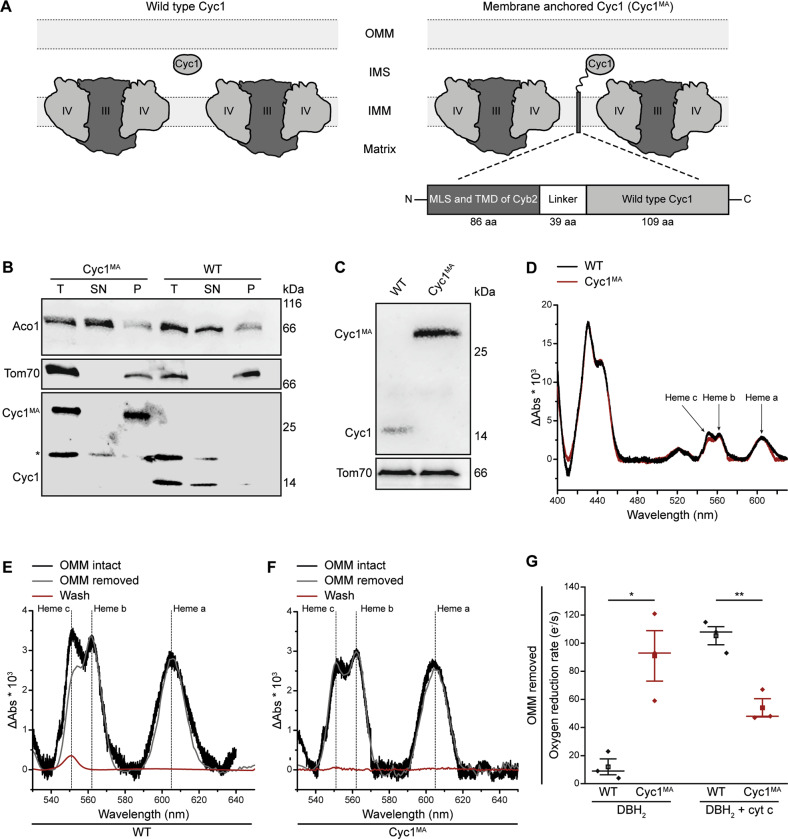
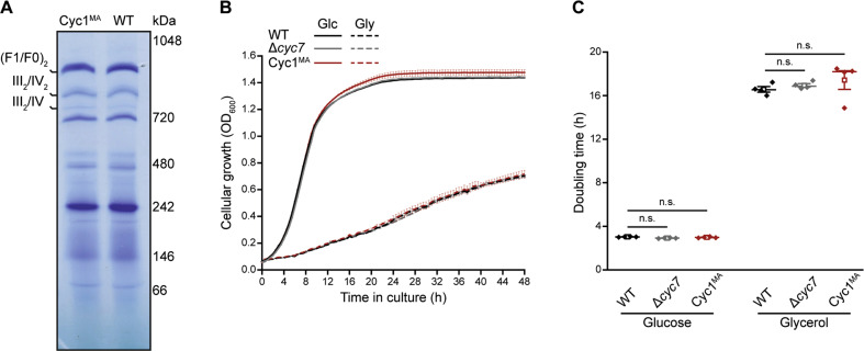
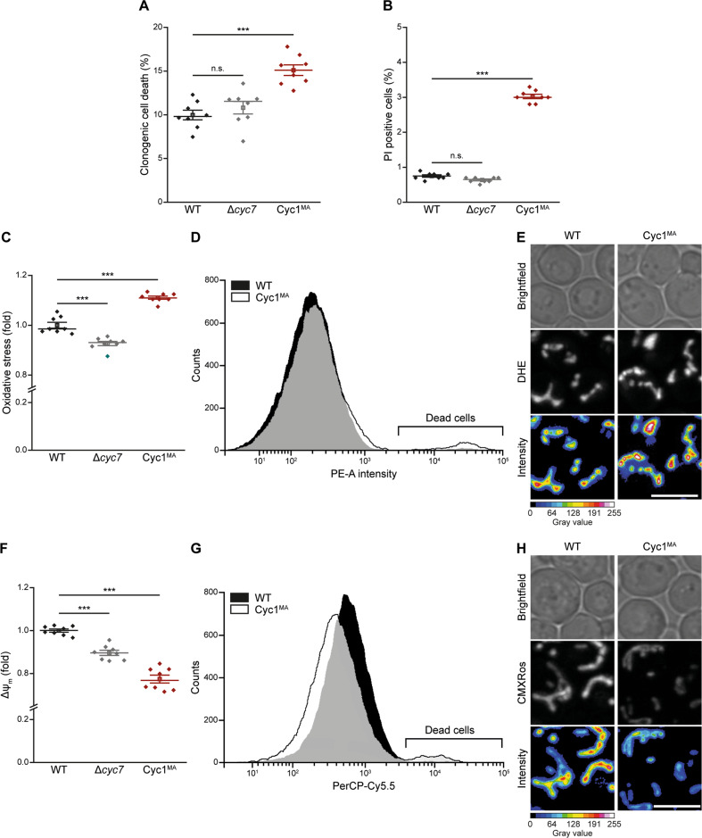
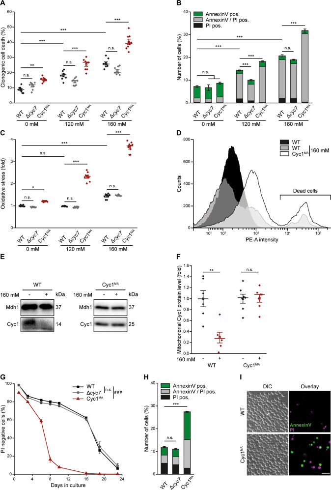
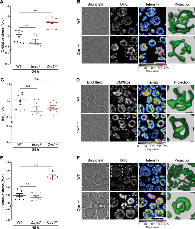
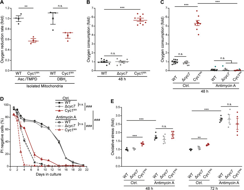
Intrinsic apoptosis as a modality of regulated cell death is intimately linked to permeabilization of the outer mitochondrial membrane and subsequent release of the protein cytochrome c into the cytosol, where it can participate in caspase activation via apoptosome formation. Interestingly, cytochrome c release is an ancient feature of regulated cell death even in unicellular eukaryotes that do not contain an apoptosome. Therefore, it was speculated that cytochrome c release might have an additional, more fundamental role for cell death signalling, because its absence from mitochondria disrupts oxidative phosphorylation. Here, we permanently anchored cytochrome c with a transmembrane segment to the inner mitochondrial membrane of the yeast Saccharomyces cerevisiae, thereby inhibiting its release from mitochondria during regulated cell death. This cytochrome c retains respiratory growth and correct assembly of mitochondrial respiratory chain supercomplexes. However, membrane anchoring leads to a sensitisation to acetic acid-induced cell death and increased oxidative stress, a compensatory elevation of cellular oxygen-consumption in aged cells and a decreased chronological lifespan. We therefore conclude that loss of cytochrome c from mitochondria during regulated cell death and the subsequent disruption of oxidative phosphorylation is not required for efficient execution of cell death in yeast, and that mobility of cytochrome c within the mitochondrial intermembrane space confers a fitness advantage that overcomes a potential role in regulated cell death signalling in the absence of an apoptosome.


Conflict of interest statement

The authors declare that they have no conflict of interest.

Figures

Fig. 1
Fig. 1. Generation of a yeast model with membrane-anchored cytochrome c.
a Scheme of respiratory supercomplexes in yeast, with wild type cytochrome c (left panel) and genetically engineered membrane-anchored cytochrome c (Cyc1MA; right panel). For construction of Cyc1MA, the mitochondrial localization sequence (MLS) and the transmembrane domain (TMD) of cytochrome b2 (Cyb2) from Saccharomyces cerevisiae was chromosomally integrated at the N-terminus of the CYC1 gene together with the linker sequence of Rhodobacter sphaeroides cytochrome cγ. b Immunoblot analysis of submitochondrial fractionation with lysates from isolated mitochondria of wild type (WT) and Cyc1MA cells. Total lysates (T) were separated via ultracentrifugation and the supernatant (SN) and pellet (P) fractions were applied for immunoblotting. Blots were probed with antibodies against aconitase (Aco1), Cyc1 and Tom70. The asterisk indicates a cross reaction of the Cyc1 antibody. c Immunoblot analysis of isolated mitochondria from WT and Cyc1MA strains. Blots were probed with antibodies against Cyc1 and the outer mitochondrial membrane protein Tom70. df Reduced-minus-oxidized difference spectra of WT and Cyc1MA strains. Spectra were normalized to the maxima of the α band at 605 nm. d Direct comparison of spectra from WT and Cyc1MA strains. e Spectra from WT mitochondria before (black) and after (grey) removal of the outer mitochondrial membrane (OMM), as well as from respective wash fraction (red). f Spectra from Cyc1MA mitochondria before (black) and after (grey) removal of the outer mitochondrial membrane (OMM), as well as from respective wash fraction (red). g Measurement of decylubiquinol (DBH2)-driven coupled bc1 complex and cytochrome c oxidase activity in isolated mitochondria devoid of the outer mitochondrial membrane (OMM) of WT as well as CYC7 deletion strains harbouring a membrane-anchored form of Cyc1 (Cyc1MA). Where indicated, yeast cytochrome c (cyt c) was added. The oxygen reduction rate was calculated as electrons per second (e/s) per cytochrome c oxidase. Mean (square) ± s.e.m., median (centre line) and single data points (n = 3) are depicted. *P < 0.05; **P < 0.01.
Fig. 2
Fig. 2. Characterization of yeast cells expressing membrane-anchored cytochrome c.
a Blue native electrophoresis of isolated mitochondria from WT and Cyc1MA strains. Gel was stained with Coomassie and respiratory supercomplexes are highlighted. b, c Cellular growth of strains described in a, analysed via automatic measurement of optical density (OD600) every 30 min with a Bioscreen CTM growth curve system either on glucose (Glc) or glycerol (Gly) as carbon source, respectively. Growth curves b as well as doubling time in exponential growth phase c are presented. Line graph in b shows mean ± s.e.m. For box plot in c, mean (square) ± s.e.m., median (centre line) and single data points (n = 4) are depicted. n.s. not significant.
Fig. 3
Fig. 3. Loss of cytochrome c mobility enhances cell death of exponentially growing yeast cells.
a Clonogenic death of wild type (WT), CYC7 deletion (Δcyc7), as well as CYC7 deletion strains harbouring a membrane-anchored form of Cyc1 (Cyc1MA), determined by counting colony-forming units after plating 500 cells of indicated strains on YEPD agar plates. b Flow cytometric quantification of loss of membrane integrity as indicated by propidium iodide (PI) staining of cells described in a. ce Analysis of oxidative stress, determined by the reactive oxygen species-driven conversion of non-fluorescent dihydroethidium (DHE) to fluorescent ethidium (Eth) of cells described in a. c Flow cytometric quantification of mean fluorescence intensities is shown as fold values of WT cells. d Dead cells, accumulating Eth due to a loss of membrane integrity, were excluded from the analysis. e Z-projections of representative confocal micrographs of DHE-stained cells are depicted. fh Quantification and visualization of mitochondrial transmembrane potential (Δψm) of cells described above. f Flow cytometrically quantified mean fluorescence intensities of Mitotracker CMXRos-stained cells are presented as fold values of WT cells. g Dead cells, accumulating the fluorescent dye due to a loss of membrane integrity were excluded from the analysis. h Z-projections of representative confocal micrographs of Mitotracker CMXRos-stained cells are depicted. Mean (square) ± s.e.m., median (centre line) and single data points (n = 8) are depicted. Data point indicated in turquoise was identified as an outlier by using the 2.2-fold interquartile range labelling rule. n.s. not significant, ***P < 0.001; scale bars represent 5 µm.
Fig. 4
Fig. 4. Reduced mobility of cytochrome c sensitizes to regulated cell death stimuli and reduces chronological lifespan of yeast.
a Clonogenic death of wild type (WT) and CYC7 deletion strains (Δcyc7), as well as CYC7 deletion strain harbouring a membrane-anchored form of Cyc1 (Cyc1MA) during exponential growth. Cells were treated with indicated concentrations of acetic acid for 1 h and colony-forming units were quantified after plating 500 cells of indicated strains on YEPD agar plates. b Flow cytometric quantification of AnnexinV/propidium iodide (PI) co-staining of cells described in a. c, d Analysis of oxidative stress, indicated by the reactive oxygen species-driven conversion of non-fluorescent dihydroethidium (DHE) to fluorescent ethidium (Eth) of cells described in a. c Flow cytometric quantification of mean fluorescence intensities is shown as fold values of WT cells. d Dead cells, accumulating Eth due to a loss of membrane integrity, were excluded from the analysis. e, f Immunoblot analysis of isolated mitochondria from WT and Cyc1MA strains. Cells were treated with 160 mM acetic acid for 1 h directly before isolation of mitochondria (+) or left untreated (−). Blots were probed with antibodies against Cyc1 and Mdh1 (mitochondrial malate dehydrogenase) as loading control. Relative levels were calculated after normalization to Mdh1 levels. Representative immunoblots (e) as well as densitometric quantification of mitochondrial Cyc1 levels (f) are shown. g Chronological lifespan of indicated strains, evaluated by the flow cytometric quantification of PI negative cells. h, i Quantification of cell death via AnnexinV/PI co-staining of cells as described in b after 48 h. h Flow cytometric quantification and i representative epifluorescence micrographs are presented. Bar charts in b, h and line graph in g show mean ± s.e.m. (n = 8). For box plots, mean (square) ± s.e.m., median (centre line) and single data points (a, c: n = 8; f: n = 6 for wild type and n = 7 for Cyc1MA) are depicted. Simple main effects are visualized as n.s. not significant, *P < 0.05, **P < 0.01 and ***P < 0.001; main effects are presented as n.s. not significant and ###P < 0.001 in g; scale bar indicates 10 µm.
Fig. 5
Fig. 5. Stationary yeast cells with membrane-tethered cytochrome c show altered mitochondrial morphology and increased oxidative stress.
a, b Analysis of oxidative stress, determined by the reactive oxygen species-driven conversion of non-fluorescent dihydroethidium (DHE) to fluorescent ethidium (Eth) of wild type (WT) and CYC7 deletion strains (Δcyc7), as well as CYC7 deletion strain harbouring a membrane-anchored form of Cyc1 (Cyc1MA) after 24 h. a Flow cytometric quantification of mean fluorescence intensities is shown as fold values of WT cells. Dead cells, accumulating Eth due to a loss of membrane integrity, were excluded from the analysis. b Z-projections of representative confocal micrographs of DHE-stained cells are depicted. c, d Determination of mitochondrial transmembrane potential (Δψm) via Mitotracker CMXRos staining of cells as described above. c Flow cytometrically quantified mean fluorescence intensity is presented as fold values of WT cells. Dead cells, accumulating the fluorescent dye, due to a loss of membrane integrity, were excluded from the analysis. d Z-projections of representative confocal micrographs of Mitotracker CMXRos-stained cells are depicted. For analysis of Δψm after 48 h, please see Supplementary Fig. 1. e, f Evaluation of oxidative stress as described in a after 48 h in culture. e Flow cytometric quantification and f representative confocal micrographs are shown. Mean (square) ± s.e.m., median (centre line) and single data points (n = 8) are depicted. n.s. not significant, ***P < 0.001; scale bars indicate 5 µm.
Fig. 6
Fig. 6. Reduced mobility of cytochrome c decreases respiratory chain activity and induces compensatory upregulation of respiration.
a Measurement of Ascorbate (Asc) and N,N,N′,N′-tetremethyl-p-phenylenediamine hydrochloride (TMPD)-driven cytochrome c oxidase (COX) activity and decylubiquinol (DBH2)-driven coupled bc1 complex and COX activity in isolated mitochondria of logarithmically growing wild type (WT) cells as well as the CYC7 deletion strain harbouring a membrane-anchored form of Cyc1 (Cyc1MA). Activity was calculated as electron per second per COX and was normalized to WT cells in order to present the oxygen reduction rate as fold value. b Cellular oxygen consumption of WT, CYC7 deletion (Δcyc7) and Cyc1MA strains after 48 h. Measured oxygen consumption was normalized to living cells and subsequently presented as fold of WT cells. c Measurement of cellular oxygen consumption as described in b. Cells were treated after 24 h with 50 µM antimycin A (dissolved in ethanol) and with equivalent amounts of ethanol as a control (Ctrl.). Normalization was performed as described above. d Chronological lifespan of indicated strains, evaluated by the flow cytometric quantification of PI negative cells. Cells were treated with antimycin A as described in c. e Analysis of oxidative stress, determined by the reactive oxygen species-driven conversion of non-fluorescent dihydroethidium (DHE) to fluorescent ethidium (Eth) of cells treated with antimycin A and respective controls as described in c. Measurement was performed after 48 and 72 h. Mean (square) ± s.e.m., median (centre line) and single data points (n = 3 in a and n = 8 in b, c) are depicted for dot plots in (ac, e). Data points indicated in turquoise were identified as outliers by using the 2.2-fold interquartile range labelling rule. Line graph in d shows mean ± s.e.m. (n = 4). Simple main effects are visualized as n.s. not significant, **P < 0.01 and ***P < 0.001; Main effects are presented as n.s. not significant and ###P < 0.001.

Similar articles

Cited by

References

    1. Hüttemann M, et al. The multiple functions of cytochrome c and their regulation in life and death decisions of the mammalian cell: From respiration to apoptosis. Mitochondrion. 2011;11:369–381. - PMC - PubMed
    1. Ott M, Robertson JD, Gogvadze V, Zhivotovsky B, Orrenius S. Cytochrome c release from mitochondria proceeds by a two-step process. Proc. Natl Acad. Sci. USA. 2002;99:1259–1263. - PMC - PubMed
    1. Ott M, Zhivotovsky B, Orrenius S. Role of cardiolipin in cytochrome c release from mitochondria. Cell Death Differ. 2007;14:1243–1247. - PubMed
    1. Burke PV, Raitt DC, Allen LA, Kellogg EA, Poyton RO. Effects of oxygen concentration on the expression of cytochrome c and cytochrome c oxidase genes in yeast. J. Biol. Chem. 1997;272:14705–14712. - PubMed
    1. Galluzzi L, et al. Molecular mechanisms of cell death: recommendations of the Nomenclature Committee on Cell Death 2018. Cell Death Differ. 2018;25:486–541. - PMC - PubMed

Publication types

LinkOut - more resources