. 2020 Dec;46(7):707-721.
doi: 10.1111/nan.12659. Epub 2020 Sep 24.

Hippocampal subfield pathologic burden in Lewy body diseases vs. Alzheimer's disease

Affiliations

Hippocampal subfield pathologic burden in Lewy body diseases vs. Alzheimer's disease

D G Coughlin et al. Neuropathol Appl Neurobiol. 2020 Dec.

Abstract

Aims: Lewy body diseases (LBD) are characterized by alpha-synuclein (SYN) pathology, but comorbid Alzheimer's disease (AD) pathology is common and the relationship between these pathologies in microanatomic hippocampal subfields is understudied. Here we use digital histological methods to test the association between hippocampal SYN pathology and the distribution of tau and amyloid-beta (Aβ) pathology in LBD and contrast with AD subjects. We also correlate pathologic burden with antemortem episodic memory testing.

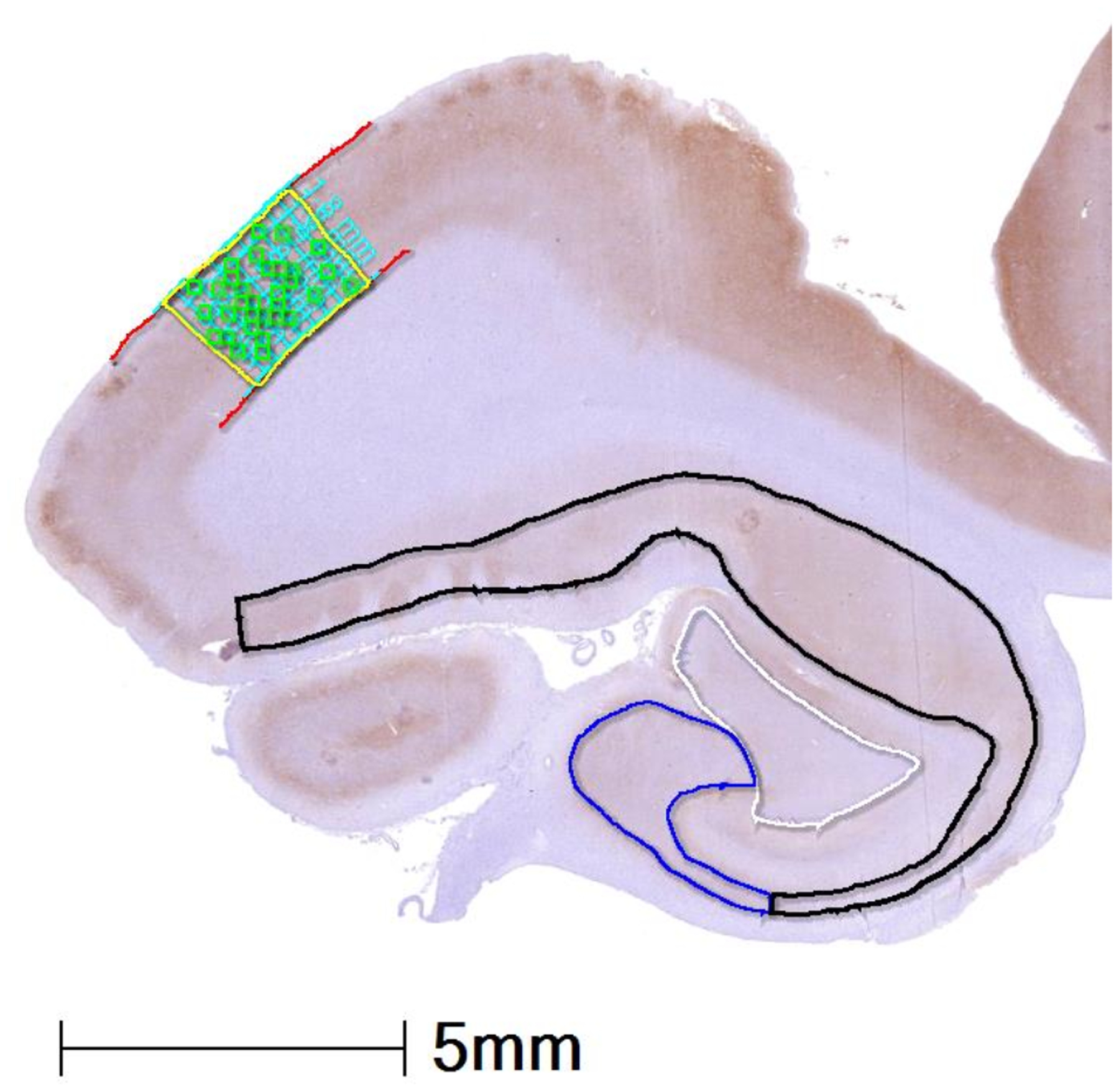
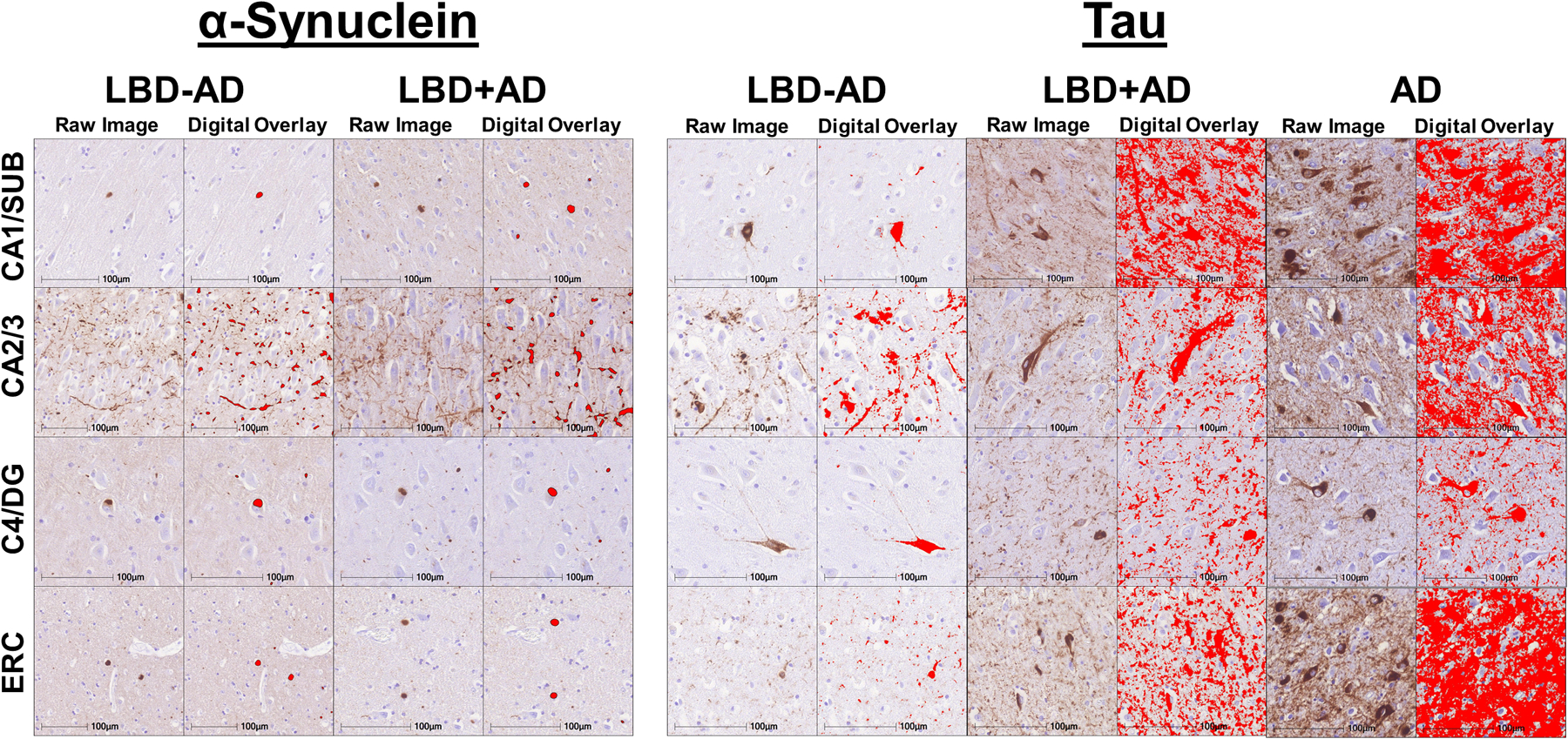
Methods: Hippocampal sections from 49 autopsy-confirmed LBD cases, 30 with no/low AD copathology (LBD - AD) and 19 with moderate/severe AD copathology (LBD + AD), and 30 AD patients were stained for SYN, tau, and Aβ. Sections underwent digital histological analysis of subfield pathological burden which was correlated with antemortem memory testing.

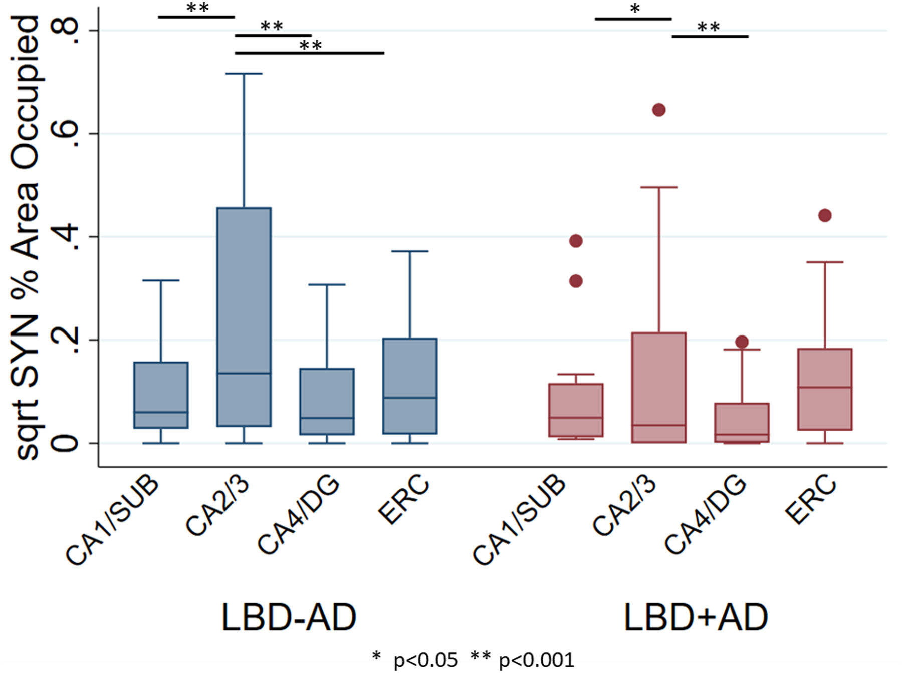
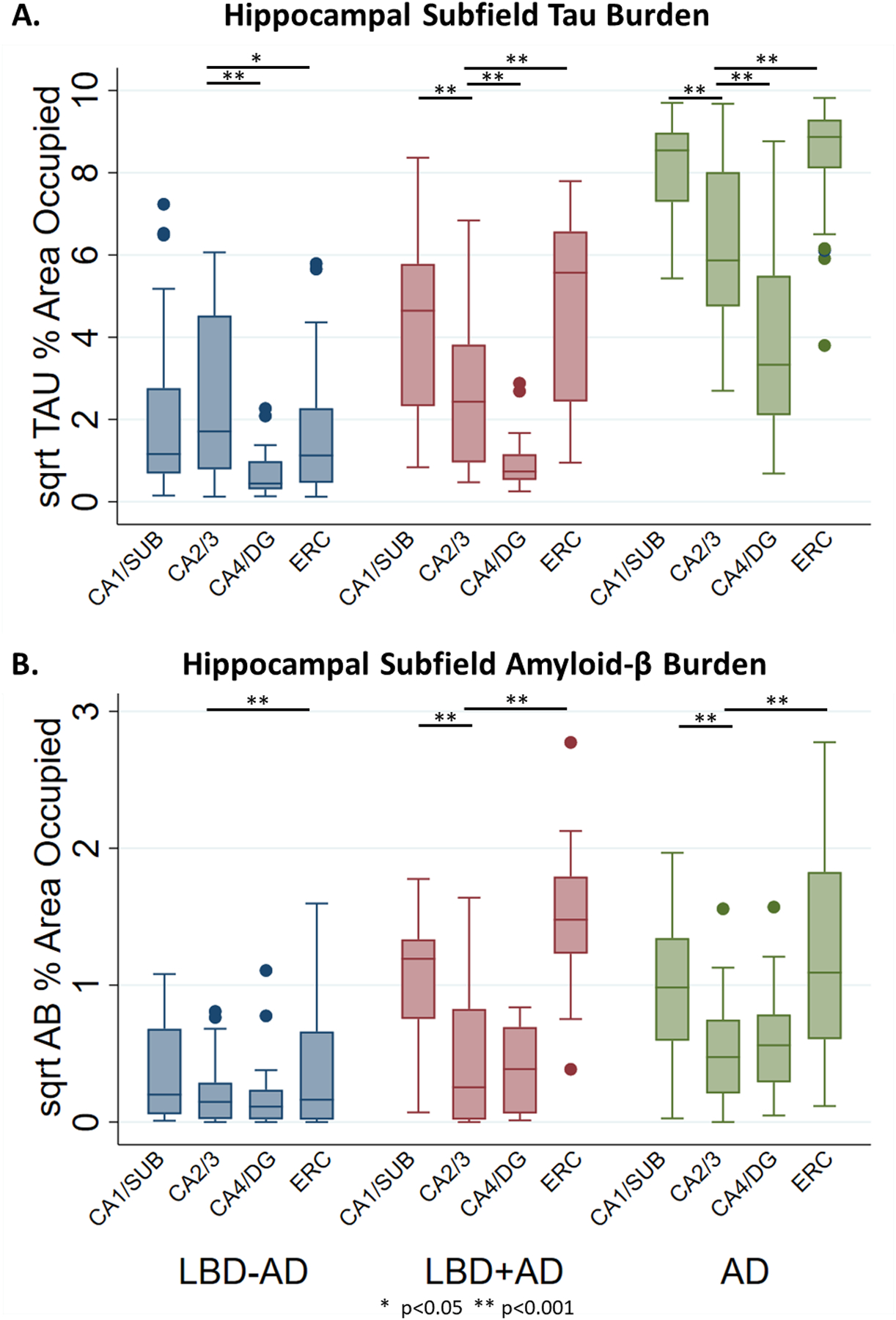
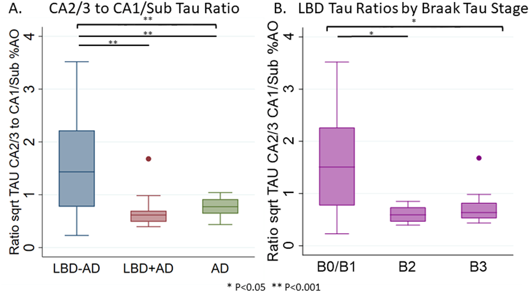
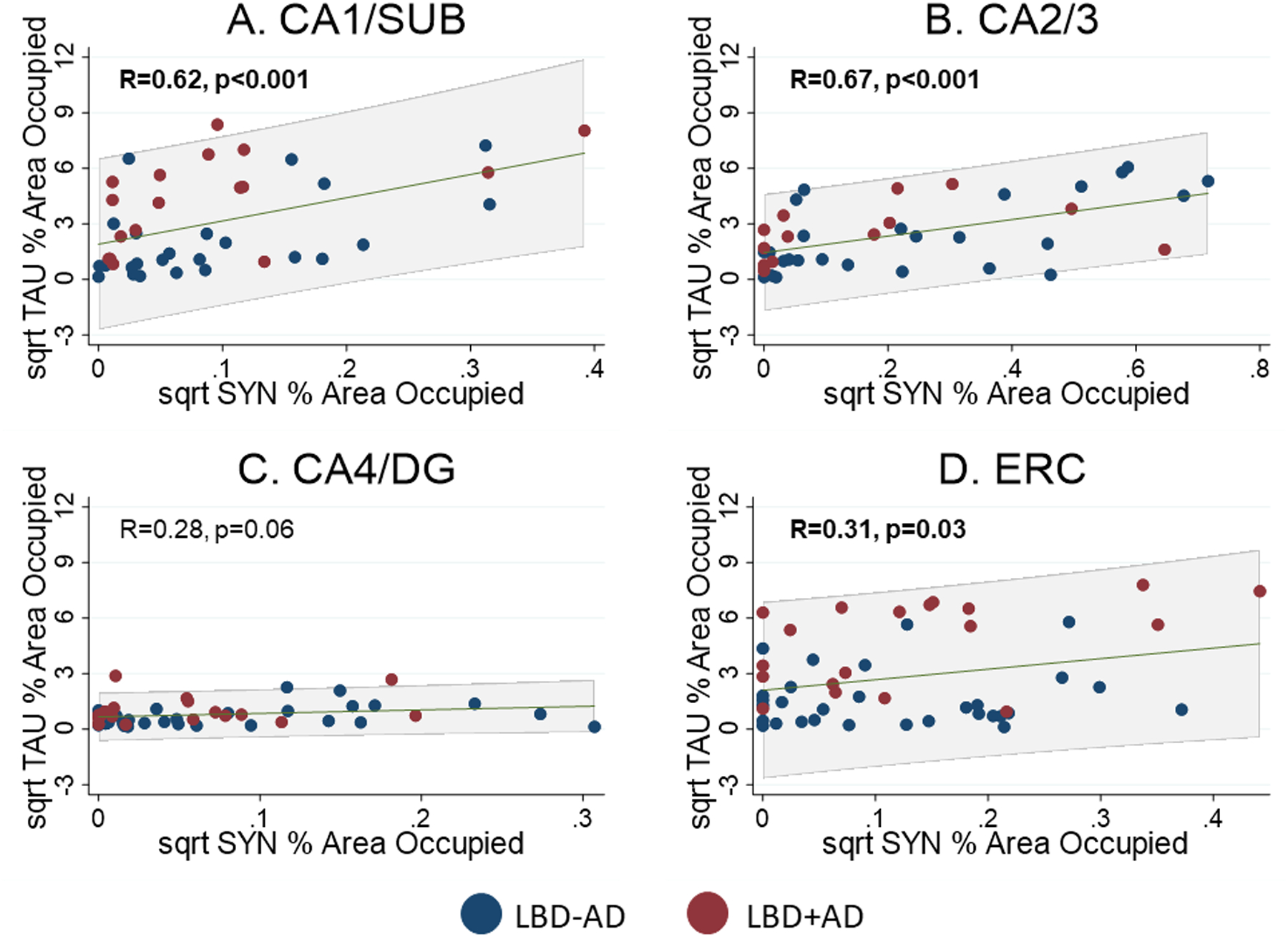
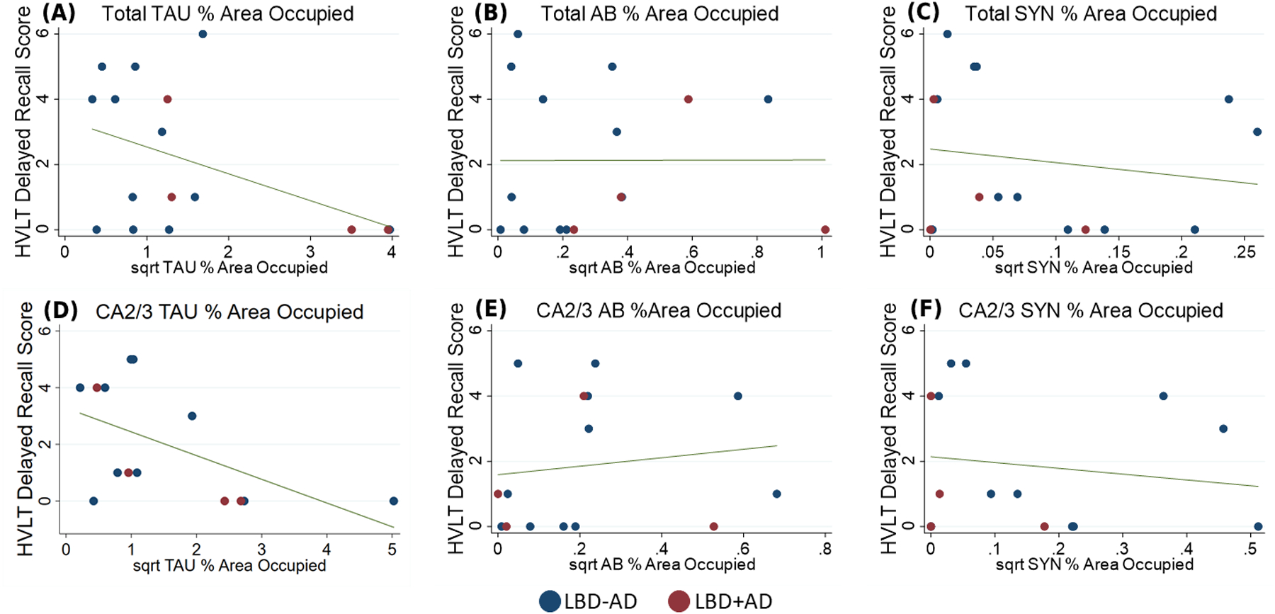
Results: LBD - AD and LBD + AD had similar severity and distribution of SYN pathology (P > 0.05), CA2/3 being the most affected subfield (P < 0.02). In LBD, SYN correlated with tau across subfields (R = 0.49, P < 0.001). Tau burden was higher in AD than LBD + AD (P < 0.001), CA1/subiculum and entorhinal cortex (ERC) being most affected regions (P = 0.04 to <0.01). However, tau pathology in LBD - AD was greatest in CA2/3, which was equivalent to LBD + AD. Aβ severity and distribution was similar between LBD + AD and AD. Total hippocampal tau and CA2/3 tau was inversely correlated with memory performance in LBD (R = -0.52, -0.69, P = 0.04, 0.009).

Conclusions: Our findings suggest that tau burden in hippocampal subfields may map closely with the distribution of SYN pathology in subfield CA2/3 in LBD diverging from traditional AD and contribute to episodic memory dysfunction in LBD.

Keywords: Alzheimer’s disease; Lewy body diseases; hippocampus; neuropathology; synuclein; tau.


Figures

Figure 1
Figure 1. Hippocampal Subfield Segmentation
Representative image of hippocampal anatomy with example of manually segmented hippocampal subfields from an LBD+AD case stained for tau (AT8). CA4/DG: white, CA2/3: blue, CA1/SUB: black, ERC: yellow with grey matter outlined in red, orthogonal transecting lines in blue, and random tiles which were averaged for %AO measurement in green. Abbreviations: CA1: cornu ammonis field 1, SUB: subiculum, CA2/3 cornu ammonis field 2 and 3, CA4: cornu ammonis field 4, DG: dentate gyrus, ERC: entorhinal cortex.
Figure 2
Figure 2. Digital Histology Detection Overlay
Representative photomicrographs in different hippocampal subfields in LBD-AD, LBD+AD, and AD cases stained for SYN (SYN303) and tau (AT8) The first column for each group shows raw images and the second column for each group depicts digital detection of pathology (%AO- red overlay).
Figure 3
Figure 3. Hippocampal Subfield Synuclein Burden in LBD-AD and LBD+AD
Box-plots depict median, interquartile range and range of square root of %AO of SYN pathology in hippocampal subfields of LBD-AD (blue) and LBD+AD (red) cases. Abbreviations: CA1/SUB: average cornu ammonis field 1 and subiculum, CA2/3 average cornu ammonis field 2 and 3, CA4/DG: average cornu ammonis field 4 and dentate gyrus, ERC: entorhinal cortex. Similar patterns of SYN deposition are observed across groups. * p<0.05, ** p<0.001 from linear mixed effects models comparing region.
Figure 4
Figure 4. Hippocampal Subfield Tau and Aβ Burden in LBD-AD, LBD+AD, and AD
Box-plots depict median, interquartile range and range of square root of %AO of pathology in hippocampal subfields of LBD-AD (blue), LBD+AD (red), and AD (green) cases for A) Tau %AO in LBD-AD, LBD+AD, and AD and B) Aβ %AO in LBD-AD, LBD+AD, and AD. Similar patterns of tau deposition are observed between LBD+AD and AD although AD consistently has higher degree of tau deposition. LBD-AD shows a pattern of tau deposition similar to SYN deposition. LBD+AD and AD have similar distribution and severity of Aβ pathology Abbreviations: CA1/SUB: average cornu ammonis field 1 and subiculum, CA2/3 average cornu ammonis field 2 and 3, CA4/DG: average cornu ammonis field 4 and dentate gyrus, ERC: entorhinal cortex. * p<0.05, ** p<0.001 from linear mixed effects models.
Figure 5
Figure 5. Ratio of CA2/3 Tau to CA1/SUB Tau
Box-plots depict median, interquartile range and range of the ratio of the square root of tau %AO in CA2/3 to CA1/SUB in A) LBD-AD (blue), LBD+AD (red), and AD (green) and B) in the entirety of the LBD cohort by Braak tau stage. Brackets indicate ANOVA differences across all groups, while lines indicate significant differences between groups. * p<0.05 ** p<0.001.
Figure 6
Figure 6. Regional Correlation between SYN and tau Burdens in LBD
Scatterplots showing relationship between square root SYN %AO and tau %AO in A) CA1/SUB, B) CA2/3, C) CA4/DG, and D) ERC. Blue points are from LBD-AD cases and Red points indicated LBD+AD cases. Linear prediction is shown in green with 95% confidence intervals shown in grey shaded region. Significant correlations were observed in CA1/SUB and CA2/3 and ERC. Abbreviations: CA1/SUB: average cornu ammonis field 1 and subiculum, CA2/3 average cornu ammonis field 2 and 3, CA4/DG: average cornu ammonis field 4 and dentate gyrus, ERC: entorhinal cortex.
Figure 7
Figure 7. HVLT Delayed Recall Scores and Tau, Aβ, and SYN %AO Burden
Hopkins Verbal Learning Task Delayed Recall Score plotted against a) square root total tau %AO and b) square root total Aβ %AO c) square root total SYN %AO d) square root CA2/3 tau %AO and e) square root CA2/3 Aβ %AO f) square root CA2/3 SYN %AO Blue: LBD-AD, Red: LBD+AD, Line: predicted linear fit.

Comment in

References

    1. Irwin DJ, Lee VM-Y, Trojanowski JQ. Parkinson’s disease dementia: convergence of [alpha]-synuclein, tau and amyloid-[beta] pathologies. Nat Rev Neurosci 2013; 14: 626–36 - PMC - PubMed
    1. Adamowicz DH, Roy S, Salmon DP, Galasko DR, Hansen LA, Masliah E, Gage FH. Hippocampal alpha-Synuclein in Dementia with Lewy Bodies Contributes to Memory Impairment and Is Consistent with Spread of Pathology. J Neurosci 2017; 37: 1675–84 - PMC - PubMed
    1. Dickson DW, Ruan D, Crystal H, Mark M, Davies P, Kress Y, Yen S-H. Hippocampal degeneration differentiates diffuse Lewy body disease (DLBD) from Alzheimer’s disease: light and electron microscopic immunocytochemistry of CA2–3 neurites specific to DLBD. Neurology 1991; 41: 1402- - PubMed
    1. Braak H, Del Tredici K, Rub U, de Vos RA, Jansen Steur EN, Braak E. Staging of brain pathology related to sporadic Parkinson’s disease. Neurobiol Aging 2003; 24: 197–211 - PubMed
    1. Galvin JE, Uryu K, Lee VM-Y, Trojanowski JQ. Axon pathology in Parkinson’s disease and Lewy body dementia hippocampus contains α-, β-, and γ-synuclein. Proceedings of the National Academy of Sciences 1999; 96: 13450–5 - PMC - PubMed

Publication types