. 2020 Oct;39(4):315-330.
doi: 10.14366/usg.20072. Epub 2020 Aug 25.

Clinical applications of Doppler ultrasonography for thyroid disease: consensus statement by the Korean Society of Thyroid Radiology

Affiliations

Clinical applications of Doppler ultrasonography for thyroid disease: consensus statement by the Korean Society of Thyroid Radiology

Jin Chung et al. Ultrasonography. 2020 Oct.

Abstract

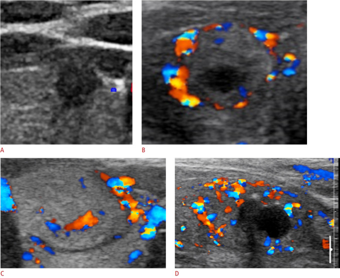
Doppler ultrasonography (US) is widely used for the differential diagnosis of thyroid nodules, metastatic cervical lymph nodes in patients with thyroid cancer, and diffuse parenchymal disease, as well as for guidance in various US-guided procedures, including biopsy and ablation. However, controversies remain regarding the appropriate use and interpretation of Doppler US. Therefore, the Korean Society of Thyroid Radiology organized a taskforce to develop a consensus statement on the clinical use of Doppler US for thyroid disease. The review and recommendations in this article are based on a comprehensive analysis of the current literature and the consensus of experts.

Keywords: Doppler ultrasound; Thyroid.


Conflict of interest statement

No potential conflict of interest relevant to this article was reported.

Figures

Fig. 1.
Fig. 1.. Vascular patterns of thyroid nodules on color Doppler ultrasonography.
The patterns are categorized into four types: type 1, absence of thyroid nodule vascularity (A); type 2, perinodular vascularity only (presence of circumferential vascularity at the margin of a thyroid nodule) (B); type 3, mild intranodular vascularity with or without perinodular vascularity (vascularity <50%) (C); type 4, marked intranodular vascularity with or without perinodular vascularity (vascularity >50%) (D).
Fig. 2.
Fig. 2.. A 49-year-old woman with benign follicular adenoma.
A. Transverse color Doppler sonography shows increased perinodular vascularity. B. Transverse power Doppler sonography shows more increased perinodular vascularity than the color Doppler sonography.
Fig. 3.
Fig. 3.. A 53-year-old woman with minimally invasive follicular carcinoma.
A. Transverse gray-scale sonography shows an isoechoic mass in the right thyroid. B. Predominant internal flow is seen in the thyroid nodule on a transverse color Doppler sonography.
Fig. 4.
Fig. 4.. A 50-year-old woman with Hashimoto thyroiditis diagnosed as diffuse thyroid disease on sonography.
Transverse (A) and longitudinal (B) gray-scale sonography show decreased echogenicity, coarse echotexture, a normal anteroposterior diameter of the thyroid gland, and lobulated margins. Longitudinal color Doppler sonography (C) shows increased parenchymal vascularity of the thyroid.
Fig. 5.
Fig. 5.. A 31-year-old man with Graves disease diagnosed as diffuse thyroid disease on sonography.
Transverse (A) and longitudinal (B) gray-scale sonography show decreased echogenicity, coarse echotexture, an increased anteroposterior diameter of the thyroid gland, and smooth margins. A qualitative visual assessment of thyroid parenchymal vascularity on longitudinal color Doppler sonography (C, D) revealed thyroid inferno (C), while quantitative blood flow measurement revealed a peak systolic velocity (PSV) of 53.1 cm/sec in the right superior thyroid artery (D). EDV, end diastolic velocity; MDV, mean diastolic velocity.
Fig. 6.
Fig. 6.. Ultrasound (US)-guided core needle biopsy of a thyroid nodule.
A. Before the procedure, the vessels along the approach route were carefully evaluated by Doppler US. B. Using a freehand technique, a core needle was directed from the isthmus toward the nodule while avoiding the vascular structures. C. After the tip of the biopsy needle was advanced into the edge of the nodule, the stylet and cutting cannula of the needle were sequentially fired.
Fig. 7.
Fig. 7.. Feeding artery of a thyroid nodule.
Before thyroid radiofrequency ablation, the main feeding artery from the superior thyroid artery is identified on spectral Doppler ultrasonography.
Fig. 8.
Fig. 8.. An under-ablated nodule portion on Doppler ultrasonography.
On gray-scale (A) and Doppler ultrasonography (B), most of the nodule shows low echogenicity without vascularity (arrows), thereby suggesting an ablated portion, but the medial-posterior portion of the nodule appears isoechoic with vascularity (arrowheads), suggesting the presence of an under-ablated portion.

References

    1. White DN. Johann Christian Doppler and his effect: a brief history. Ultrasound Med Biol. 1982;8:583–591. - PubMed
    1. Iared W, Shigueoka DC, Cristofoli JC, Andriolo R, Atallah AN, Ajzen SA, et al. Use of color Doppler ultrasonography for the prediction of malignancy in follicular thyroid neoplasms: systematic review and meta-analysis. J Ultrasound Med. 2010;29:419–425. - PubMed
    1. Hahn SY, Shin JH, Na DG, Ha EJ, Ahn HS, Lim HK, et al. Ethanol ablation of the thyroid nodules: 2018 consensus statement by the Korean Society of Thyroid Radiology. Korean J Radiol. 2019;20:609–620. - PMC - PubMed
    1. Kim JH, Baek JH, Lim HK, Ahn HS, Baek SM, Choi YJ, et al. 2017 Thyroid radiofrequency ablation guideline: Korean Society of Thyroid Radiology. Korean J Radiol. 2018;19:632–655. - PMC - PubMed
    1. Lee YH, Baek JH, Jung SL, Kwak JY, Kim JH, Shin JH, et al. Ultrasound-guided fine needle aspiration of thyroid nodules: a consensus statement by the Korean Society of Thyroid Radiology. Korean J Radiol. 2015;16:391–401. - PMC - PubMed

LinkOut - more resources