. 2020 Nov 13;295(46):15662-15676.
doi: 10.1074/jbc.RA120.015552. Epub 2020 Sep 6.

Astrocyte-specific deletion of the transcription factor Yin Yang 1 in murine substantia nigra mitigates manganese-induced dopaminergic neurotoxicity

Affiliations

Astrocyte-specific deletion of the transcription factor Yin Yang 1 in murine substantia nigra mitigates manganese-induced dopaminergic neurotoxicity

Edward Pajarillo et al. J Biol Chem. .

Abstract

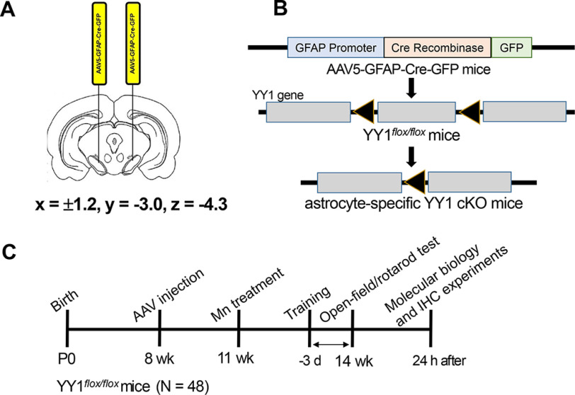
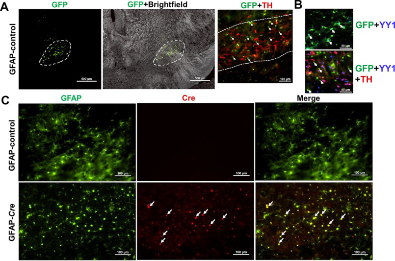
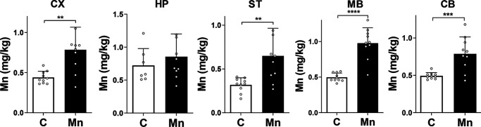
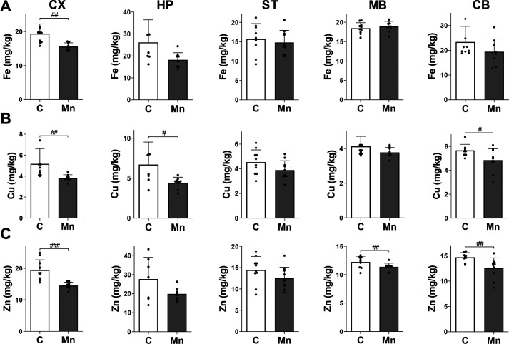
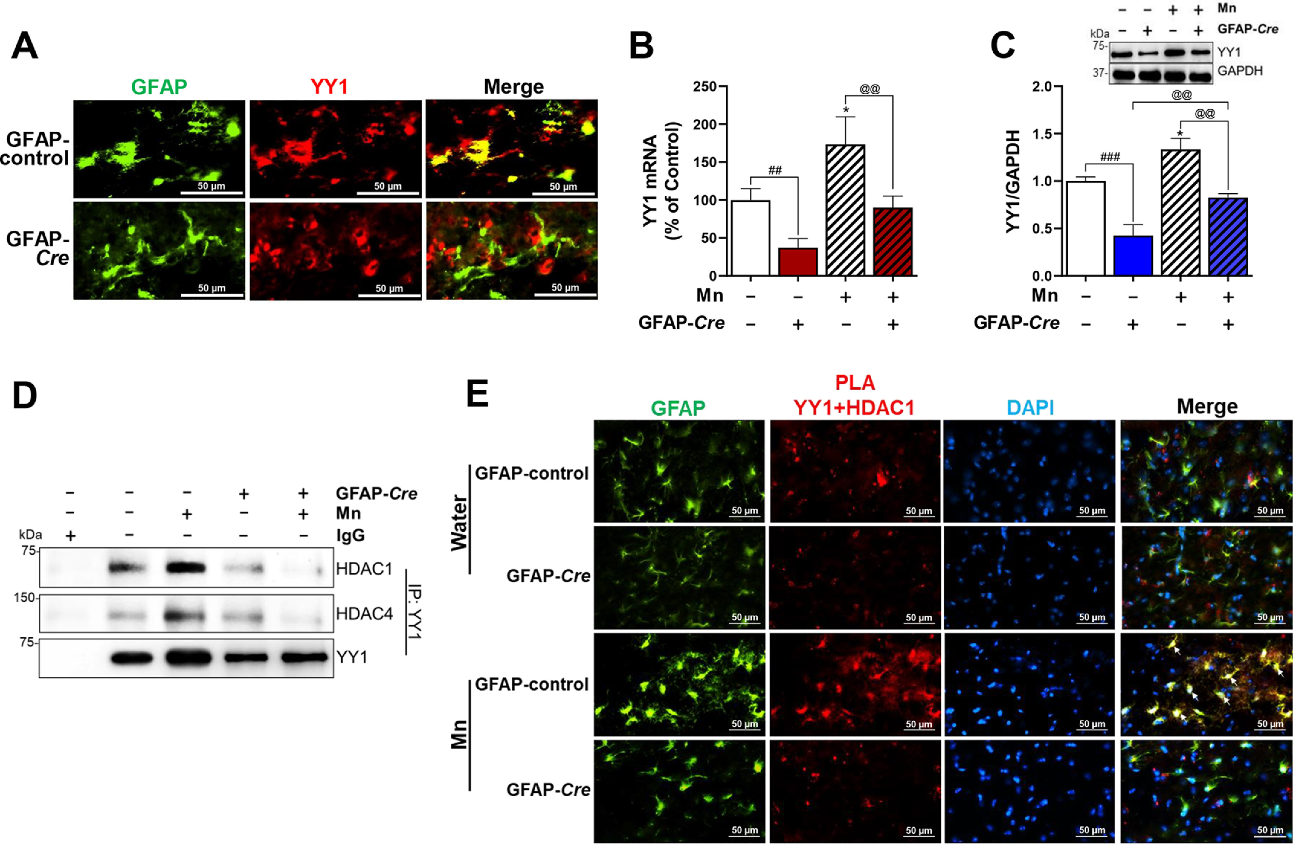
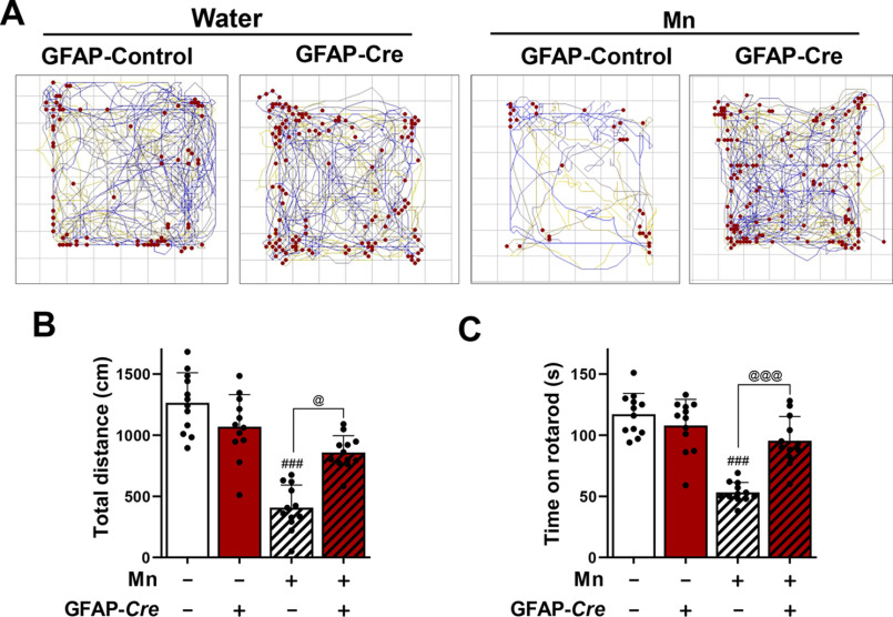
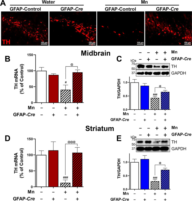
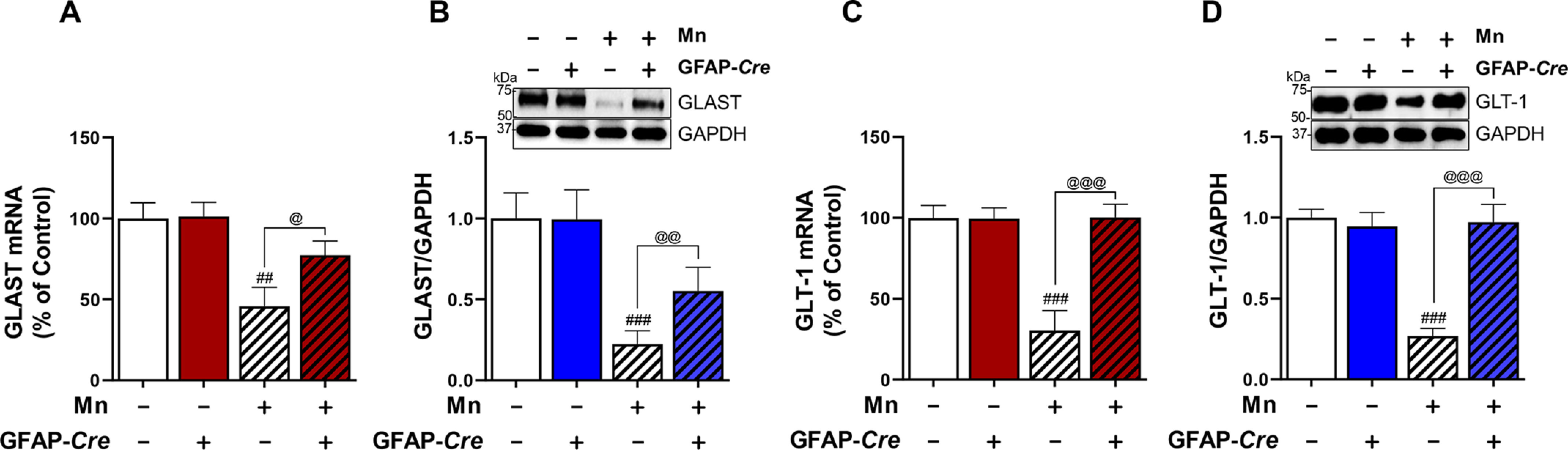
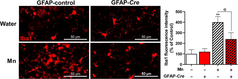
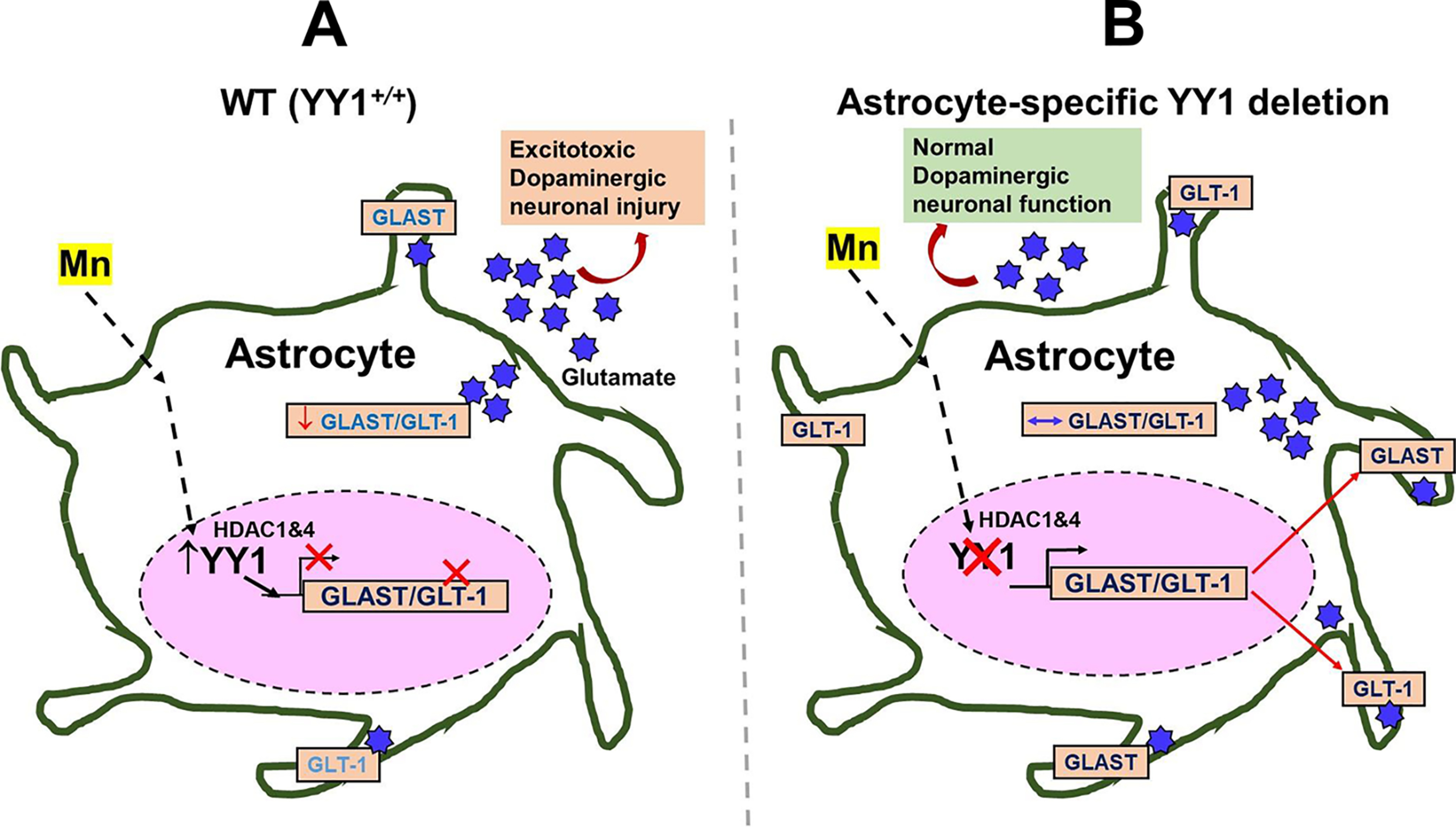
Manganese (Mn)-induced neurotoxicity resembles Parkinson's disease (PD), but the mechanisms underpinning its effects remain unknown. Mn dysregulates astrocytic glutamate transporters, GLT-1 and GLAST, and dopaminergic function, including tyrosine hydroxylase (TH). Our previous in vitro studies have shown that Mn repressed GLAST and GLT-1 via activation of transcription factor Yin Yang 1 (YY1). Here, we investigated if in vivo astrocytic YY1 deletion mitigates Mn-induced dopaminergic neurotoxicity, attenuating Mn-induced reduction in GLAST/GLT-1 expression in murine substantia nigra (SN). AAV5-GFAP-Cre-GFP particles were infused into the SN of 8-week-old YY1 flox/flox mice to generate a region-specific astrocytic YY1 conditional knockout (cKO) mouse model. 3 weeks after adeno-associated viral (AAV) infusion, mice were exposed to 330 μg of Mn (MnCl2 30 mg/kg, intranasal instillation, daily) for 3 weeks. After Mn exposure, motor functions were determined in open-field and rotarod tests, followed by Western blotting, quantitative PCR, and immunohistochemistry to assess YY1, TH, GLAST, and GLT-1 levels. Infusion of AAV5-GFAP-Cre-GFP vectors into the SN resulted in region-specific astrocytic YY1 deletion and attenuation of Mn-induced impairment of motor functions, reduction of TH-expressing cells in SN, and TH mRNA/protein levels in midbrain/striatum. Astrocytic YY1 deletion also attenuated the Mn-induced decrease in GLAST/GLT-1 mRNA/protein levels in midbrain. Moreover, YY1 deletion abrogated its interaction with histone deacetylases in astrocytes. These results indicate that astrocytic YY1 plays a critical role in Mn-induced neurotoxicity in vivo, at least in part, by reducing astrocytic GLAST/GLT-1. Thus, YY1 might be a potential target for treatment of Mn toxicity and other neurological disorders associated with dysregulation of GLAST/GLT-1.

Keywords: GLASTGLT-1; Parkinson disease; adeno-associated viral (AAV); adeno-associated viral vector; animal model; astrocyte; dopaminergic neurotoxicity; glutamate; manganese; manganese Yin Yang 1; tyrosine hydroxylase.


Conflict of interest statement

Conflict of interest—The authors declare that they have no conflicts of interest with the contents of this article..

Figures

Figure 1.
Figure 1.
Schematic diagram of the experimental paradigm. A, coordinates of stereotaxic injection and bilateral infusion of AAV vectors into the SN of the mouse brain. B, genomic deletion of astrocytic YY1 by GFAP-Cre expression in astrocytes of the SN regions of YY1-loxP mice using a Cre-lox method. To induce astrocyte-specific YY1 deletion, AAV5-GFAP-Cre-GFP vectors were infused into the SN, using AAV5-GFAP-GFP vectors as control vectors. C, Mn exposure (30 mg/kg, intranasal instillation, daily for 3 weeks) after bilateral infusion of AAV5 particles into the SN regions of the mouse brain, as described under “Experimental procedures.” 3 weeks after AAV5 infusion, MnCl2 was administered intranasally into both nostrils alternately and distilled water was used as a vehicle.
Figure 2.
Figure 2.
Validation of AAV vector infusion into the murine SN and Cre recombinase expression in astrocytes. 3 weeks after AAV vector infusion, the mouse brain was perfused and coronal sections were stained with antibodies for GFAP and Cre as described under “Experimental procedures.” A, cells expressing GFP (green, 4 × magnification) depict the site of AAV vector infusion in SN regions where TH-expressing dopaminergic neurons are localized (red, TH; green, GFP; ×10 magnification). White arrows, GFP-expressing astrocytes in SN. B, YY1 is co-expressed with GFP-expressing astrocytes (top panel), and the bottom panel shows expression of YY1 (blue), TH-expressing dopaminergic neurons (red) and GFP-expressing astrocytes (green) in SN 3 weeks after AAV5-GFAP-GFP (GFAP-control) vector infusion (×20 magnification). White arrows show YY1 co-expressing with GFP. C, coronal sections immunostained with GFAP and Cre antibodies showed co-localized expression of GFAP (green) and Cre (red) in the SN region, indicating that Cre was expressed in astrocytes (×20 magnification). White arrows show Cre co-expressing with GFAP.
Figure 3.
Figure 3.
Mn exposure via intranasal instillation increased Mn levels in various regions of the mouse brain. Mice were treated with MnCl2 (30 mg/kg, daily) for 3 weeks via intranasal instillation. Twenty-four hours after the last Mn treatment, mouse brain tissues were dissected, and Mn levels in each region were measured by ICP-MS/MS, as described under “Experimental procedures.” CX, frontal cortex; HP, hippocampus; ST, striatum; MB, midbrain; CB, cerebellum. **, p < 0.01; ***, p < 0.001; ****, p < 0.0001 compared with each other by Student's t test; n = 7-10. Data are expressed as mean ± S.D.
Figure 4.
Figure 4.
Mn elevation decreases accumulation of other divalent metals (Fe, Cu, and Zn) in the brain. A–C, after mice were treated daily with MnCl2 (30 mg/kg) for 3 weeks via intranasal instillation, levels of Fe (A), Cu (B), and Zn (C) were measured in various regions of the mouse brain by ICP-MS as described under “Experimental procedures.” CX, frontal cortex; HP, hippocampus; ST, striatum; MB, midbrain; CB, cerebellum. #, p < 0.05; ##, p < 0.01; ###, p < 0.001, compared with each other by Student's t test; n = 7–10. Data are expressed as mean ± S.D.
Figure 5.
Figure 5.
Astrocytic YY1 deletion by infusion of AAV-GFAP-Cre-GFP viral vectors into the SN attenuates Mn-increased YY1 expression and YY1 interaction with HDACs. A, after AAV infusion, coronal sections were stained with antibodies for GFAP and YY1, followed by IHC as described under “Experimental procedures.” Cells stained with GFAP (green) depict astrocytes and YY1 (red) indicates YY1 expression (×40 magnification). B and C, after Mn treatment, midbrain samples were extracted for total RNA and protein, followed by qPCR and Western blotting, as described under “Experimental procedures.” YY1 mRNA (B) and protein (C) levels in the midbrain were measured. GAPDH was used as loading controls of mRNA and protein. D, midbrain samples were tested for interaction of YY1 with HDAC1 and/or HDAC4 by co-IP as described under “Experimental procedures.” E, interaction of YY1 and HDAC1 in the SN of the mouse brain was determined by proximity ligation assay (×40 magnification). GFAP (green), YY1-HDAC1 proximity ligation signal (red), and DAPI (blue). *, p < 0.05 compared with the controls; ##, p < 0.01; ###, p < 0.001; @@, p < 0.01 compared with each other (one-way ANOVA followed by Tukey's post hoc test; n = 3). Data are expressed as mean ± S.D.
Figure 6.
Figure 6.
Deletion of astrocytic YY1 in SN attenuates Mn-induced impairment of locomotor activity and motor coordination. After Mn exposure (MnCl2, 30 mg/kg, intranasal instillation, daily for 3 weeks), locomotor activity and motor coordination were measured as described under “Experimental procedures.” A, the traces show one mouse's movement, and red dots depict vertical activity as a representative. B, total distance traveled, and C, time spent on the rotating rod. ###, p < 0.001 compared with the controls. @, p < 0.05; @@@, p < 0.001 compared with each other (one-way ANOVA followed by Tukey's post hoc test; n = 12). Data are expressed as mean ± S.D.
Figure 7.
Figure 7.
Deletion of astrocytic YY1 attenuates Mn-induced decrease of TH expression in SN/midbrain and striatum. A, after Mn exposure (MnCl2, 30 mg/kg, intranasal instillation, daily for 3 weeks), mice were perfused, and coronal sections of brain tissues were immunostained with TH antibody as described under “Experimental procedures.” The expression of TH protein was shown as red fluorescence signals (TRITC) in the SN of the mouse brain (×10 magnification). B–E, after Mn treatment, midbrain (B and C) and striatal (D and E) regions were processed for TH mRNA and protein levels by qPCR and Western blotting, respectively, as described under “Experimental procedures.” GAPDH was used as a loading control. *, p < 0.05; #, p < 0.05; ###, p < 0.001 compared with GFAP-GFP/vehicle; @, p < 0.05; @@@, p < 0.001 compared with each other (one-way ANOVA followed by Tukey's post hoc test; n = 3). Data are expressed as mean ± S.D.
Figure 8.
Figure 8.
Astrocytic YY1 mediates Mn-induced down-regulation of astrocytic glutamate transporters in the midbrain of mice. After Mn exposure (MnCl2, 30 mg/kg, intranasal instillation, daily for 3 weeks), midbrain tissues of mice were processed to measure mRNA and protein levels by qPCR and Western blotting, respectively, as described under “Experimental procedures.” A and B, Mn decreased mRNA and protein levels of GLAST, whereas astrocytic YY1 deletion attenuated these Mn effects on GLAST in the midbrain region. C and D, Mn decreased mRNA and protein levels of GLT-1, whereas astrocytic YY1 deletion attenuated these Mn effects on GLT-1 in the midbrain region. GAPDH was used as a loading control. ##, p < 0.01; ###, p < 0.001 compared with the controls. @, p < 0.05; @@, p < 0.01; @@@, p < 0.01 compared with each other (one-way ANOVA followed by Tukey's post hoc test; n = 3). Bar graphs are expressed as mean ± S.D.
Figure 9.
Figure 9.
Astrocytic YY1 deletion attenuates Mn-induced microglial activation in the SN of the mouse brain. After Mn exposure (MnCl2, 30 mg/kg, intranasal instillation, daily for 3 weeks), mice were perfused, and coronal sections of brain tissues were immunostained with Iba1 antibody as described under “Experimental procedures.” The expression of Iba1 protein was shown as red fluorescent signals in the SN of the mouse brain (×40 magnification). Mn increased Iba1 fluorescence intensity in the SN of the mouse brain, whereas astrocytic YY1 deletion attenuated these Mn effects. **, p < 0.05 compared with the controls. @, p < 0.05 compared with each other (one-way ANOVA followed by Tukey's post hoc test; n = 3). Data are expressed as mean ± S.D.
Figure 10.
Figure 10.
Schematic diagram of the proposed mechanism for YY1's role in Mn-induced GLAST and GLT-1 dysregulation in astrocytes. A, Mn-induced activation of YY1, which forms a repressor complex with HDACs, in astrocytes decreases levels of glutamate transporters GLAST/GLT-1 in the plasma membranes, resulting in extracellular glutamate accumulation leading to dopaminergic toxicity. B, the deletion of astrocytic YY1 attenuates the Mn-induced decrease in GLAST and GLT-1 by inhibiting YY1-HDAC activity. Because Mn-induced dysregulation of astrocytic glutamate transporters (GLAST and GLT-1) is closely associated with glutamate accumulation in the synaptic cleft, this mechanism could lead to Mn neurotoxicity in the SN region of the mouse brain, where dopaminergic cell bodies are located. The findings suggest that the deletion of astrocytic YY1 can be a critical target to attenuate Mn-induced dopaminergic toxicity.

References

    1. Keen C. L., Ensunsa J. L., Watson M. H., Baly D. L., Donovan S. M., Monaco M. H., and Clegg M. S. (1999) Nutritional aspects of manganese from experimental studies. Neurotoxicology 20, 213–223 - PubMed
    1. Kwakye G. F., Paoliello M. M., Mukhopadhyay S., Bowman A. B., and Aschner M. (2015) Manganese-induced Parkinsonism and Parkinson's disease: shared and distinguishable features. Int. J. Environ. Res. Public Health 12, 7519–7540 10.3390/ijerph120707519 - DOI - PMC - PubMed
    1. Bowler R. M., Kornblith E. S., Gocheva V. V., Colledge M. A., Bollweg G., Kim Y., Beseler C. L., Wright C. W., Adams S. W., and Lobdell D. T. (2015) Environmental exposure to manganese in air: Associations with cognitive functions. Neurotoxicology 49, 139–148 10.1016/j.neuro.2015.06.004 - DOI - PMC - PubMed
    1. Dlamini W. W., Nelson G., Nielsen S. S., and Racette B. A. (2020) Manganese exposure, parkinsonian signs, and quality of life in South African mine workers. Am. J. Ind. Med. 63, 36–43 10.1002/ajim.23060 - DOI - PMC - PubMed
    1. Kullar S. S., Shao K., Surette C., Foucher D., Mergler D., Cormier P., Bellinger D. C., Barbeau B., Sauvé S., and Bouchard M. F. (2019) A benchmark concentration analysis for manganese in drinking water and IQ deficits in children. Environ. Int. 130, 104889 10.1016/j.envint.2019.05.083 - DOI - PubMed

Publication types

MeSH terms

LinkOut - more resources