. 2021 Jan;288(1):160-174.
doi: 10.1111/febs.15556. Epub 2020 Sep 25.

Sarcolemma wounding activates dynamin-dependent endocytosis in striated muscle

Affiliations

Sarcolemma wounding activates dynamin-dependent endocytosis in striated muscle

Joel R McDade et al. FEBS J. 2021 Jan.

Abstract

Plasma membrane repair is an evolutionarily conserved mechanism by which cells can seal breaches in the plasma membrane. Mutations in several proteins with putative roles in sarcolemma integrity, membrane repair, and membrane transport result in several forms of muscle disease; however, the mechanisms that are activated and responsible for sarcolemma resealing are not well understood. Using the standard assays for membrane repair, which track the uptake of FM 1-43 dye into adult skeletal muscle fibers following laser-induced sarcolemma disruption, we show that labeling of resting fibers by FM1-43 prior to membrane wounding and the induced FM1-43 dye uptake after sarcolemma wounding occurs via dynamin-dependent endocytosis. Dysferlin-deficient muscle fibers show elevated dye uptake following wounding, which is the basis for the assertion that membrane repair is defective in this model. Our data show that dynamin inhibition mitigates the differences in FM1-43 dye uptake between dysferlin-null and wild-type muscle fibers, suggesting that elevated wound-induced FM1-43 uptake in dysferlin-deficient muscle may actually be due to enhanced dynamin-dependent endocytosis following wounding, though dynamin inhibition had no effect on dysferlin trafficking after wounding. By monitoring calcium flux after membrane wounding, we show that reversal of calcium precedes the sustained, slower increase of dynamin-dependent FM1-43 uptake in WT fibers, and that dysferlin-deficient muscle fibers have persistently increased calcium after wounding, consistent with its proposed role in resealing. These data highlight a previously unappreciated role for dynamin-dependent endocytosis in wounded skeletal muscle fibers and identify overactive dynamin-dependent endocytosis following sarcolemma wounding as a potential mechanism or consequence of dysferlin deficiency.

Keywords: dynamin; dysferlin; endocytosis; membrane repair; membrane transport; skeletal muscle.


Conflict of interest statement

Conflicts of Interest: The authors have no conflicts to declare

Figures

Figure 1.
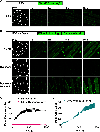
Figure 1.. Resting adult skeletal muscle fibers take up FM1-43 via a dynamin-dependent mechanism. FM1-43 labeling occurs over several minutes in adult skeletal muscle fibers.
FM1-43 uptake was assessed in untreated, resting adult C57BL/6 skeletal muscle fibers (A) and in resting adult skeletal muscle fibers following pre-treatment with DMSO or dynamin inhibitor dynasore (B). Untreated and DMSO treated fibers take up extracellular FM1-43 with maximal labeling occurring within 10min (A and B top, respectively, quantified in C). Pre-treatment with 80μM dynasore completely inhibits FM1-43 uptake in resting skeletal muscle fibers (B, middle, quantified in C). Dynasore-treated muscle fibers take up FM1-43 following removal of dynasore from the extracellular solution (C bottom, quantified D). The red scale bar indicates 50μm. Statistical comparisons were performed using an unpaired, two-tailed Student’s t-test. Statistical significance (p<0.05) between DMSO and Dynasore is denoted by (*) for each timepoint under the horizontal bar. Error bars represent the standard deviation of the mean. Data is summarized from three independent loading plates.
Figure 2.
Figure 2.
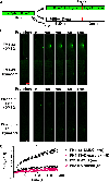
Wound-induced FM1-43 uptake is dynamin dependent in adult skeletal muscle fibers. C57BL/6 muscle fibers were loaded with FM1-43 normally, treated with DMSO +/− 80μM dynasore and subjected to laser-induced wounding (A). Dynasore treatment (middle B, magenta squares in C) results in significantly reduced wound-induced FM1-43 uptake compared to DMSO treated control cells (top B, black circles in C). FM1-43 uptake in dynasore-washout cells was not significantly different than DMSO treated controls, indicating a reversible effect of dynasore treatment on wound-induced FM1-43 uptake in skeletal muscle cells (bottom B, teal triangles C). These results were independently confirmed using the same experimental setup (A) with an additional dynamin inhibitor, Dyngo4a, again showing that dynamin inhibition results in significantly reduced wound-induced FM1-43 uptake compared to DMSO controls, which is reversible upon Dyngo4A washout (C). Statistical comparisons were performed using a Two-Way ANOVA followed by posthoc Student’s t-tests. Statistical significance (p<0.05) between DMSO and Dynasore/Dyngo4A is denoted by (*) and between Dynasore/Dyngo4a and Washout by (#) for each timepoint under horizontal bar. N.S. designates no statistical difference (p>0.05) between DMSO and Washout conditions in both C and D. Error bars represent the standard deviation of the mean. One representative experiment of three independent replicates (B-C) or two independent replicates (D) is shown.
Figure 3.
Figure 3.. FM1-43 uptake after wounding is dependent upon extracellular FM1-43.
Isolated skeletal muscle fibers from adult C57BL/6 mice were loaded with 2.5μM FM1-43 for ~10min and wounded in the presence or absence of Dynasore (A). Washout conditions were removed of extracellular FM1-43 and cells then wounded in the presence or absence of dynasore. Representative images of all conditions show that FM1-43 washout severely decreases FM1-43 fluorescence after wounding, however there is a rapid accumulation of dye at the wound that is uninhibited by Dynasore treatment (B, quantified, C). Statistical comparisons were performed using an unpaired, two-tailed Student’s t-test (A) or Two-Way ANOVA followed by posthoc Student’s t-tests (C). The threshold for statistical significance was not met in A (p>0.05), but statistical significance (p<0.05) between FM1-43+DMSO and all other traces is denoted by (*) for each timepoint under the horizontal bar. Error bars represent the standard deviation of the mean. One representative experiment of two independent replicates is shown.
Figure 4.
Figure 4.. Dynamin is required for increased wound-induced uptake of FM1-43 in dysferlin-null muscle fibers.
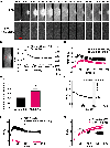
Basal FM1-43 uptake is unchanged in dysferlin-null A/J muscle fibers compared to A/WySnJ wild-type controls(A). DMSO-treated, dysferlin null muscle fibers (representative images B, magenta triangles in C) take up more FM1-43 dye following wounding than wild-type controls (representative images B, black circles in C). Dynasore treatment significantly reduces wound-induced FM1-43 uptake in wild-type and dysferlin-null muscle fibers (representative images B, open black cirlces and open magenta triangles in C, respectively). Statistical comparisons were performed using Student’s t-tests. Statstical significance (p<0.05) between A/J DMSO and A/J Dyna is denoted by (*) and between A/WySnJ DMSO and A/WySnJ Dyna by (#) and between A/WySnJ DMSO and A/J DMSO by (^) for each timepoint under horizontal bar. N.S. designates no statistical difference (p>0.05) between A/WySnJ Dyna and A/J Dyna. The dysf-pHGFP TG reporter mouse provides a real-time assessment of dysferlin localization in adult skeletal muscle fibers (D). Recruitment of dysferlin to lesions (white arrowhead E, quantified F) and endocytosis of dysferlin following wounding (red arrowhead E, quantified G) are unchanged in dynasore-treated cells, indicating that dynamin activity is not required for dysferlin transport following wounding. Statistical comparisons were made using an unpaired, two-tailed Student’s t-tests and did not meet the threshold of significance (p>0.05). Error bars represent the standard deviation of the mean. One representative experiment of three independent replicates (A), one independent replicate (B-C), and two independent replicates (D-G) is shown.
Figure 5.
Figure 5.. Calcium influx into the cell as measured by Fluo-4 relative fluorescence shows membrane repair is a much faster process than as previously indicated by FM1-43 wounding studies.
Isolated wild-type C57BL/6 muscle fibers were loaded with 3μM Fluo-4 for 1hr and then wounded with a high powered laser, representative images shown (top A). Calcium increases at the wound site were quantified, described by the white box in B, which showed that wounding causes a rapid increase in calcium at the wound site (one representative trace shown B, summary data from one representative experiment C). Calcium increases at the wound are followed by an increase in cytoplasmic calcium at sites distal to the wound (red box in B, and magenta open circles C). Calcium at the wound peaks and reverses at the wound at approximately 40s post wound (solid line B, summary data in D), but cytoplasmic calcium reversal is significantly delayed to approximately 60s (D) A bolus of 10mM caffeine to wounded fibers, one representative trace shown, stimulates calcium release from the SR that exceeds peak calcium after wounding (E). Removing calcium from extracellular media and chelating any trace calcium with 1mM EGTA prevents the increase in cytosolic calcium following wounding (representative images A bottom, quantified F). While calcium flux reverses within the first minute following wounding, the concentration of FM1-43 rises in the cell for more than 2 minutes, and then continues to increase at a constant rate even after 2 minutes (G). Statistical compaisons were performed using an unpaired, two-tailed Student’s t-test. Statistical significance (p<0.05) is denoted by (*). Error bars represent the standard error of the mean. One representative experiment of three independent replicates is shown.
Figure 6.
Figure 6.. Dynamin inhibition does not affect calcium influx after membrane wounding.
Isolated wild-type C57BL/6 muscle fibers were loaded with 3μM Fluo-4 for 1hr and treated either with a vehicle (DMSO, top panel) or 80μM Dynasore (bottom panel) prior to wounding shows calcium influx after wounding is uninhibited by Dynasore treatment (representative images A, quantified B). Quantification of the time at which the calcium signal peaks and reverses across 6 independent replicates shows Dynasore treated cells have a faster time to peak calcium compared to vehicle control (C). Statistical comparisons were performed using a Two-Way ANOVA followed by posthoc Student’s t-tests. Statistical significance (p<0.05) between Dynasore and both PSS and Washout is denoted by (*). Error bars represent the standard error of the mean.
Figure 7.
Figure 7.. Dysferlin-deficiency muscle fibers have persistently elevated calcium after membrane wounding.
Isolated muscle fibers from dysferlin-deficient BLA/J mice and wild-type littermate controls (C57BL/6) were loaded with 3μM Fluo-4 for 1hr and wounded with a high-powered laser (representative images A). Calcium concentrations after wounding are increased in dysferlin-deficient fibers (individual traces B, summary of one experiment C), Peak calcium was normalized to WT peak calcium and pooled among three independent replicates shown that peak calcium influx is increased in dysferlin-deficient cells (D), but the time to calcium reversal is similar in WT and dysferlin-deficient cells (solid line D, summary E). The steady state calcium at the end of imaging for three independent replicates was normalized to WT steady state calcium, which is also greater in dysferlin deficient cells (F). Statistical compaisons were performed using an unpaired, two-tailed Student’s t-test. Statistical significance (p<0.05) between WT and BLA/J is denoted by (*). Error bars represent the standard error of the mean.
Figure 8.
Figure 8.. General model of dynamin-mediated endocytosis and dysferlin-mediated membrane repair in skeletal muscle.
In normal muscle, sarcolemma wounding leads to localized calcium influx and activation of dysferlin-mediated membrane repair, preventing over-activation of dynamin-mediated endocytosis (A). In dysferlin-deficient muscle wherein dysferlin-mediated repair is impaired, prolonged calcium influx through unsealed lesions leads to increased activation of dynamin-dependent endocytosis which may compensate for impaired dysferlin-MMR or contribute to disease pathology (B).

References

    1. Allen DG, Whitehead NP & Froehner SC (2016) Absence of Dystrophin Disrupts Skeletal Muscle Signaling: Roles of Ca2+, Reactive Oxygen Species, and Nitric Oxide in the Development of Muscular Dystrophy, Physiological reviews. 96, 253–305. - PMC - PubMed
    1. Claflin DR & Brooks SV (2008) Direct observation of failing fibers in muscles of dystrophic mice provides mechanistic insight into muscular dystrophy, American Journal of Physiology - Cell Physiology. 294, C651–C658. - PubMed
    1. Bashir R, Britton S, Strachan T, Keers S, Vafiadaki E, Lako M, Richard I, Marchand S, Bourg N, Argov Z, Sadeh M, Mahjneh I, Marconi G, Passos-Bueno MR, Moreira E. d. S., Zatz M, Beckmann JS & Bushby K (1998) A gene related to Caenorhabditis elegans spermatogenesis factor fer-1 is mutated in limb-girdle muscular dystrophy type 2B, Nature Genetics. 20, 37–42. - PubMed
    1. Bansal D, Miyake K, Vogel SS, Groh S, Chen C-C, Williamson R, McNeil PL & Campbell KP (2003) Defective membrane repair in dysferlin-deficient muscular dystrophy, Nature. 423, 168–172. - PubMed
    1. Lennon NJ, Kho A, Bacskai BJ, Perlmutter SL, Hyman BT & Brown RH (2003) Dysferlin Interacts with Annexins A1 and A2 and Mediates Sarcolemmal Wound-healing, Journal of Biological Chemistry. 278, 50466–50473. - PubMed

Publication types

MeSH terms