. 2020 Nov 9;12(1):1800897.
doi: 10.1080/19490976.2020.1800897.

Impact and consequences of intensive chemotherapy on intestinal barrier and microbiota in acute myeloid leukemia: the role of mucosal strengthening

Affiliations

Impact and consequences of intensive chemotherapy on intestinal barrier and microbiota in acute myeloid leukemia: the role of mucosal strengthening

Thomas Hueso et al. Gut Microbes. .

Abstract

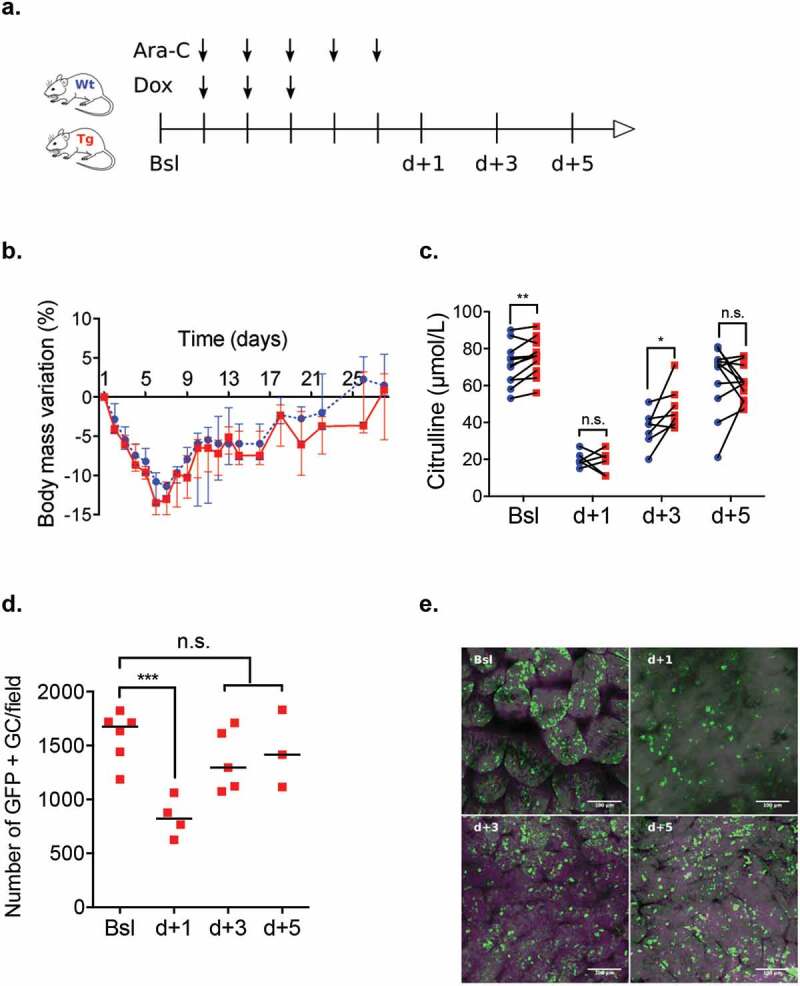
Induction chemotherapy (7 + 3 regimen) remains the gold standard for patients with acute myeloid leukemia (AML) but is responsible for gut damage leading to several complications such as bloodstream infection (BSI). We aimed to investigate the impact of induction chemotherapy on the intestinal barrier of patients with AML and in wild-type mice. Next, we assessed the potential benefit of strengthening the mucosal barrier in transgenic mice releasing a recombinant protein able to reinforce the mucus layer (Tg222). In patients, we observed a decrease of plasma citrulline, which is a marker of the functional enterocyte mass, of short-chain fatty acids and of fecal bacterial load, except for Escherichia coli and Enterococcus spp., which became dominant. Both the α and β-diversities of fecal microbiota decreased. In wild-type mice, citrulline levels decreased under chemotherapy along with an increase of E. coli and Enterococcus spp load associated with concomitant histologic impairment. By comparison with wild-type mice, Tg222 mice, 3 days after completing chemotherapy, had higher citrulline levels, a faster healing epithelium, and preserved α-diversity of their intestinal microbiota. This was associated with reduced bacterial translocations. Our results highlight the intestinal damage and the dysbiosis induced by the 7 + 3 regimen. As a proof of concept, our transgenic model suggests that strengthening the intestinal barrier is a promising approach to limit BSI and improve AML patients' outcome.

Keywords: Intestinal barrier; acute leukemia; chemotherapy; microbiota; mucus.


Figures

Figure 1.
Figure 1.
(a) Body mass variation and plasma citrulline level in patients before induction (T0), during aplasia (T1) and after hematological recovery (T2). (b) Quantitative PCR for aerotolerant and anaerobic bacteria in patients’ feces. Bacteroides, Bifidobacterium, Akkermansia spp, C. coccoides and C. leptum represented dominant groups. Streptococcus spp, Enterococcus spp, E. coli and Lactobacillus spp represented sub-dominant groups. p < .05 is considered significant, n.s.: not significant. *p < .05; **p < .01; ***p < .001.
Figure 2.
Figure 2.
(a) Alpha-diversity in patients’ stool microbiome, represented by the Shannon index and Chao-1 index, before induction (T0), during aplasia (T1) and after hematological recovery (T2). p < .05 is considered significant. (b) A subset of OTUs, grouped by family, with a raw count at least of 300 was kept. Bray-Curtis distance and UPGMA algorithm were used to perform pairwise distance between samples and hierarchical clustering, respectively. The heatmap highlighted a clustering of two groups dominated by Bifidobacteriaceae, Lachnospiraceae and Ruminococcaceae at T0 progressively replaced by Enterococcaceae or Enterobacteriaceae at T1 and T2.
Figure 3.
Figure 3.
(a). Induction chemotherapy in a wild-type mouse model that consisted of a combined administration of aracytine (AraC) for five days and doxorubicin (Dox) for three days injected intraperitoneally. Baseline (Bsl) corresponded to the time before chemotherapy administration; d + 1, d + 3, and d + 5 corresponded to one day, three days and five days after the completion of chemotherapy. (b) Overall survival for different chemotherapies: AraC (5 days) + Dox (3 days). Triangle: AraC (150 mg/kg/d) + Dox (3 mg/kg/d); circle: AraC (200 mg/kg/d) + Dox (3 mg/kg/d); and square: AraC (200 mg/kg/d) + Dox (6 mg/kg/d). c – g. A model with AraC 150 mg/kg/d and Dox 3 mg/kg/d was elected resulting in decrease of blood count body weight and plasma citrulline level (n = 10 to 15 at each time). Wt: wild-type B6D2F1; WBC: white blood count; Lym: lymphocyte; Gra: granulocytes; RBC: red blood count. p < .05 is considered significant, * p < .05; **p < .01; ***p < .001.
Figure 4.
Figure 4.
(a). Histological analyses of terminal ileum mucosa after HE and ABPAS staining, (b) Representative immunostaining of PCNA and apoptosis along the ileal villus-crypt axis (in green) at d + 1 and d + 3 and d + 5. (n = 7 to 10). Other cells were counterstained with Hoechst 33258 (in blue). HE: Hematoxylin/eosin; ABPAS: Alcian blue/periodic-acid Schiff; PCNA: proliferating cell nuclear antigen; TUNEL: TDT-mediated dUTP-biotin nick end-labeling; Lu: lumen. p < .05 significant, *p < .05; **p < .01.
Figure 5.
Figure 5.
(a) Induction chemotherapy model (AraC (150 mg/kg.d) + Dox (3 mg/kg/d)) in Wt and Tg mice. (b – c) Comparison of weight loss and plasma citrulline level of Wt (blue circles) and Tg mice (red squares) at baseline (Bsl) and d + 1, d + 3 and d + 5 after chemotherapy completion. (d – e) Tracking of GFP-tagged transgene using epifluorescence microscopy (in green) in the ileal lumen of Tg mice from Bsl to d + 5. Ara-C: aracytine; Dox: doxorubicin; GC: Goblet cells;. p < .05 is considered significant, n.s., not significant. *p < .05; **p < .01.
Figure 6.
Figure 6.
(a) Transgenic and wild type mice received chemotherapy regimen. Control group received PBS (included 2 Tg and 2 Wt mice). All mice were sacrificed at d + 3. (b-c) Histological analyses with HE and ABPAS staining and representative immunostaining with PCNA and apoptosis along the ileal villus-crypt axis (in green) at d + 3 after completion of induction chemotherapy. Other cells were counterstained with Hoechst 33258 (in blue).(d) Metabarcoding sequencing analyses of alpha diversity in ileum adherent microbiota at d + 3 in Wt and Tg mice and in chemotherapy and control groups. Ara-C: aracytine; Dox: doxorubicin; PBS: phosphate-buffered saline; HE: Hematoxylin/eosin; ABPAS: Alcian blue/periodic-acid Schiff; PCNA: proliferating cell nuclear antigen; TUNEL: TDT-mediated dUTP-biotin nick end-labeling; Lu: lumen. p < .05 is considered significant; n.s., not significant.
Figure 6.
Figure 6.
(Continued).
Figure 7.
Figure 7.
Oral gavage of 10 CFU of Salmonella Typhimurium (S. Typhimurium) was conducted the last day of the chemotherapy regimen in Wt, Tg and controlled (Ctrl) mice. (b) Comparison of intestinal translocation of S. Typhimurium in Wt (blue circles), Tg (red squares) and Ctrl mice (green triangles) in liver spleen and feces at d + 2 after oral gavage. Ara-C: aracytine; Dox: doxorubicin; PBS: phosphate-buffered saline. CFU: Colony forming unit. p < .05 is considered significant, n.s., not significant. *p < .05; **p < .01; ***p < .001.

References

    1. Lee JH, Kim H, Joo YD, Lee WS, Bae SH, Young Zang D, Kwon J, Kim MK, Lee J, Lee GW, et al. Prospective randomized comparison of idarubicin and high-dose daunorubicin in induction chemotherapy for newly diagnosed acute myeloid leukemia. J Clin Oncol. 2017;35(24):2754–17. doi:10.1200/JCO.2017.72.8618. - DOI - PubMed
    1. Kolonen A, Sinisalo M, Huttunen R, Syrjanën J, Aittoniemi J, Huhtala H, Sankelo M, Rintala H, Räty R, Jantunen E, et al. Bloodstream infections in acute myeloid leukemia patients treated according to the Finnish Leukemia Group AML-2003 protocol – a prospective nationwide study. Infect Dis (Auckl). 2017;49(11–12):799–808. doi:10.1080/23744235.2017.1347814. - DOI - PubMed
    1. Conn JR, Catchpoole EM, Runnegar N, Mapp SJ, Markey KA.. Low rates of antibiotic resistance and infectious mortality in a cohort of high-risk hematology patients: A single center, retrospective analysis of blood stream infection. PLoS One. 2017;12(5):1–13. doi:10.1371/journal.pone.0178059. - DOI - PMC - PubMed
    1. Ubeda C, Taur Y, Jenq RR, Equinda MJ, Son T, Samstein M, Viale A, Socci ND, van den Brink MRM, Kamboj M, et al. Vancomycin-resistant Enterococcus domination of intestinal microbiota is enabled by antibiotic treatment in mice and precedes bloodstream invasion in humans. J Clin Invest. 2010;120(12):4332–4341. doi:10.1172/JCI43918. - DOI - PMC - PubMed
    1. Wrzosek L, Miquel S, Noordine ML, Bouet S, Joncquel Chevallier-Curt M, Robert V, Philippe C, Bridonneau C, Cherbuy C, Robbe-Masselot C, et al. Bacteroides thetaiotaomicron and Faecalibacterium prausnitzii influence the production of mucus glycans and the development of goblet cells in the colonic epithelium of a gnotobiotic model rodent. BMC Biol. 2013;11:61. doi:10.1186/1741-7007-11-61. - DOI - PMC - PubMed

MeSH terms

Substances