. 2020 Sep 7;19(1):138.
doi: 10.1186/s12943-020-01253-y.

Circular RNA CDR1as disrupts the p53/MDM2 complex to inhibit Gliomagenesis

Affiliations

Circular RNA CDR1as disrupts the p53/MDM2 complex to inhibit Gliomagenesis

Jiacheng Lou et al. Mol Cancer. .

Abstract

Background: Inactivation of the tumor suppressor p53 is critical for pathogenesis of glioma, in particular glioblastoma multiforme (GBM). MDM2, the main negative regulator of p53, binds to and forms a stable complex with p53 to regulate its activity. Hitherto, it is unclear whether the stability of the p53/MDM2 complex is affected by lncRNAs, in particular circular RNAs that are usually abundant and conserved, and frequently implicated in different oncogenic processes.

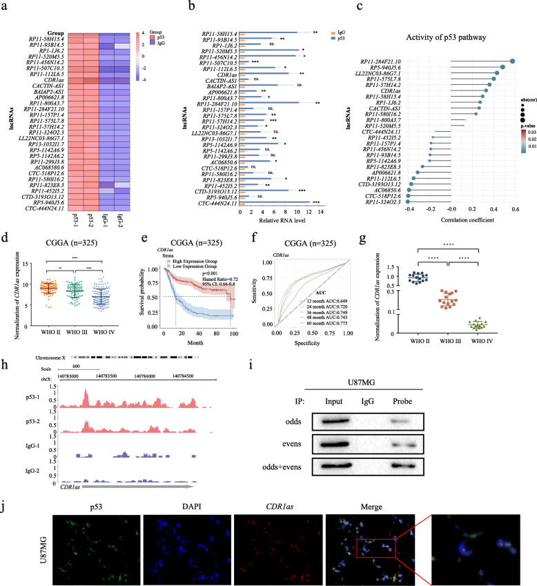
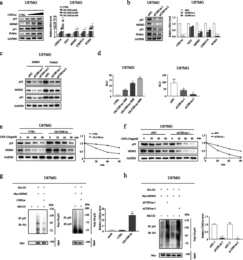
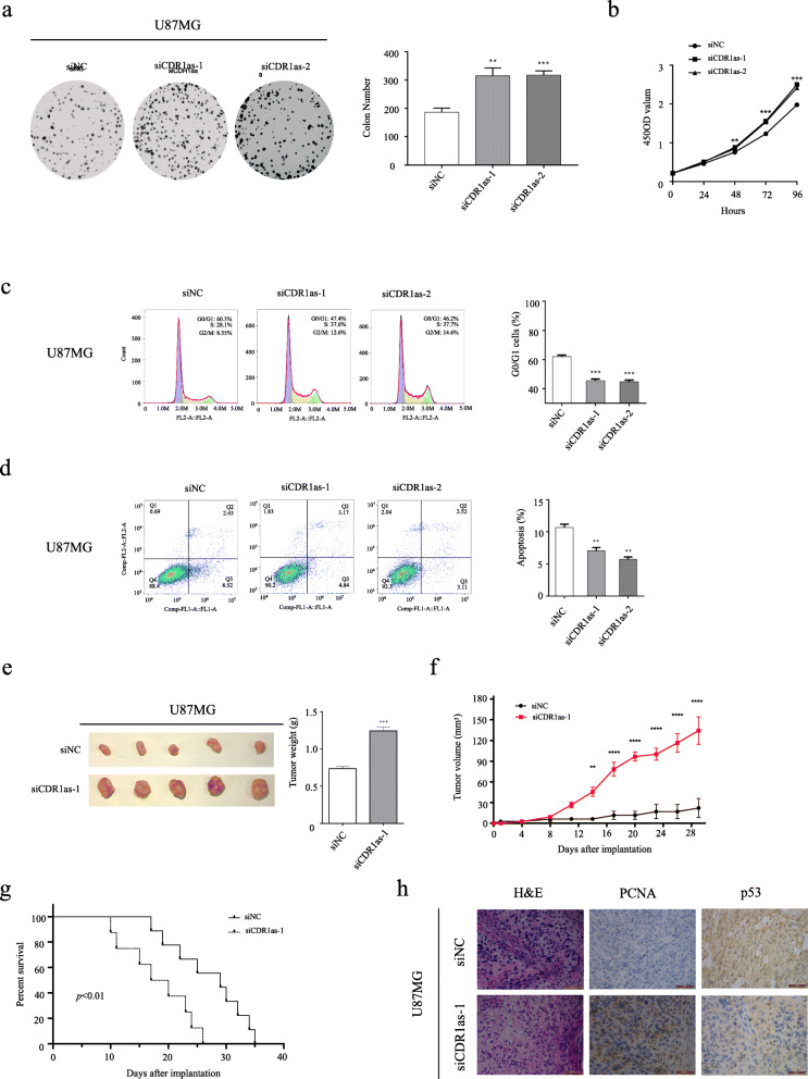
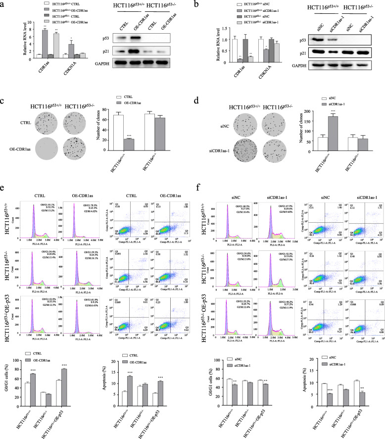
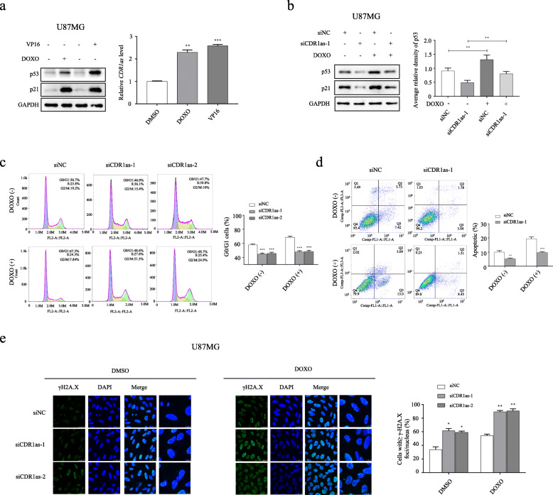
Methods: RIP-seq and RIP-qPCR assays were performed to determine the most enriched lncRNAs (including circular RNAs) bound by p53, followed by bioinformatic assays to estimate the relevance of their expression with p53 signaling and gliomagenesis. Subsequently, the clinical significance of CDR1as was evaluated in the largest cohort of Chinese glioma patients from CGGA (n = 325), and its expression in human glioma tissues was further evaluated by RNA FISH and RT-qPCR, respectively. Assays combining RNA FISH with protein immunofluorescence were performed to determine co-localization of CDR1as and p53, followed by CHIRP assays to confirm RNA-protein interaction. Immunoblot assays were carried out to evaluate protein expression, p53/MDM2 interaction and p53 ubiquitination in cells in which CDR1as expression was manipulated. After AGO2 or Dicer was knocked-down to inhibit miRNA biogenesis, effects of CDR1as on p53 expression, stability and activity were determined by immunoblot, RT-qPCR and luciferase reporter assays. Meanwhile, impacts of CDR1as on DNA damage were evaluated by flow cytometric assays and immunohistochemistry. Tumorigenicity assays were performed to determine the effects of CDR1as on colony formation, cell proliferation, the cell cycle and apoptosis (in vitro), and on tumor volume/weight and survival of nude mice xenografted with GBM cells (in vivo).

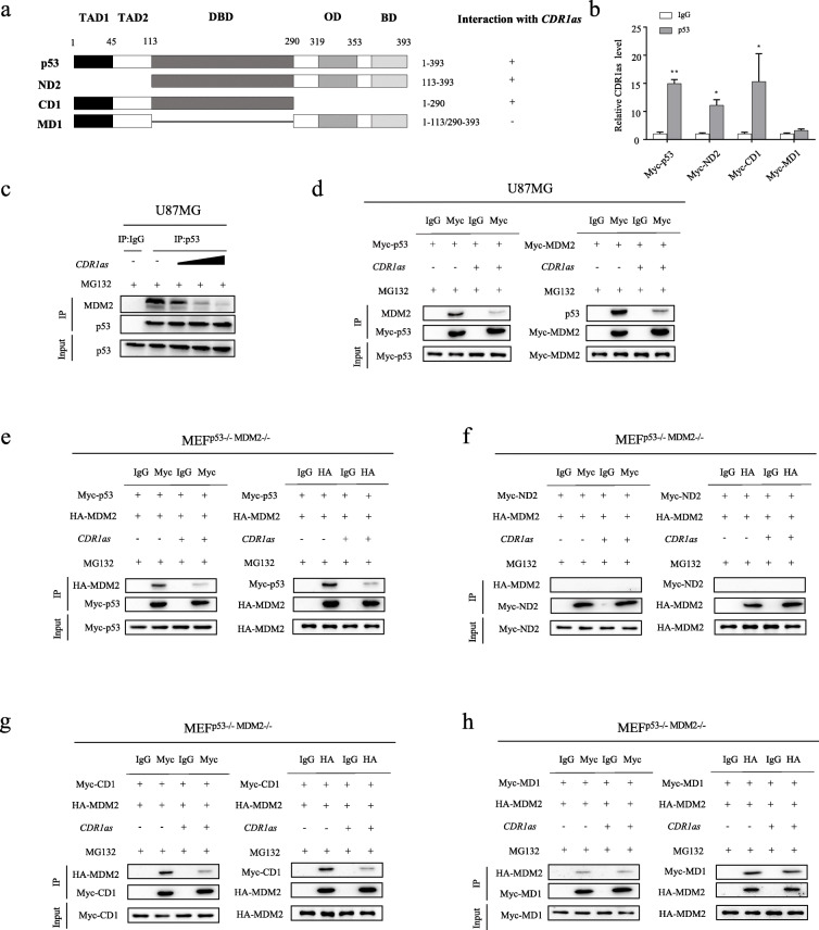
Results: CDR1as is found to bind to p53 protein. CDR1as expression decreases with increasing glioma grade and it is a reliable independent predictor of overall survival in glioma, particularly in GBM. Through a mechanism independent of acting as a miRNA sponge, CDR1as stabilizes p53 protein by preventing it from ubiquitination. CDR1as directly interacts with the p53 DBD domain that is essential for MDM2 binding, thus disrupting the p53/MDM2 complex formation. Induced upon DNA damage, CDR1as may preserve p53 function and protect cells from DNA damage. Significantly, CDR1as inhibits tumor growth in vitro and in vivo, but has little impact in cells where p53 is absent or mutated.

Conclusions: Rather than acting as a miRNA sponge, CDR1as functions as a tumor suppressor through binding directly to p53 at its DBD region to restrict MDM2 interaction. Thus, CDR1as binding disrupts the p53/MDM2 complex to prevent p53 from ubiquitination and degradation. CDR1as may also sense DNA damage signals and form a protective complex with p53 to preserve p53 function. Therefore, CDR1as depletion may play a potent role in promoting tumorigenesis through down-regulating p53 expression in glioma. Our results broaden further our understanding of the roles and mechanism of action of circular RNAs in general and CDR1as in particular, and can potentially open up novel therapeutic avenues for effective glioma treatment.

Keywords: CDR1as; DNA damage; Glioma; MDM2; p53.


Conflict of interest statement

The authors have no commercial or other associations that might pose a conflict of interest.

Figures

Fig. 1
Fig. 1
p53 physically interacts with CDR1as indicating glioma prognosis. a Heatmap of 30 most enriched lncRNAs binding to p53 protein determined by RIP-seq. b Validation of 30 candidate lncRNAs binding to p53 protein by RIP-qPCR. c Plots of the correlation between the scores of p53 pathway gene sets and expression of candidate lncRNAs in glioma samples in the CGGA cohort. d CDR1as expression in glioma with different WHO grades in the CGGA cohort. e, f Kaplan-Meier curves of the overall survival (e) and ROC curves (f) of glioma patients in the CGGA cohort. g RT-qPCR assays of CDR1as expression in glioma samples collected by ourselves. h Mapping of RIP-seq reads back to genomic locus of CDR1as. i Validation of the p53-CDR1as interaction by CHIRP. j Co-localization analysis of p53 and CDR1as in U87MG cells using protein IF and RNA FISH assays respectively. ns, no significance; *p < 0.05; **p < 0.01; ***p < 0.001; ****p < 0.0001
Fig. 2
Fig. 2
CDR1as up-regulates expression of p53 protein by inhibiting its ubiquitination in U87MG cells. a Western blot analysis of p53 and its targets (left); and validation of RNA levels of CDR1as, TP53, MDM2, CDKN1A and PUMA by RT-qPCR (right) in U87MG cells transfected with increasing concentrations of plasmid encoding CDR1as. b Western blot analysis of p53 and its targets (left); and validation of RNA levels of CDR1as, TP53, MDM2, CDKN1A and PUMA by RT-qPCR (right) in U87MG cells transfected with different siCDR1as or siNC. c Western blot analysis of p53 and its targets in CDR1as knocked-down U87MG cells treated with Nutlin3 or DMSO. d Luciferase reporter assays for p53 transcription activity in U87MG cells transfected with increasing concentrations of plasmid encoding CDR1as (left); and in U87MG cells transfected with different siCDR1as or siNC (right). e, f Immunoblot of p53 and MDM2 protein and quantification of p53 relative level at the indicated time in U87MG cells transfected with plasmid encoding CDR1as or control plasmid (e); and in U87MG cells transfected with siCDR1as-1 or siNC (f) after CHX treatment to block protein synthesis. g Immunoblot of p53 ubiquitination in U87MG cells co-transfected with the plasmids encoding HA-ubiquitin (HA-Ub), Myc-MDM2 and CDR1as after MG132 treatment (left); and validation of CDR1as expression by RT-qPCR (right). h Immunoblot of p53 ubiquitination in CDR1as knocked-down (or siNC treated) U87MG cells transfected with the plasmids encoding HA-Ub and Myc-MDM2 after MG132 treatment (left); and validation of CDR1as expression by RT-qPCR (right). *p < 0.05; **p < 0.01; ***p < 0.001; ****p < 0.0001
Fig. 3
Fig. 3
CDR1as directly binds with the DBD region of p53 and disrupts the p53/MDM2 complex. a A schema showing four constructs containing full-length or different domains of p53. b RIP-qPCR analysis of CDR1as binding with the indicated p53 constructs. c IP analysis of interaction between MDM2 and p53 in U87MG cells transfected with increasing concentrations of plasmid encoding CDR1as. d IP analysis of interaction between MDM2 and p53 in U87MG cells co-transfected with plasmids encoding CDR1as, Myc-p53 or Myc-MDM2. e-h IP analysis of interaction between MDM2 and the indicated p53 constructs in MEF DKO (p53−/−; MDM2−/−) cells co-transfected with plasmids encoding CDR1as, HA-MDM2, and the indicated p53 constructs. *p < 0.05; **p < 0.01
Fig. 4
Fig. 4
CDR1as suppresses gliomagenesis of U87MG cells in vitro and in vivo. a-d Colony formation assays (a), cell proliferation assays (b), flow cytometric cell cycle assays (c) and flow cytometric apoptosis assays (d) for U87MG cells treated with different siCDR1as or siNC. e Excised tumors from nude mice xenografted with U87MG cells treated with siCDR1as-1 or siNC. f Volume of xenografted tumors derived from U87MG cells treated with siCDR1as-1 or siNC. g Kaplan-Meier curves of the overall survival of nude mice xenografted with U87MG cells treated with siCDR1as-1 or siNC. h IHC assays for xenografted tumors derived from U87MG cells stained with H&E, PCNA antibody and p53 antibody respectively. *p < 0.05; **p < 0.01; ***p < 0.001; **** p < 0.0001
Fig. 5
Fig. 5
CDR1as functions in a p53-dependent manner. a, b Ectopic expression (a), and knock-down (b) of CDR1as in HCT116 cells in which p53 is intact (HCT116p53+/+) or absent (HCT116p53−/−). c, d Colony formation assays for HCT116p53+/+ cells and HCT116p53−/− cells in which CDR1as is ectopically expressed (c) or knocked down (d). e Flow cytometric cell cycle assays (left) and apoptosis assays (right) for the indicated cells transfected with CDR1as expressing plasmid (or control plasmid). f Flow cytometric cell cycle assays (left) and apoptosis assays (right) for the indicated cells transfected with siCDR1as-1 (or siNC). *p < 0.05; **p < 0.01; ***p < 0.001
Fig. 6
Fig. 6
CDR1as serves as a protective machinery to preserve p53 function against DNA damage in U87MG cells. a Western blot analysis of p53 and p21 expression (left); and RT-qPCR analysis of CDR1as expression (right) in U87MG cells after 48 h treatment with DOXO, VP16 or DMSO (as control). b Western blot analysis of p53 and p21 expression (left); and densitometric analysis of p53 expression normalized to GAPDH (right) in U87MG cells transfected with siCDR1as-1 or siNC after 48 h treatment of DOXO or DMSO. c Flow cytometric analysis of cell cycle in U87MG cells transfected with different siCDR1as or siNC after 48 h treatment of DOXO or DMSO. d Flow cytometric analysis of apoptosis in U87MG cells transfected with siCDR1as-1 or siNC after 48 h treatment of DOXO or DMSO. e IF analysis of γH2A.X (DNA damage marker) in U87MG cells transfected with different siCDR1as or siNC after 48 h treatment of DOXO or DMSO (left); quantification of number of γH2A.X positive cells with equal or more than 10 γH2A.X foci/nucleus (right). *p < 0.05; ** p < 0.01; ***p < 0.001

References

    1. Ostrom QT, Cioffi G, Gittleman H, Patil N, Waite K, Kruchko C, Barnholtz-Sloan JS. CBTRUS statistical report: primary brain and other central nervous system tumors diagnosed in the United States in 2012–2016. Neuro Oncol. 2019;21(Supplement_5):v1–v100. - PMC - PubMed
    1. Khasraw M, Lassman AB. Advances in the treatment of malignant gliomas. Curr Oncol Rep. 2010;12(1):26–33. - PubMed
    1. Kastenhuber ER, Lowe SW. Putting p53 in context. Cell. 2017;170(6):1062–1078. - PMC - PubMed
    1. Vogelstein B, Lane D, Levine AJ. Surfing the p53 network. Nature. 2000;408(6810):307–310. - PubMed
    1. Research CGA. N., comprehensive genomic characterization defines human glioblastoma genes and core pathways. Nature. 2008;455(7216):1061–1068. - PMC - PubMed

Publication types