Review
. 2020 Oct;63(10):1990-1998.
doi: 10.1007/s00125-020-05205-5. Epub 2020 Sep 7.

Metabolic and functional specialisations of the pancreatic beta cell: gene disallowance, mitochondrial metabolism and intercellular connectivity

Affiliations
Review

Metabolic and functional specialisations of the pancreatic beta cell: gene disallowance, mitochondrial metabolism and intercellular connectivity

Guy A Rutter et al. Diabetologia. 2020 Oct.

Abstract

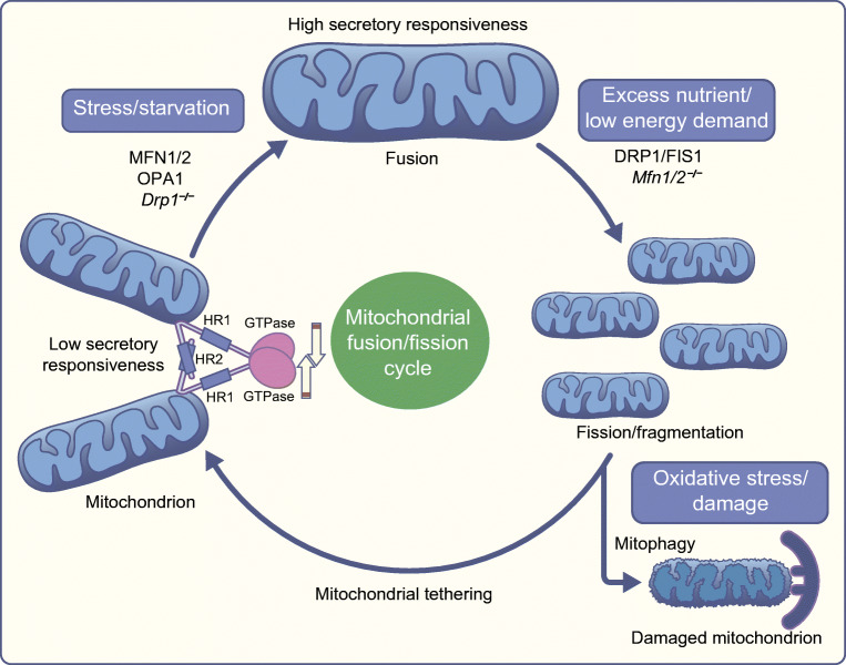
All forms of diabetes mellitus involve the loss or dysfunction of pancreatic beta cells, with the former predominating in type 1 diabetes and the latter in type 2 diabetes. Deeper understanding of the coupling mechanisms that link glucose metabolism in these cells to the control of insulin secretion is therefore likely to be essential to develop new therapies. Beta cells display a remarkable metabolic specialisation, expressing high levels of metabolic sensing enzymes, including the glucose transporter GLUT2 (encoded by SLC2A2) and glucokinase (encoded by GCK). Genetic evidence flowing from both monogenic forms of diabetes and genome-wide association studies for the more common type 2 diabetes, supports the importance for normal glucose-stimulated insulin secretion of metabolic signalling via altered ATP generation, while also highlighting unsuspected roles for Zn2+ storage, intracellular lipid transfer and other processes. Intriguingly, genes involved in non-oxidative metabolic fates of the sugar, such as those for lactate dehydrogenase (LDHA) and monocarboxylate transporter-1 ([MCT-1] SLC16A1), as well as the acyl-CoA thioesterase (ACOT7) and others, are selectively repressed ('disallowed') in beta cells. Furthermore, mutations in genes critical for mitochondrial oxidative metabolism, such as TRL-CAG1-7 encoding tRNALeu, are linked to maternally inherited forms of diabetes. Correspondingly, impaired Ca2+ uptake into mitochondria, or collapse of a normally interconnected mitochondrial network, are associated with defective insulin secretion. Here, we suggest that altered mitochondrial metabolism may also impair beta cell-beta cell communication. Thus, we argue that defective oxidative glucose metabolism is central to beta cell failure in diabetes, acting both at the level of single beta cells and potentially across the whole islet to impair insulin secretion. Graphical abstract.

Keywords: Beta cells; Disallowed genes; Insulin secretion; Interconnectivity; Mitochondria; Review; Type 2 diabetes.


Figures

None
Graphical abstract
Fig. 1
Fig. 1
Signalling mechanisms and the role of disallowed genes in beta cell insulin secretion in response to glucose (GSIS). See the main text for further details. GTP is proposed to stimulate insulin release in the cytosol. Products of disallowed genes involved in insulin secretion are represented in red. Lack of lactate dehydrogenase (LDH) and monocarboxylate transporter-1 (MCT-1/SLC16A1) prevents the conversion and extracellular entry, respectively, of lactate and pyruvate which would otherwise prompt inappropriate insulin release. NEFA are activated to FA-CoA in the cytoplasm and can access the mitochondria through carnitine palmitoyltransferase I (CPT-1), where β-oxidation generates Ac-CoA that incorporates into the TCA cycle to potentially enhance insulin secretion. In the cytosol, a glycerolipid/NEFA cycle (GL/NEFA), fatty acids (FA) are esterified with glucose-derived glycerol-3-phosphate (Gro3P) to generate monoacylglycerol (MAG), which enhances insulin release. NEFA could potentially (grey dotted arrow) be released from the beta cell and agonise free fatty acid receptor 1 (FFAR1/GPR40). Low ACOT7 limits the FA-CoA hydrolysis that would result in a lower FA-CoA/NEFA ratio in the cytoplasm or mitochondria. This could affect β-oxidation, the GL/NEFA cycle and the activation of FFAR1 and thus prevent undesired secretory granule release. Examples of transcription factors contributing to gene disallowance are depicted in blue (RFX6, PAX6) and miRNAs are shown in red (miR-29a/b). Ac-CoA, Acyl-CoA; GK, Glucokinase; Pyr, pyruvate; SCS-GTP, succinyl-CoA synthetase; TCA, tricarboxylate cycle. This figure is available as part of a downloadable slideset
Fig. 2
Fig. 2
Putative roles for proteins controlling mitochondrial shape and dynamics in beta cells. See the text for further discussion. The outer mitochondrial membrane (OMM) GTPases MFN1 and MFN2 are responsible for the fusion of these membranes on two adjacent mitochondria, while optic atrophy 1 (OPA1), drives inner mitochondrial membrane (IMM) fusion. Heptad repeat domains 2 (HR2) are essential for the initial tethering between adjacent mitochondria, while hydrolysis of the GTPase domain is needed for fusion completion. The latter allows the transfer of mitochondrial membrane components, metabolites and normal mtDNA copies. Elongated mitochondria, with high secretory responsiveness, will undergo fission with the support of DRP1 and FIS1. Fragmentation is an essential process involved in isolating dysfunctional mitochondrial units or mutant mtDNA copies from the mitochondrial network. During mitochondrial division, organelles moderately malfunctioning or damaged (depolarised) due to oxidative stress will undergo autophagy, a process also referred to as mitophagy. Functional mitochondria will instead either remain fragmented (low secretory responsiveness) during high nutrient supply conditions or will fuse with neighbouring organelles when the cell is under high energy demand (starvation). Studies showed that deletion or silencing of Drp1 (Drp1/) or Mfn1 and Mfn2 (Mfn1/2/), affect insulin secretion and glucose homeostasis in mice. This figure is available as part of a downloadable slideset
Fig. 3
Fig. 3
Single cell RNA-seq analysis of islets from the zebrafish (Danio rario) to identify putative hub/leader cells. Cluster analysis was performed based on the co-expression of high Gck, but low Ins1 levels in a subset corresponding to ~10% of all cells. (a) Heatmap showing the top 20 genes defining the putative hub cells. Hub and follower cells are defined as ‘1’ and ‘0’, respectively. (b) Statistically over-represented Gene Ontology (GO) Biological Process (BP) terms in genes upregulated in putative hub cells. FDR, false discovery rate. Adapted from [59] with permission from Springer Nature, ©2019. This figure is available as part of a downloadable slideset
Fig. 4
Fig. 4
Beta cell network model depicting impairment of insulin secretion following external or genetic alterations to mitochondrial fusion protein expression. Leader (hub) beta cells (green) coordinate pulsatile insulin secretion and signal propagation across an islet through signalling routes such as the gap junction protein connexin 36. Diabetogenic insults or genetic deletion/lowered expression of proteins involved in the mitochondrial fusion process cause the mitochondrial network to rapidly fragment and no fusion occurs while these proteins are absent. This will also lead to progressive reduction in insulin secretion, loss of beta to beta cell interconnection, and development of type 2 diabetes, and may conceivably contribute to secretory insufficiency in type 1 diabetes [60] in some circumstances. For simplicity, non-beta cells are omitted from the islet diagram (left-hand panel). Red structures (no central nucleus) represent capillaries. This figure is available as part of a downloadable slideset

References

    1. Henquin JC, Dufrane D, Nenquin M. Nutrient control of insulin secretion in isolated normal human islets. Diabetes. 2006;55(12):3470–3477. doi: 10.2337/db06-0868. - DOI - PubMed
    1. Milner RD. The secretion of insulin from foetal and postnatal rabbit pancreas in vitro in response to various substances. J Endocrinol. 1969;44(2):267–272. doi: 10.1677/joe.0.0440267. - DOI - PubMed
    1. Rutter GA, Pullen TJ, Hodson DJ, Martinez-Sanchez A. Pancreatic β-cell identity, glucose sensing and the control of insulin secretion. Biochem J. 2015;466:202–218. doi: 10.1042/BJ20141384. - DOI - PubMed
    1. McCulloch LJ, van de Bunt M, Braun M, Frayn KN, Clark A, Gloyn AL. GLUT2 (SLC2A2) is not the principal glucose transporter in human pancreatic beta cells: implications for understanding genetic association signals at this locus. Mol Genet Metab. 2011;104(4):648–653. doi: 10.1016/j.ymgme.2011.08.026. - DOI - PubMed
    1. Rorsman P, Ashcroft FM. Pancreatic β-cell electrical activity and insulin secretion: of mice and men. Physiol Rev. 2018;98(1):117–214. doi: 10.1152/physrev.00008.2017. - DOI - PMC - PubMed

Publication types