Review
. 2020 Oct;63(10):2022-2029.
doi: 10.1007/s00125-020-05185-6. Epub 2020 Sep 7.

Functional changes in beta cells during ageing and senescence

Affiliations
Review

Functional changes in beta cells during ageing and senescence

Cristina Aguayo-Mazzucato. Diabetologia. 2020 Oct.

Abstract

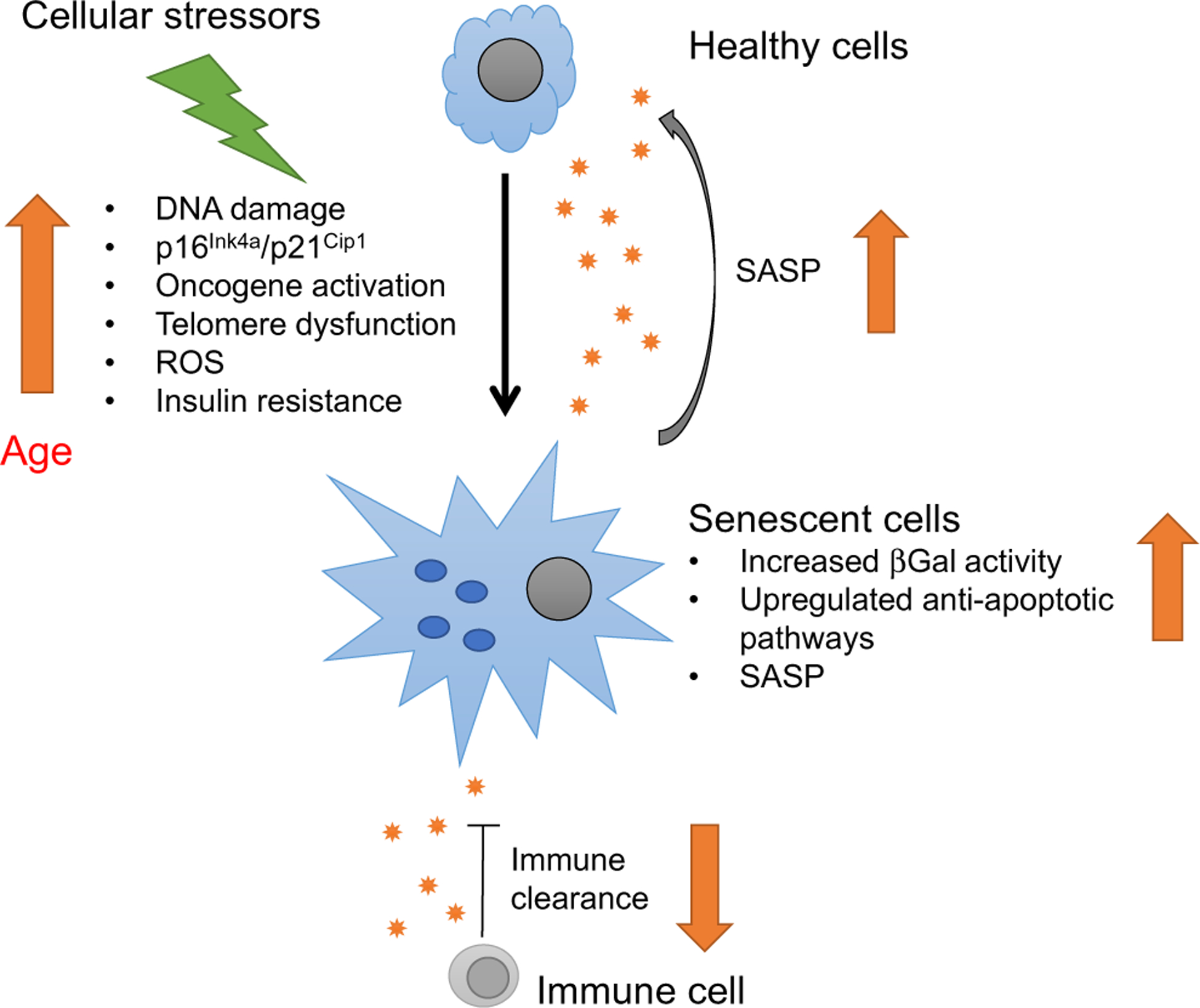
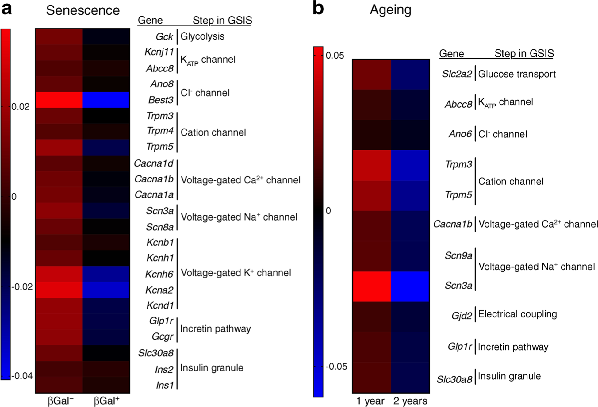
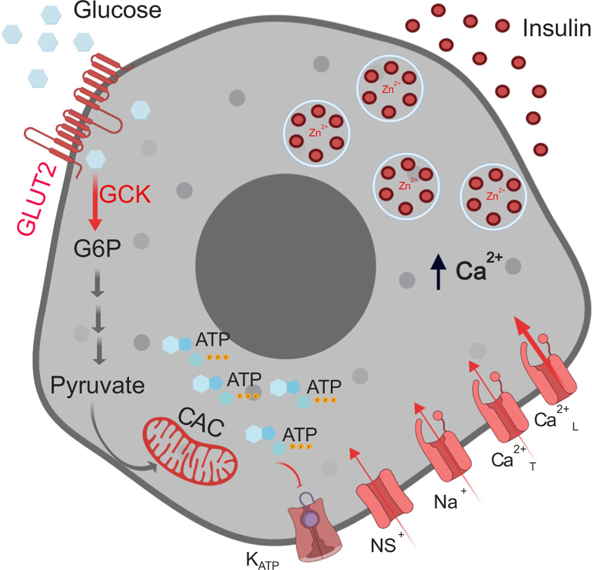
Insulin secretion from beta cells is crucial for maintaining euglycaemia and preventing type 2 diabetes, a disease correlated with ageing. Therefore, understanding the functional changes that beta cell function undergoes with age can reveal new therapeutic targets and strategies to delay or revert the disease. Herein, a systematic review of the literature agrees that, as humans age, their beta cell function declines, independently of peripheral insulin resistance, BMI and waist circumference. Rodent studies reveal that, with age, basal insulin secretion increases with either no change or an increase in stimulated insulin secretion, but the biological significance of this is unclear. The accumulation of senescent beta cells could explain some of these functional changes: transcriptional analysis of senescent and aged beta cells revealed parallel downregulation of several steps along the pathway linking glucose stimulation and insulin secretion. Moreover, specific deletion of senescent cells (senolysis) improved residual beta cell function, gene expression profile and blood glucose levels. In conclusion, cellular senescence could underlie the functional decline of beta cells during ageing and could represent a novel and promising approach for recovering insulin secretion. Graphical abstract.

Keywords: Ageing; Beta cells; Function; Human; Insulin secretion; Review; Rodents; Senescence; Senolysis; Type 2 diabetes.


Figures

Fig. 1
Fig. 1
Cellular senescence is a stress response (to DNA damage, ER stress, oncogene activation that induces expression of senescence markers and effectors p16Ink4a and p21Cip1) that leads to a lack of proliferation in response to growth stimuli, increased activity of senescence-associated (SA-βGal) and to the secretion of an array of proteins specific to each cell type known as SASP. SASP proteins include soluble and insoluble factors, such as chemokines, cytokines and extracellular matrix remodelling factors. SASP can induce dysfunction in surrounding cells and promote their entry into senescence, i.e. they have a ‘contagious-like’ effect. Some SASP proteins can recruit immune cells for clearance of senescent cells. Since senescence is a cellular stress response, it can occur at any time, but with age, cellular stressors increase and the immune response decreases, leading to an accumulation of senescent cells.
Fig. 2
Fig. 2
Beta cell gene expression changes due to senescence and chronological ageing are similar and reveal downregulation of key players in the stimulus–secretion coupling mechanism. (a) Islets isolated from 7–8-month-old C56Bl/6 J mice were FACS sorted into non-senescent (βGal) and senescent (βGal+) for RNA-Seq; Mean expression is shown for n = 7 sets of paired samples. Data reported in this panel have been deposited in NCBI Gene Expression Omnibus (GEO accession number GSE121539) [2]. (b) Microarray data of purified beta cells of MIP-GFP mice 1 and 2 years of age. Mean expression is shown for n = 4 for 1 year of age, n = 3 for 2 years of age. Data reported in this panel have been deposited in the NCBI GEO database under accession number GSE72753 [16].
Fig. 3
Fig. 3
Glucose stimulus–insulin secretion coupling mechanism in beta cells. As extracellular glucose concentrations increase, glucose uptake via GLUT2 increases glucose metabolism via glycolysis (glucokinase-GCK), the citric acid cycle (CAC) and oxidative phosphorylation, increasing ATP production. This, in turn, leads to closure of the KATP channels, which, along with continued conductance through non-selective cationic channels (NS+), results in membrane depolarisation. When the membrane potential reaches a certain threshold, voltage-gated sodium (Na+) channels and T-type Ca2+ channels (Ca2+T) open increasing the membrane potential further to activate the L-type Ca2+ channels (Ca2+L). The associated Ca2+ influx triggers exocytosis of insulin-containing secretory granules. Shown in the different shades of red are components decreased in aged and senescent cells that could result in decreased insulin secretion. G6P, glucose 6-phosphate. Created in BioRender.com.

References

    1. Koopman RJ, Mainous AG 3rd, Diaz VA, Geesey ME (2005) Changes in age at diagnosis of type 2 diabetes mellitus in the United States, 1988 to 2000. Ann Fam Med 3(1):60–63. 10.1370/afm.214 - DOI - PMC - PubMed
    1. Aguayo-Mazzucato C, Andle J, Lee TB Jr et al. (2019) Acceleration of beta cell aging determines diabetes and senolysis improves disease outcomes. Cell Metab 30(1):129–142 e124. 10.1016/j.cmet.2019.05.006 - DOI - PMC - PubMed
    1. Thompson PJ, Shah A, Ntranos V, Van Gool F, Atkinson M, Bhushan A (2019) Targeted elimination of senescent beta cells prevents type 1 diabetes. Cell Metab 29(5):1045–1060 e1010. 10.1016/j.cmet.2019.01.021 - DOI - PubMed
    1. Xiao J, Weng J, Ji L et al. (2014) Worse pancreatic beta-cell function and better insulin sensitivity in older Chinese without diabetes. J Gerontol A Biol Sci Med Sci 69(4):463–470. 10.1093/gerona/glt104 - DOI - PubMed
    1. Hirose H, Takayama M, Iwao Y, Kawabe H (2016) Effects of aging on visceral and subcutaneous fat areas and on homeostasis model assessment of insulin resistance and insulin secretion capacity in a comprehensive health checkup. J Atheroscler Thromb 23(2):207–215. 10.5551/jat.30700 - DOI - PubMed

Publication types