. 2020 Sep 7;70(1):39.
doi: 10.1186/s12576-020-00766-x.

Mitogen-activated protein kinase p38 modulates pacemaker ion channels differentiation in P19-derived pluripotent cells

Affiliations

Mitogen-activated protein kinase p38 modulates pacemaker ion channels differentiation in P19-derived pluripotent cells

Mingqi Zheng et al. J Physiol Sci. .

Abstract

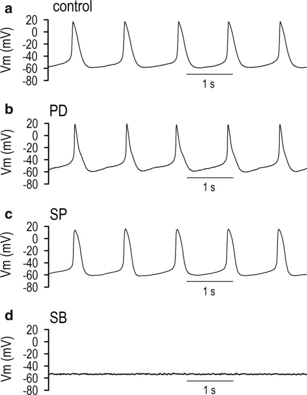
Signal regulators during early cardiogenetic differentiation for the cellular automaticity are largely unknown. Our investigations were designed to clarify the role of transcription factors and their modulators in P19-derived cardiomyocytes to the expression of cardiac pacemaker ion channels. Transcription factors Csx/Nkx2.5 and GATA4 but not MEF2C were markedly inhibited by p38 MAP kinase inhibition in a distinct manner; expression but not phosphorylation of GATA4 was reduced by inhibition of p38 MAP kinase actions. In the presence of an ERK1/2,5 inhibitor PD98059 or a JNK MAP kinase inhibitor SP600125, P19 cells successfully differentiated into cardiomyocytes displaying spontaneous beatings with expression of three types of pacemaker ion channels. We demonstrate that acquisition of cellular automaticity and the expression of pacemaker ion channels are regulated by the transcription factors, Csx/Nkx2.5 and GATA4, through intracellular signals including p38 MAP kinase in the process of P19-derived pluripotent cells differentiation into cardiomyocytes.

Keywords: Cardiogenesis; Csx/Nkx2.5; GATA4; MEF2C; P19CL6; p38MAP kinase.


Conflict of interest statement

All authors declare that they have no conflict of interest.

Figures

Fig. 1
Fig. 1
Action potentials in differentiating P19 cells. a Representative action potentials (APs) from a single isolated beating cardiomyocytes derived from P19 cells on differentiation day 14 with vehicle (control). Note that the APs exhibit distinct spontaneous diastolic depolarization with the maximum diastolic potential (MDP) of − 58.2 mV. b–d Representative action potentials (membrane potentials) in P19-derived cardiomyocytes on differentiation day 14 in the presence of 10 μM PD98059 (b), 10 μM SP600125 (c), and 10 μM SB203580 (d) recorded by the same protocol described in a
Fig. 2
Fig. 2
Current traces of ICa.L, ICa.T and hyperpolarization-activated inward current (If) in P19-derived cardiomyocytes and Effects of MAPK inhibitors. a Representative current traces of ICa.L, ICa.T and If from a single isolated beating cardiomyocytes derived from P19 cells on differentiation day 14 with vehicle (control). ICa.T was recorded by subtracting the current traces obtained by VHP of − 50 mV from the current traces obtained by VHP of − 100 mV. If was obtained from VHP of − 40 mV to various test potentials ranging from − 50 to − 140 mV in 10 mV increment. b–d Representative current traces of ICa.L, ICa.T and If in P19-derived cardiomyocytes on differentiation day 14 in the presence of 10 μM PD98059 (b), 10 μM SP600125 (c), and 10 μM SB203580 (d). eg The graphic representation of the current (I)–voltage (V) relationships of ICa.L (a), ICa.T (b), and conductance (G)–voltage (V) relationship of If (g). h The maximum chord conductance of ICa.L (Gmax, Ca.L) obtained at the potential range from + 20 mV to + 40 mV. i The maximum chord conductance of ICa.T (Gmax, Ca.T) obtained at the potential range from − 10 mV to + 10 mV. j The maximum chord conductance of If (Gmax, f) obtained at the potential range from − 120 to − 150 mV. Current amplitude of ICa.L and ICa.T were assessed by means of time-dependent components of each trace. *p < 0.05 vs. control, **p < 0.01 vs. control. Filled circle, control (n = 17–37); filled square, 10 μM PD98059 (n = 12–17); filled upward triangle, 10 μM SP600125 (n = 9–11); filled downward triangle, 10 μM SB203580 (n = 21–27)
Fig. 3
Fig. 3
Cardiac-specific transcription factors expression in P19-derived cardiomyocytes. a RT-PCR products of Csx/Nkx2.5, GATA4, and MEF2C prepared from P19-derived cardiomyocytes on differentiation day 0 to 7 and day 14 with vehicle (control). b Time course of mRNA expression of Csx/Nkx2.5 (filled circle), GATA4 (filled diamond) and MEF2C (filled upward triangle) normalized to GAPDH mRNA in the differentiation period. c Fractional mRNA expression of Csx/Nkx2.5, GATA4 and MEF2C on day 14, normalized to those on day 4. RT-PCR products (d, g, j) and their time course (e, h, k) of Csx/Nkx2.5, GATA4, and MEF2C prepared from P19-derived cardiomyocytes on differentiation day 0 to 7 and day 14 in the presence of 10 μM PD98059, 10 μM SP600125, and 10 μM SB203580. f, i, l Fractional mRNA expression of Csx/Nkx2.5, GATA4 and MEF2C on day 14, normalized to those on day 4 in the presence of PD98059 (f), SP600125 (i), and SB203580 (l)
Fig. 4
Fig. 4
Effects of MAPK inhibition on cardiac-specific transcription factors expression. Quantitative analyses in RT-PCR for three transcription factors Csx/Nkx2.5 (a), GATA4 (b), and MEF2C (c), were performed on the differentiation day 14 with or without MAP kinase inhibitors (control): PD98059 (10 μM), SP600125 (10 μM) and SB203580 (10 μM). Each expression level was normalized to the ratio in control cells on the differentiation day 14. Representative PCR products are shown in inset (upper) with the reference gene GAPDH (below). Each mRNA product was normalized to that of GAPDH. Data were calculated and normalized to those in the control condition (without MAP kinase inhibitor). *p < 0.05 vs. control, **p < 0.01 vs. control
Fig. 5
Fig. 5
Effects of MAPK inhibition on p38 and phospho-p38 proteins expression. a Time-dependent changes of p38 and phospho-p38 proteins extracted from P19-derived cardiomyocytes assessed by Western blot analysis. b p38 and phospho-p38 proteins expression on the differentiation day 14 with or without MAP kinase inhibitors: PD98059 (10 μM), SP600125 (10 μM) and SB203580 (10 μM). The results in bar graphs are expressed as the ratio of phosphorylated proteins to total proteins. **p < 0.01 vs. day 0 or Me2SO (−). †p < 0.01 vs. Me2SO (+)
Fig. 6
Fig. 6
Effects of p38 MAPK inhibition on Csx/Nkx2.5 and GATA4 proteins expression. A p38 MAPK inhibitor SB203580 reduced expression of Csx/Nkx2.5 (a), GATA4 (b) and phospho-GATA4 proteins (c) in a concentration-dependent manner assessed by Western blot analysis in P19-derived cardiomyocytes. d The ratio of phospho-GATA4 proteins to total GATA4 proteins with or without SB203580 (1–10 μM) assessed by data in panels (b, c). *p < 0.01 vs. SB (−) at day 10

References

    1. Irisawa H, Brown HF, Giles W. Cardiac pacemaking in the sinoatrial node. Physiol Rev. 1993;73:197–227. - PubMed
    1. Cho HS, Takano M, Noma A. The electrophysiological properties of spontaneously beating pacemaker cells isolated from mouse sinoatrial node. J Physiol. 2003;550:169–180. - PMC - PubMed
    1. Hagiwara N, Irisawa H, Kameyama M. Contribution of two types of calcium currents to the pacemaker potentials of rabbit sino-atrial node cells. J Physiol. 1988;395:233–253. - PMC - PubMed
    1. DiFrancesco D. The contribution of the “pacemaker” current (If) to generation of spontaneous activity in rabbit sino-atrial node myocytes. J Physiol. 1991;434:23–40. - PMC - PubMed
    1. Boyett MR, Honjo H, Kodama I. The sinoatrial node, a heterogeneous pacemaker structure. Cardiovasc Res. 2000;47:658–687. - PubMed

MeSH terms