

Review
doi: 10.1136/postgradmedj-2020-138137.
Epub 2020 Sep 7.
Lung ultrasound in the COVID-19 pandemic
Affiliations
- PMID: 32895294
- PMCID: PMC10016966
- DOI: 10.1136/postgradmedj-2020-138137
Item in Clipboard
Review
Lung ultrasound in the COVID-19 pandemic
Postgrad Med J.
2021 Jan.
Abstract
Lung ultrasound has been described for over a decade and international protocols exist for its application. It is a controversial area among pulmonologists and has had more uptake with emergency as well as intensive care physicians. We discuss the basics and evidence behind the use of lung ultrasound in respiratory failure, and what role we see it playing in the current 2019 novel coronavirus pandemic.
Keywords: Adult thoracic medicine; Respiratory infections; Thoracic medicine; Ultrasonography.
© Author(s) (or their employer(s)) 2021. No commercial re-use. See rights and permissions. Published by BMJ.
Conflict of interest statement
Competing interests: None declared.
Figures
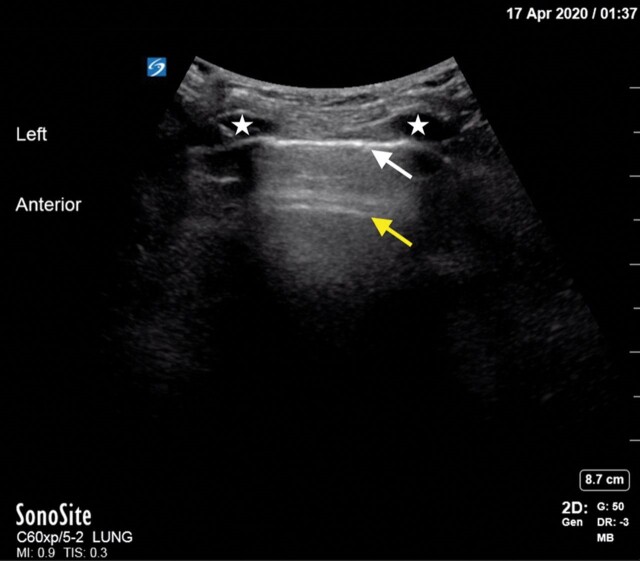
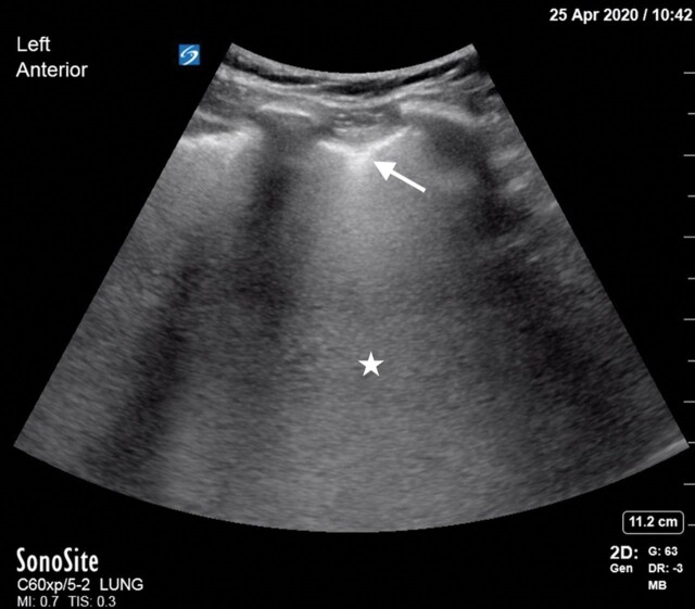
USS image showing pleural line (white arrow), A line (yellow arrow), ribs (white star).
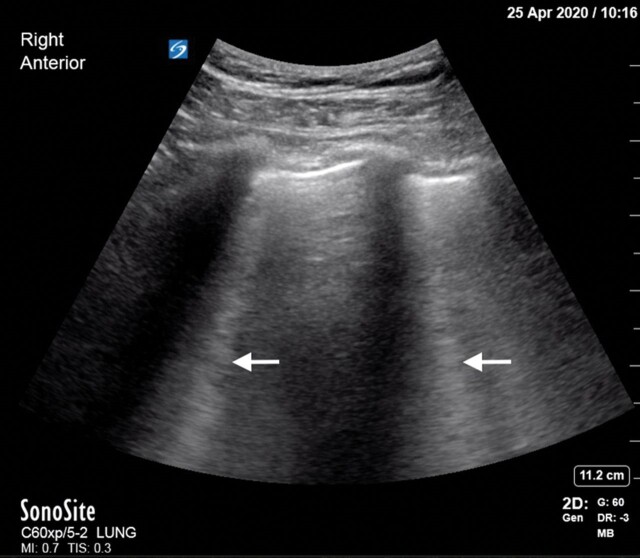
USS image showing multiple B lines.
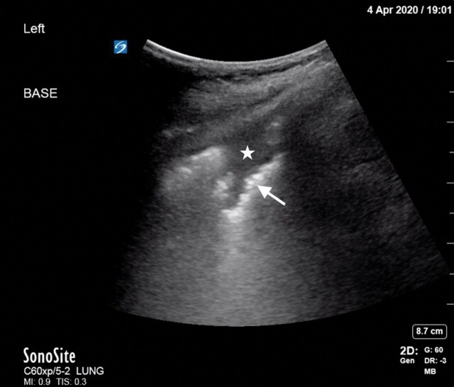
USS image showing shred sign with fractal line (white arrow), consolidated lung (white star).
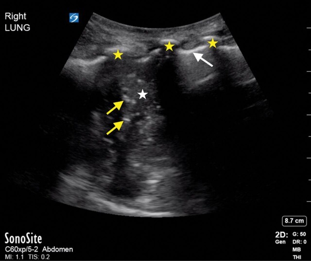
USS image showing right lung consolidation with hepatisation (white star), air bronchograms (yellow arrows); pleural line (white arrow), ribs (yellow star).
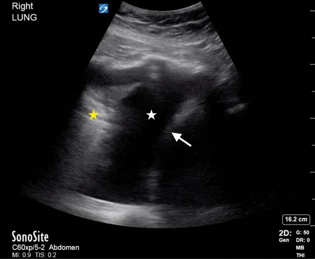
USS image showing right lung effusion (white star), atelectatic lung (yellow star) and diaphragm (white arrow).
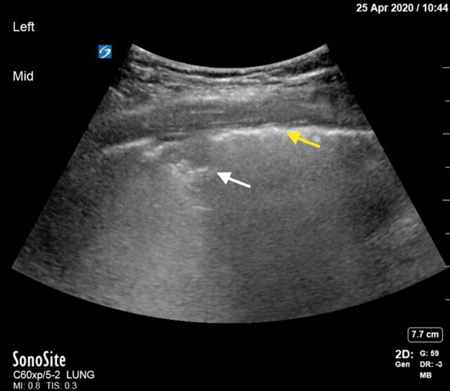
USS image of Covid-19 patient with sub pleural consolidation (white arrow) and irregular pleural line (yellow arrow).
USS image of Covid-19 patient showing Covid with white lung (white star) and sub pleural consolidation (white arrow).
References
-
- World Health Organisation. Coronavirus (COVID-19) Situation dashboard [online]. 2020. Available https://www.who.int/emergencies/diseases/novel-coronavirus-2019/situatio... (accessed 28 Aug 2020)https://www.who.int/emergencies/diseases/novel-coronavirus-2019/situatio... (accessed 28 Aug 2020)
-
- NHS England and NHS Improvement coronavirus. Speciality Guides [online]. 2020. Available https://www.england.nhs.uk/coronavirus/secondary-care/other-resources/sp... (accessed 28 Aug 2020)https://www.england.nhs.uk/coronavirus/secondary-care/other-resources/sp... (accessed 28 Aug 2020)
-
- Tian S, Hu W, Niu L, et al. Pulmonary pathology of early-phase 2019 novel coronavirus (COVID-19) pneumonia in two patients with lung cancer. J Thorac Oncol Adv 2020;15:700–4.doi: 10.1016%2Fj.jtho.2020.02.010 - PMC - PubMed
Publication types
MeSH terms
LinkOut - more resources
Full Text Sources
Medical
