Review
. 2020 Nov;16(11):618-635.
doi: 10.1038/s41582-020-0395-6. Epub 2020 Sep 7.

International consensus recommendations on the diagnostic work-up for malformations of cortical development

Affiliations
Review

International consensus recommendations on the diagnostic work-up for malformations of cortical development

Renske Oegema et al. Nat Rev Neurol. 2020 Nov.

Abstract

Malformations of cortical development (MCDs) are neurodevelopmental disorders that result from abnormal development of the cerebral cortex in utero. MCDs place a substantial burden on affected individuals, their families and societies worldwide, as these individuals can experience lifelong drug-resistant epilepsy, cerebral palsy, feeding difficulties, intellectual disability and other neurological and behavioural anomalies. The diagnostic pathway for MCDs is complex owing to wide variations in presentation and aetiology, thereby hampering timely and adequate management. In this article, the international MCD network Neuro-MIG provides consensus recommendations to aid both expert and non-expert clinicians in the diagnostic work-up of MCDs with the aim of improving patient management worldwide. We reviewed the literature on clinical presentation, aetiology and diagnostic approaches for the main MCD subtypes and collected data on current practices and recommendations from clinicians and diagnostic laboratories within Neuro-MIG. We reached consensus by 42 professionals from 20 countries, using expert discussions and a Delphi consensus process. We present a diagnostic workflow that can be applied to any individual with MCD and a comprehensive list of MCD-related genes with their associated phenotypes. The workflow is designed to maximize the diagnostic yield and increase the number of patients receiving personalized care and counselling on prognosis and recurrence risk.


Conflict of interest statement

The authors declare no competing interests.

Figures
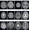
Fig. 1
Fig. 1. MRI scans showing common malformations of cortical development. The brain was scanned in the axial plane unless otherwise stated.
a | Normal brain on T1-weighted images. b | Normal brain on T2-weighted images. c | Primary microcephaly with a small brain. d | Abnormally large brain (megalencephaly) with abnormal appearance of the perisylvian cortex (arrows point to small gyri suggestive of polymicrogyria). e | Bilateral nodular heterotopia (arrows) situated along the ventricular walls. f | Lissencephaly spectrum with agyria–severe pachygyria (arrows). g | Lissencephaly spectrum with subcortical band heterotopia visible as a thick band isointense to the cortex (asterisks). h | Generalized thickened cortex with broad gyri and white matter abnormalities consistent with cobblestone complex (arrows). i | Bilateral frontoparietal polymicrogyria with abnormally small gyri and shallow sulci (arrows). j | Coronal scan showing schizencephaly, characterized by a cleft lined by grey matter extending from the cortex to the ventricle (arrow). k | Abnormally oriented sulci of varying depth with normal cortical thickness (arrows). l | Focal cortical dysplasia with blurring of the grey–white matter boundary and hyperintensity of the white matter on T2-weighted imaging (arrow).
Fig. 2
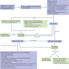
Fig. 2. Diagnostic workflow for MCDs.
This step-by-step diagnostic approach was formulated by Neuro-MIG. The main diagnostic steps are in purple-lined boxes. aSeek expert review. bIncluding prenatal and perinatal history. cIncludes construction of a pedigree and enquiry for consanguinity. dBased on additional features (for example, sick infant, abnormal liver function tests, retinal scarring or hearing loss), perinatal history (for example, maternal rash or fever) and/or imaging abnormalities (for example, calcifications, white matter injury or cysts). eOffer genetic counselling and segregation analysis to the patient and family members. fAffected brain tissue (if available), fibroblasts or saliva. CMV, cytomegalovirus; GOUS, gene of uncertain significance; MCD, malformation of cortical development; NGS, next-generation sequencing; OFC, occipitofrontal circumference, VOUS, variant of uncertain significance.

References

    1. Guerrini R, Dobyns WB. Malformations of cortical development: clinical features and genetic causes. Lancet Neurol. 2014;13:710–726. doi: 10.1016/S1474-4422(14)70040-7. - DOI - PMC - PubMed
    1. Coryell J, et al. Neuroimaging of early life epilepsy. Pediatrics. 2018;142:e20180672. doi: 10.1542/peds.2018-0672. - DOI - PMC - PubMed
    1. Barkovich AJ, Guerrini R, Kuzniecky RI, Jackson GD, Dobyns WB. A developmental and genetic classification for malformations of cortical development: update 2012. Brain. 2012;135:1348–1369. doi: 10.1093/brain/aws019. - DOI - PMC - PubMed
    1. de Wit MC, et al. Cortical brain malformations: effect of clinical, neuroradiological, and modern genetic classification. Arch. Neurol. 2008;65:358–366. - PubMed
    1. Wiszniewski W, et al. Comprehensive genomic analysis of patients with disorders of cerebral cortical development. Eur. J. Hum. Genet. 2018;26:1121–1131. doi: 10.1038/s41431-018-0137-z. - DOI - PMC - PubMed

MeSH terms