Review
. 2020 Oct:109:101686.
doi: 10.1016/j.ssnmr.2020.101686. Epub 2020 Aug 27.

Tailoring NMR experiments for structural characterization of amorphous biological solids: A practical guide

Affiliations
Review

Tailoring NMR experiments for structural characterization of amorphous biological solids: A practical guide

John E Kelly et al. Solid State Nucl Magn Reson. 2020 Oct.

Abstract

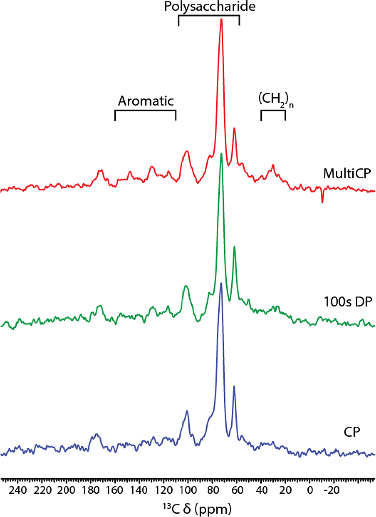
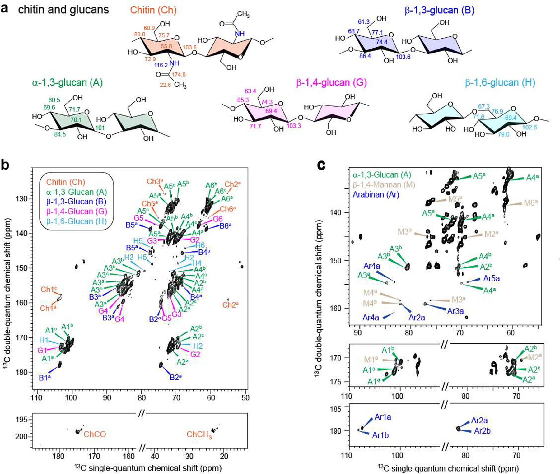
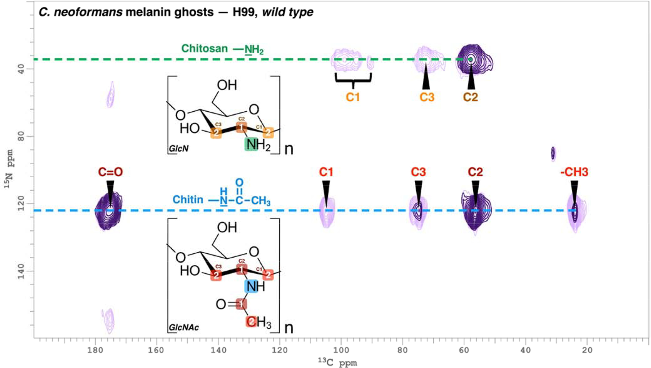
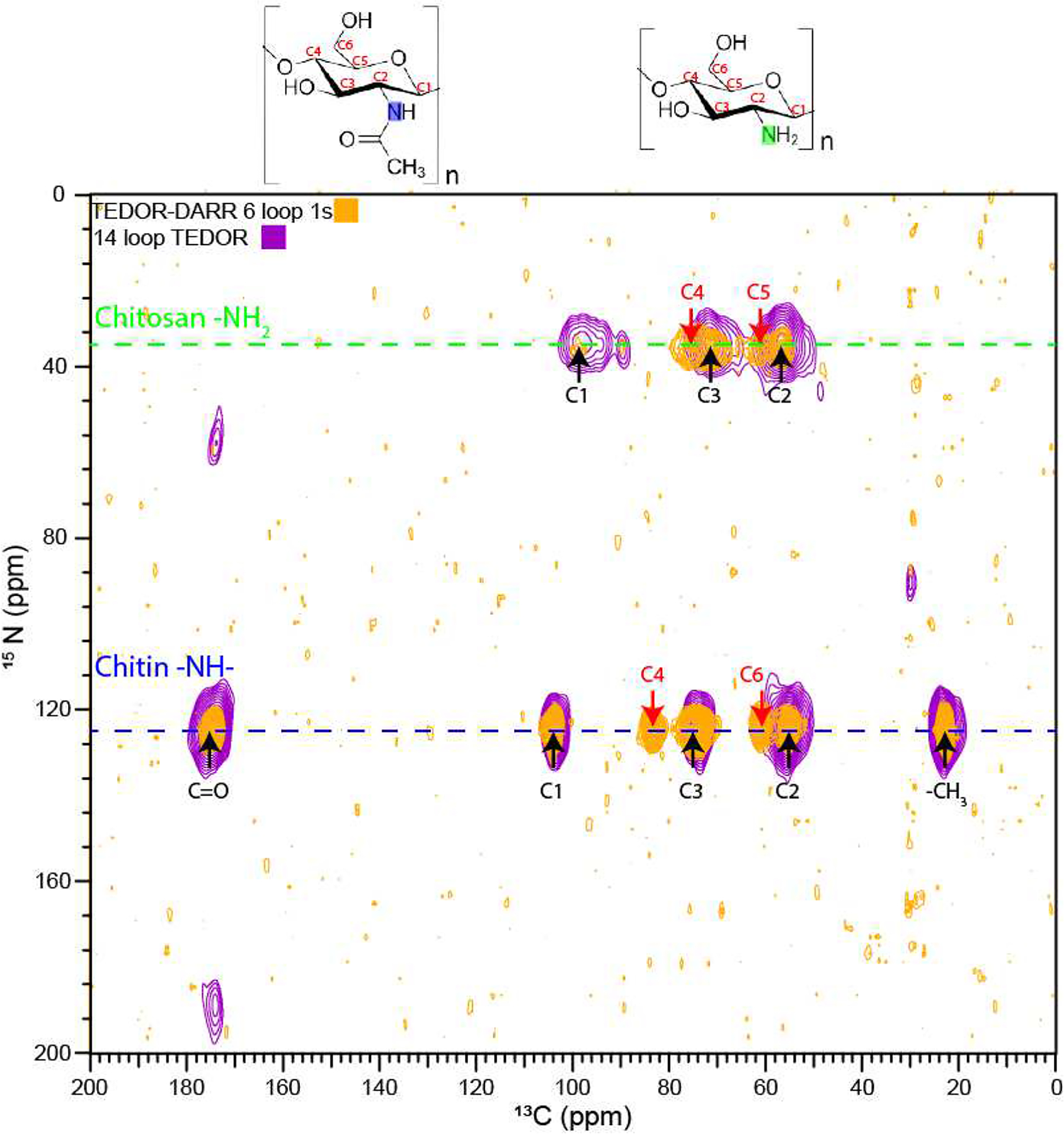
Many interesting solid-state targets for biological research do not form crystalline structures; these materials include intrinsically disordered proteins, plant biopolymer composites, cell-wall polysaccharides, and soil organic matter. The absence of aligned repeating structural elements and atomic-level rigidity presents hurdles to achieving structural elucidation and obtaining functional insights. We describe strategies for adapting several solid-state NMR methods to determine the molecular structures and compositions of these amorphous biosolids. The main spectroscopic problems in studying amorphous structures by NMR are over/under-sampling of the spin signals and spectral complexity. These problems arise in part because amorphous biosolids typically contain a mix of rigid and mobile domains, making it difficult to select a single experiment or set of acquisition conditions that fairly represents all nuclear spins in a carbon-based organic sample. These issues can be addressed by running hybrid experiments, such as using direct excitation alongside cross polarization-based methods, to develop a more holistic picture of the macromolecular system. In situations of spectral crowding or overlap, the structural elucidation strategy can be further assisted by coupling 13C spins to nuclei such as 15N, filtering out portions of the spectrum, highlighting individual moieties of interest, and adding a second or third spectral dimension to an NMR experiment in order to spread out the resonances and link them pairwise through space or through bonds. We discuss practical aspects and illustrations from the recent literature for 1D experiments that use cross or direct polarization and both homo- and heteronuclear 2D and 3D solid-state NMR experiments.

Keywords: Amorphous biosolids; Biopolymer; Correlation spectroscopy; Magic angle spinning; Solid-state NMR.


Conflict of interest statement

Declaration of competing interest The authors declare that they have no known competing financial interests or personal relationships that could have appeared to influence the work reported in this paper.

Figures

Figure 1.
Figure 1.
150 MHz 13C solid-state NMR of suberized cell walls from wound periderm of Norkotah Russet potato tubers, adapted from [28]. Bottom: 10 kHz CPMAS with a 10% linearly ramped 1H radiofrequency amplitude during a 1.0-ms contact time and a recycle delay of 3 s; middle: DPMAS with a 100-s recycle delay; top: multi-CP with 11 contact times of 1.0 ms each and separated by 1.0 s between each successive acquisition.
Figure 2.
Figure 2.
200 MHz 2D 13C CP- and DP-J-INADEQUATE spectra of A. fumigatus cell walls, adapted from [2]. a: molecular structures and representative chemical shifts of polysaccharide units; b: CP J-INADEQUATE contour plot showing through-bond connectivities for the rigid domain; c: DP J-INADEQUATE showing through-bond connectivities for polysaccharides of the mobile domain.
Figure 3.
Figure 3.
214 MHz 2D 13C PDSD spectra of Arabidopsis stems (adapted from [44]). CP-PDSD (1-s mixing) spectrum of wild type (black) stems overlaid with DP-PDSD (1.5-s mixing) spectrum of irx3 (orange) stems shows two distinct xylan conformations. The wild type two-fold xylan resonates at 105.2 ppm, and the irx3 three-fold xylan resonates at 102.6 ppm.
Figure 4.
Figure 4.
60 MHz 1D 15N CPMAS spectrum of the H99 strain of C. neoformans melanin ghosts generated from cell cultures containing [U-13C6]-D-glucose and 15N-glycine as the sole carbon and nitrogen sources, respectively (adapted from [25]). Shown are the characteristic 15N peaks corresponding to the chitin amido (123 ppm) and chitosan amino (35 ppm) nitrogens, respectively.
Figure 5.
Figure 5.
TEDOR spectra with 13C and 15N frequencies of 150 and 60 MHz, respectively for H99 C. neoformans melanin ghosts (adapted from [25]). Coherence transfer periods of 1 and 4 ms were used in separate experiments to obtain proximal carbon-nitrogen pairs separated by distances corresponding to ~1 bond length (dark purple contours) and ~2 bond lengths (light purple contours). The boxed numbers displayed on the chitin N-acetyl glucosamine (GlcNAc) and chitosan glucosamine (GlcN) monomeric units denote the number of bonds between each 13C-15N nuclear pair.
Figure 6.
Figure 6.
F3-F1 projection from a 3D TEDOR-DARR experiment obtained for H99 C. neoformans melanin ghosts with 13C and 15N frequencies of 150 and 60 MHz, respectively. A 1.6 ms coherence transfer period, corresponding to ~1 bond length, was followed by 1 s of DARR mixing. The peaks indicated with red arrows correspond to 15N-13C correlations that are not observed in the 2D TEDOR spectra of Figure 5.
Figure 7.
Figure 7.
Illustrative planes from a 3D DARR-DARR experiment obtained for wild-type A. thaliana cell walls at 150 MHz 13C (adapted from [10]). These experiments were run with preparation of the initial magnetization with either CP (tm2 = 300 ms) or DP (tm2 = 100 ms) to focus on the rigid or mobile polysaccharides, respectively. Cross-peaks marked in color correspond to intermolecular correlations.

References

    1. Zhou DH, Nieuwkoop AJ, Berthold DA, Comellas G, Sperling LJ, Tang M, Shah GJ, Brea EJ, Lemkau LR, Rienstra CM, Solid-state NMR analysis of membrane proteins and protein aggregates by proton detected spectroscopy, J. Biomol. NMR 54 (2012) 291–305. 10.1007/s10858-012-9672-z. - DOI - PMC - PubMed
    1. Kang X, Kirui A, Muszyński A, Widanage MCD, Chen A, Azadi P, Wang P, Mentink-Vigier F, Wang T, Molecular architecture of fungal cell walls revealed by solid-state NMR, Nat. Commun 9 (2018). 10.1038/s41467-018-05199-0. - DOI - PMC - PubMed
    1. Demers J-P, Fricke P, Shi C, Chevelkov V, Lange A, Structure determination of supra-molecular assemblies by solid-state NMR: Practical considerations, Prog. Nucl. Magn. Reson. Spectrosc 109 (2018) 51–78. 10.1016/j.pnmrs.2018.06.002. - DOI - PubMed
    1. McDermott A, Structure and Dynamics of Membrane Proteins by Magic Angle Spinning Solid-State NMR, Annu. Rev. Biophys 38 (2009) 385–403. 10.1146/annurev.biophys.050708.133719. - DOI - PubMed
    1. Serra O, Chatterjee S, Huang W, Stark RE, Mini-review: What nuclear magnetic resonance can tell us about protective tissues, Plant Sci. 195 (2012) 120–124. 10.1016/j.plantsci.2012.06.013. - DOI - PMC - PubMed

Publication types

MeSH terms

Substances

LinkOut - more resources