. 2020 Sep 8:26:e925361.
doi: 10.12659/MSM.925361.

Overexpression of Linc 4930556M19Rik Suppresses High Glucose-Triggered Podocyte Apoptosis, Fibrosis and Inflammation via the miR-27a-3p/Metalloproteinase 3 (TIMP3) Axis in Diabetic Nephropathy

Affiliations

Overexpression of Linc 4930556M19Rik Suppresses High Glucose-Triggered Podocyte Apoptosis, Fibrosis and Inflammation via the miR-27a-3p/Metalloproteinase 3 (TIMP3) Axis in Diabetic Nephropathy

Hong Fan et al. Med Sci Monit. .

Abstract

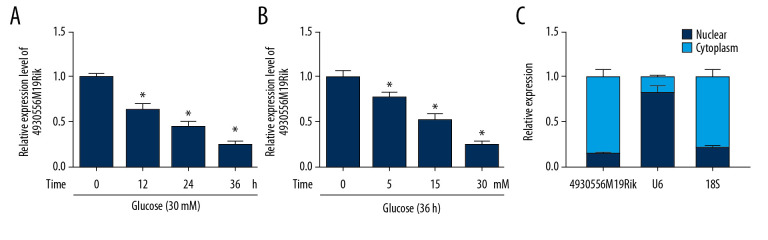
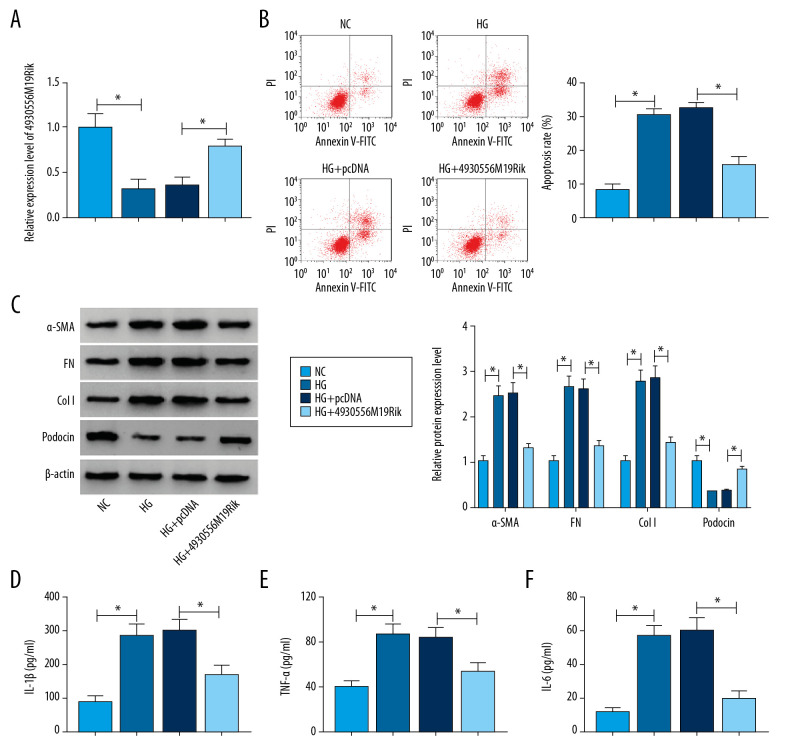
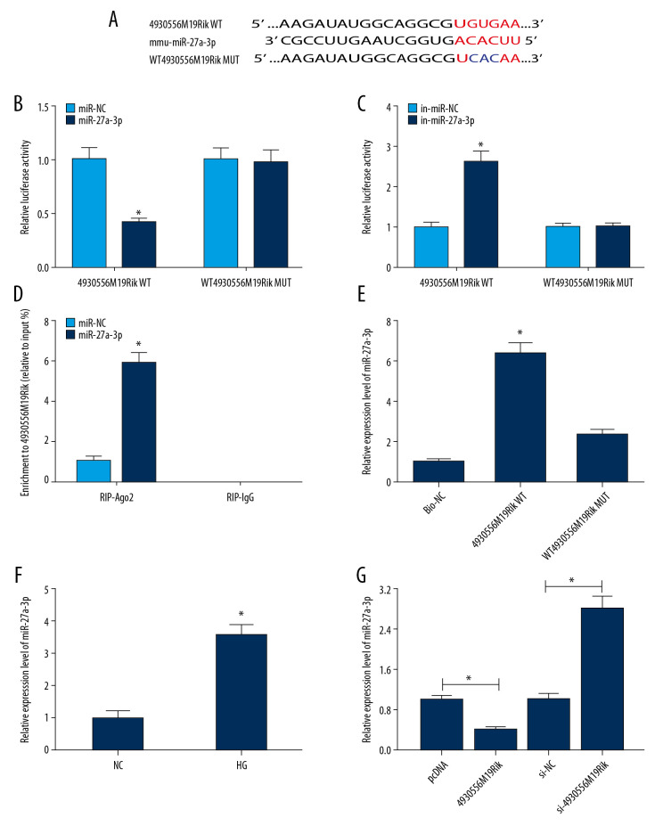
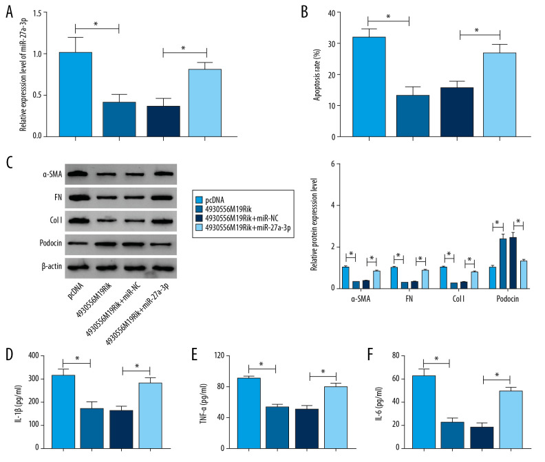
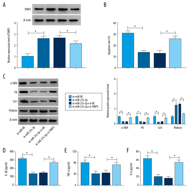
BACKGROUND Long non-coding RNAs (lncRNAs) play vital roles in development of diabetic nephropathy (DN). The goal of our study was to investigate the functional roles of long intergenic noncoding RNA (lincRNA) 4930556M19Rik in DN. MATERIAL AND METHODS A DN cell model was constructed by exposing podocytes to high glucose (HG). A subcellular fraction assay was used to determine the level of 4930556M19Rik in the nucleus and cytoplasm of podocytes. Quantitative real-time polymerase chain reaction was used to evaluate expression of 4930556M19Rik and miR-27a-3p. Western blot assay was used to assessed levels of fibrosis-related proteins, podocin, and tissue inhibitor of metalloproteinase 3 (TIMP3). Flow cytometry analysis was performed to analyze cell apoptosis. Enzyme linked immunosorbent assay was used to examine secretion of inflammatory cytokines. Dual-luciferase reporter, RIP, and RNA pull-down assays were used to verify the relationship between miR-27a-3p and 4930556M19Rik or TIMP3. RESULTS 4930556M19Rik was significantly decreased in HG-stimulated podocytes and mainly enriched in the cytoplasm of podocytes. Elevation of 4930556M19Rik hampered HG-induced cell apoptosis, fibrosis, and inflammatory in podocytes. 4930556M19Rik sponged miR-27a-3p to negatively modulate miR-27a-3p expression. MiR-27a-3p overexpression reversed the impact of 4930556M19Rik mediated cell progression in HG-induced podocytes. Moreover, TIMP3 was the target for miR-27a-3p and miR-27a-3p inhibition slowed podocyte injury by targeting TIMP3. CONCLUSIONS 4930556M19Rik overexpression slowed HG-induced podocyte injury by downregulating miR-27a-3p and upregulating TIMP3.


Figures

Figure 1
Figure 1
4930556M19Rik level was reduced in HG-stimulated podocytes. (A) Expression of 4930556M19Rik in podocytes stimulated with 30-mM glucose for 0 h, 12 h, 24 h, and 36 h was determined using qRT-PCR assay. (B) 4930556M19Rik level in podocytes induced with 0 mM, 5 mM, 15 mM and 30 mM for 36 h was detected via qRT-PCR assay. (C) Following subcellular fraction, the 4930556M19Rik level in the cytoplasmic and nuclear fractions of podocytes was examined using qRT-PCR. * P<0.05.
Figure 2
Figure 2
HG-trigged podocyte apoptosis, fibrosis and inflammation were relieved by the elevation of 4930556M19Rik. Podocytes were treated with HG (30 mM glucose), NC (5-mM glucose+24.5-mM mannitol) or transfected with pcDNA or 4930556M19Rik in HG. (A) 4930556M19Rik expression in podocytes was determined by qRT-PCR assay. (B) Apoptosis of podocytes was tested by flow cytometry analysis. (C) Protein levels of α-SMA, FN, Col I, and podocin were measured with Western blot assay. (D–F) Levels of IL-1β, TNF-α, and IL-6 were examined using ELISA kits. * P<0.05.
Figure 3
Figure 3
MiR-27a-3p was a direct target of 4930556M19Rik. (A) Complementary sequences between 4930556M19Rik and miR-27a-3p were predicted by diana_tools-lncbasev2. (B, C) Luciferase activity in podocytes co-transfected with miR-27a-3p, miR-NC, in-miR-27a-3p or in-miR-NC and 4930556M19Rik WT or 4930556M19Rik MUT was measured by dual-luciferase reporter assay. (D) After podocytes were transfected with miR-27a-3p or miR-NC, enrichment of 4930556M19Rik in Ago2 or IgG immunoprecipitation complexes was analyzed by RIP assay and qRT-PCR assay. (E) Relative expression of miR-27a-3p in podocyte lysates was determined by qRT-PCR assay following RNA pull-down assay. (F) MiR-27a-3p level in HG-induced podocytes and corresponding controls was detected using qRT-PCR assay. (G) Podocytes were transfected with pcDNA, 4930556M19Rik, si-NC or si-4930556M19Rik and then treated with HG. Next, the expression level of miR-27a-3p in podocytes was measured using qRT-PCR assay. * P<0.05.
Figure 4
Figure 4
4930556M19Rik overexpression suppressed cell apoptosis, fibrosis, and inflammatory response by binding to miR-27a-3p in HG-stimulated podocytes. Podocytes were transfected with pcDNA, 4930556M19Rik, 4930556M19Rik+miR-NC or 4930556M19Rik+miR-27a-3p followed by treatment of HG. (A) MiR-27a-3p expression in HG-induced podocytes was measured by qRT-PCR assay. (B) Apoptosis in HG-induced podocytes was tested by via flow cytometry analysis. (C) Protein levels of α-SMA, FN, Col I, and podocin in HG-induced podocytes were measured using Western blot assay. (D–F) Secretion of IL-1β, TNF-α, and IL-6 was examined with ELISA kits. * P<0.05.
Figure 5
Figure 5
TIMP3 was identified as a target gene of miR-27a-3p. (A) Potential binding sites between miR-27a-3p and TIMP3 were predicted by DianaTools-microT_CDS. (B, C) TIMP3 3′UTR WT or TIMP3 3′ UTR MUT together with miR-27a-3p, miR-NC, in-miR-27a-3p or in-miR-NC were transfected into podocytes and then the luciferase activity in podocytes was measured by dual-luciferase reporter assay. (D) The protein level of TIMP3 in HG-cultured podocytes and NC groups was measured via western blot assay. (E) After podocytes were transfected with miR-NC or miR-27a-3p and treated with HG, the level of TIMP3 protein was detected through Western blot assay. (F) Podocytes were transfected with pcDNA, 4930556M19Rik, 4930556M19Rik+miR-27a-3p or 4930556M19Rik+miR-NC and exposed to HG, then the protein level of TIMP3 was examined by western blot assay. * P<0.05.
Figure 6
Figure 6
TIMP3 knockdown reversed the suppressive roles of miR-27a-3p inhibition in cell apoptosis, fibrosis, and inflammation in HG-stimulated podocytes. Podocytes were introduced with in-miR-NC, in-miR-27a-3p, in-miR-27a-3p+si-NC or in-miR-27a-3p+si-TIMP3 followed by HG treatment. (A) The protein level of TIMP3 in HG-induced podocytes was measured by Western blot assay. (B) Cell apoptosis in HG-induced podocytes was assessed by flow cytometry analysis. (C) The protein levels of α-SMA, FN, Col I, and podocin in HG-induced podocytes were detected via Western blot assay. (D–F) Levels of IL-1β, TNF-α, and IL-6 were determined with ELISA kits. * P<0.05.

References

    1. Ha SK. ACE insertion/deletion polymorphism and diabetic nephropathy: Clinical implications of genetic information. J Diabetes Res. 2014;2014 846068. - PMC - PubMed
    1. Najafian B, Alpers CE, Fogo AB. Pathology of human diabetic nephropathy. Contrib Nephrol. 2011;170:36–47. - PubMed
    1. Alicic RZ, Johnson EJ, Tuttle KR. SGLT2 inhibition for the prevention and treatment of diabetic kidney disease: A review. Am J Kidney Dis. 2018;72:267–77. - PubMed
    1. Mercer TR, Dinger ME, Mattick JS. Long non-coding RNAs: Insights into functions. Nat Rev Genet. 2009;10:155–59. - PubMed
    1. Ma J, Zhao N, Du L, et al. Downregulation of lncRNA NEAT1 inhibits mouse mesangial cell proliferation, fibrosis, and inflammation but promotes apoptosis in diabetic nephropathy. Int J Clin Exp Pathol. 2019;12:1174–83. - PMC - PubMed