. 2020 Dec 1;66(12):1562-1572.
doi: 10.1093/clinchem/hvaa213.

Ultra-Sensitive Serial Profiling of SARS-CoV-2 Antigens and Antibodies in Plasma to Understand Disease Progression in COVID-19 Patients with Severe Disease

Affiliations

Ultra-Sensitive Serial Profiling of SARS-CoV-2 Antigens and Antibodies in Plasma to Understand Disease Progression in COVID-19 Patients with Severe Disease

Alana F Ogata et al. Clin Chem. .

Abstract

Background: Severe acute respiratory syndrome coronavirus 2 (SARS-CoV-2) has infected over 21 million people worldwide since August 16, 2020. Compared to PCR and serology tests, SARS-CoV-2 antigen assays are underdeveloped, despite their potential to identify active infection and monitor disease progression.

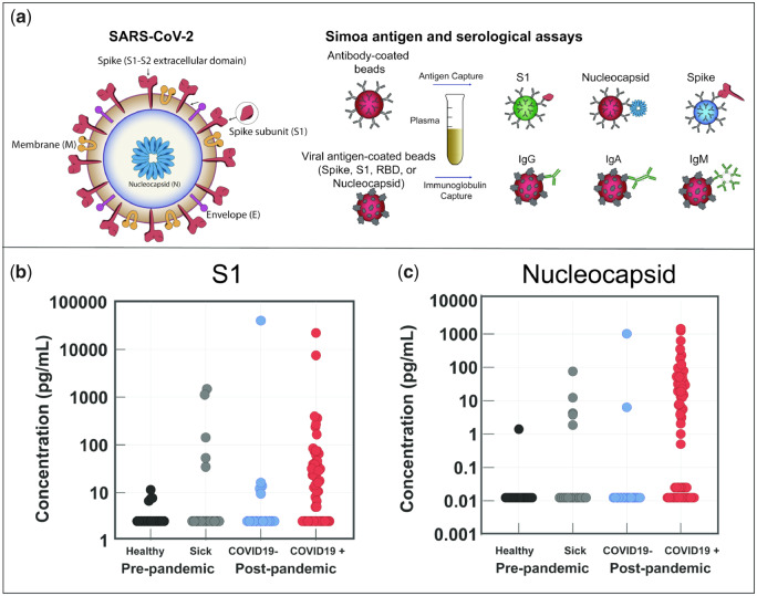
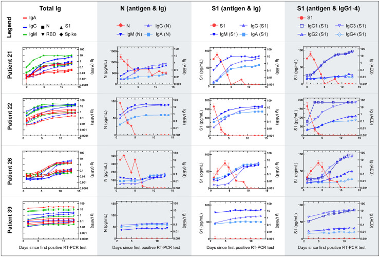
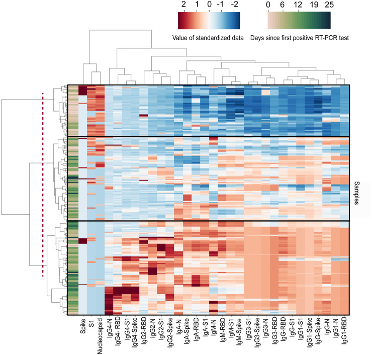
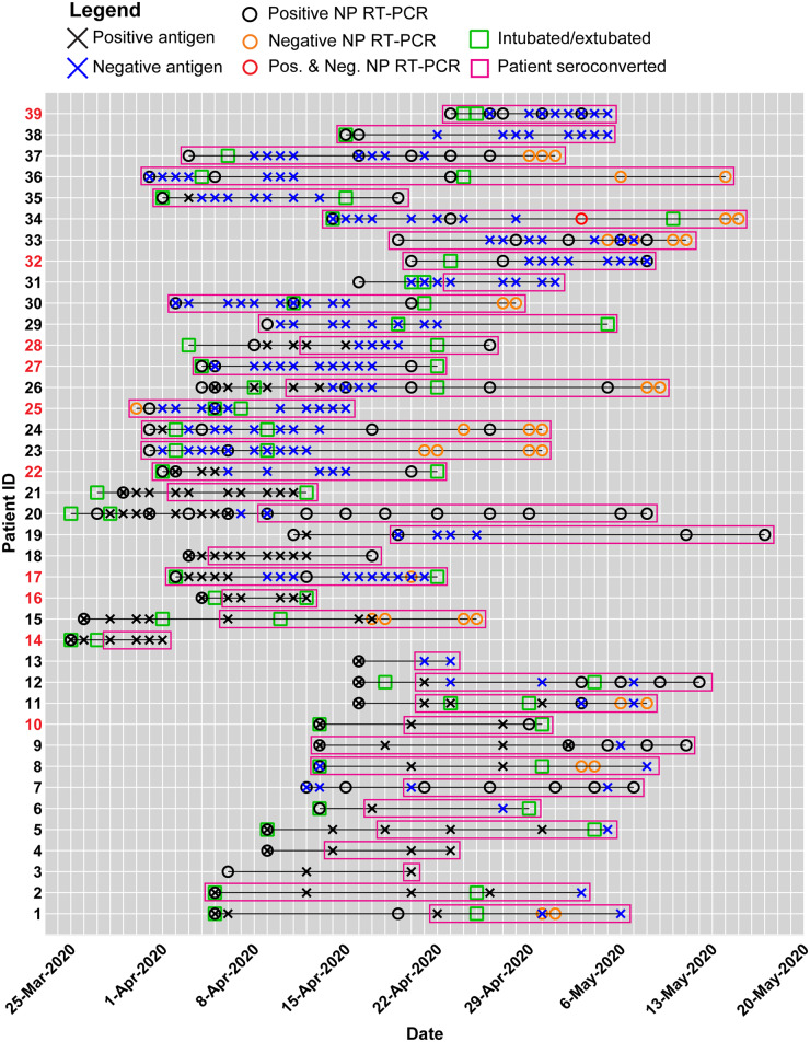
Methods: We used Single Molecule Array (Simoa) assays to quantitatively detect SARS-CoV-2 spike, S1 subunit, and nucleocapsid antigens in the plasma of patients with coronavirus disease (COVID-19). We studied plasma from 64 patients who were COVID-19 positive, 17 who were COVID-19 negative, and 34 prepandemic patients. Combined with Simoa anti-SARS-CoV-2 serological assays, we quantified changes in 31 SARS-CoV-2 biomarkers in 272 longitudinal plasma samples obtained for 39 patients with COVID-19. Data were analyzed by hierarchical clustering and were compared to longitudinal RT-PCR test results and clinical outcomes.

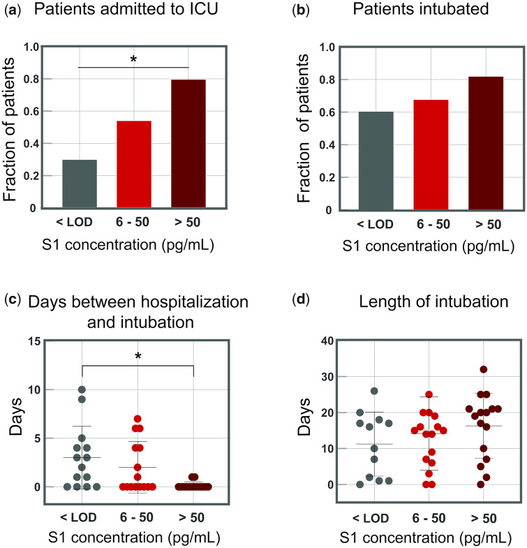
Results: SARS-CoV-2 S1 and N antigens were detectable in 41 out of 64 COVID-19 positive patients. In these patients, full antigen clearance in plasma was observed a mean ± 95% CI of 5 ± 1 days after seroconversion and nasopharyngeal RT-PCR tests reported positive results for 15 ± 5 days after viral-antigen clearance. Correlation between patients with high concentrations of S1 antigen and ICU admission (77%) and time to intubation (within 1 day) was statistically significant.

Conclusions: The reported SARS-CoV-2 Simoa antigen assay is the first to detect viral antigens in the plasma of patients who were COVID-19 positive to date. These data show that SARS-CoV-2 viral antigens in the blood are associated with disease progression, such as respiratory failure, in COVID-19 cases with severe disease.

Keywords: SARS-CoV-2; longitudinal plasma samples; serological; single molecule arrays; viral antigen.


Figures

Fig. 1
Fig. 1
SARS-CoV-2 antigen and anti-SARS-CoV-2 immunoglobulin detection in plasma. a) Schematic of Simoa detection of SARS-CoV-2 S1, spike, and N antigens and anti-SARS-CoV-2 immunoglobulins IgG, IgA, and IgM. Measurements for all antigens and immunoglobulins can be obtained from a single plasma sample (70 µL). b, c) Simoa SARS-CoV-2 antigen assay results for plasma samples collected from prepandemic healthy patients, prepandemic sick patients, and patients who tested COVID-19 negative and COVID-19 positive. Of the 64 patients, 41 who tested COVID-19 positive show detectable S1 (b) and N concentrations (c) in plasma. Each data point represents the average of 2 replicate measurements.
Fig. 2
Fig. 2
Serial data for 4 individual COVID-19 positive patients from admission to clinical recovery or death. Simoa antigen and serological results for serial plasma samples. The total IgA, IgM, and IgG levels against 4 viral antigens: nucleocapsid (N), receptor binding domain (RBD), S1, and spike, for a total of 12 interactions , N antigen concentrations with IgA, IgM, and IgG against N, S1 antigen concentrations with IgA, IgM, and IgG against S1, and S1 antigen concentrations with IgG1-4 against S1. Data points represent the mean concentration or AEB (average enzymes per bead) from 2 replicate measurements and error bars represent the standard error of the mean from 2 replicate measurements.
Fig. 3
Fig. 3
Clustergram of Simoa antigen and immunoglobulin results from 252 samples from 39 patients. Cluster map produced by Ward variance minimization algorithm. 252 samples from 39 patients were analyzed as described in the Methods section. Data were standardized after a nonlinear transformation and each row represents a single sample. Days since first RT-PCR test for each sample are presented at the far left of the clustergram. Longitudinal samples are clustered by dendrograms to the left of the clustergram. SARS-CoV-2 antigen and immunoglobulin markers are clustered by dendrograms at the top of the clustergram. The dotted line represents the cutoff that results in 3 branches: branch 1 (top), branch 2 (middle), and branch 3 (bottom). RBD: Receptor binding domain. N: Nucleocapsid.
Fig. 4
Fig. 4
Summary of clinical data and Simoa SARS-CoV-2 antigen and immunoglobulin assays for individual patients in the longitudinal analysis. A black X indicates a viral-antigen positive test, whereas a blue X indicates a viral-antigen negative test. A black circle indicates a positive NP RT-PCR test, whereas an orange circle indicates a negative NP RT-PCR test. For example, on May 3, 2020, Patient 34 received both a positive and negative NP RT-PCR result, indicated by the red circle. The intubation and extubation dates of each patient are indicated by green squares. The survival outcome for each patient is marked by the patient ID number color. Patient IDs are coded black for recovered patients who were discharged from the hospital, whereas patient IDs are coded red for deceased patients. Pink boxes represent the date of seroconversion for each patient.
Fig. 5
Fig. 5
Indicators of disease severity based on S1 concentrations in plasma for 64 COVID-19 positive patients. COVID-19 positive patients were separated into 3 groups based on S1 concentrations. The cutoff between groups 2 and 3 (50 pg/mL, 0.65 pmol/L) was chosen as 5 standard deviations above the LOD. The fraction of patients admitted to the ICU or who were intubated was calculated for each group independently. a) Fraction of COVID-19 positive patients who were immediately admitted to the ICU upon presentation to the hospital. b) Fraction of COVID-19 positive patients who were intubated during hospitalization. c) Days between date of presentation to the hospital and intubation date for intubated COVID-19 positive patients. d) The length of intubation for intubated COVID-19 positive patients. For all plots, significance indicated by the asterisks (P < 0.05).

References

    1. World Health Organization. Coronavirus disease (COVID-19) Situation Report 178. 2020. https://www.who.int/docs/default-source/coronaviruse/situation-reports/2... (Accessed August 2020).
    1. Sethuraman N, Jeremiah SS, Ryo A.. Interpreting diagnostic tests for SARS-CoV-2. JAMA 2020;323:2249–51. - PubMed
    1. Rodriguez L, Pekkarinen PT, Lakshmikanth T, et al. Systems-Level Immunomonitoring from Acute to Recovery Phase of Severe COVID-19. Cell Rep Med. 2020;1(5):100078. doi:10.1016/j.xcrm.2020.100078 - PMC - PubMed
    1. Ni L, Ye F, Cheng ML, Feng Y, Deng YQ, Zhao H, et al.Detection of SARS-CoV-2-specific humoral and cellular immunity in COVID-19 convalescent individuals. Immunity 2020;52:971–7.e3. - PMC - PubMed
    1. Amanat F, Stadlbauer D, Strohmeier S, Nguyen THO, Chromikova V, McMahon M, et al.A serological assay to detect SARS-CoV-2 seroconversion in humans. Nat Med 2020;26:1033–6. - PMC - PubMed

Publication types

MeSH terms