Figure 1.
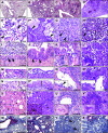
Characteristic histopathological patterns in severe acute respiratory syndrome coronavirus 2 (SARS-CoV-2)–induced pneumonia in Syrian hamsters and humans ( ° = human specimens). (A–D) In hamsters, a strongly time-dependent distribution of lesions started solely bronchially at 2 days postinfection (dpi) (A), turned into bronchointerstitial at 3 dpi (B), and into a patchy, purely interstitial pneumonia from 5 dpi onward (C), which is also characteristic in humans (D). (E and F) A consistent feature in both was acute diffuse alveolar damage, including necrosis of alveolar epithelial cells, hyaline membranes (F, arrowhead), intraalveolar macrophages (E, arrow), neutrophils (E, arrowhead), and cellular debris. (G and H) Starting at 5 dpi in hamsters, interstitial pneumonia (G) with notably high involvement of neutrophils (inset, arrowhead) and heterophils (inset, arrow) was present, similar to strong granulocytic infiltrations in humans (H, inset, arrowhead). (I and J) Marked bronchitis (I) with necrosis of bronchial epithelial cells (arrows) and intraluminal neutrophils with cellular debris (hash symbol) dominated at early time points in hamsters, followed by strong regeneration characterized by massive hyperplasia of bronchial epithelium (J, arrows indicate mitotic figures). (K and L) In both hamsters and humans, hyperplasia of alveolar type II epithelial cells with bizarre multinucleated cellular atypia (insets) was prominent. (M–O) Pleural changes included cuboidal activation of mesothelial cells (M), multifocal pleural fibrosis (N and P, bracket symbols), and pleuritis (O). (Q–V) Vascular pathology included endothelialitis (Q and R) with necrosis and desquamation of endothelial cells (Q, inset, arrowhead), alveolar edema (S and T, hash symbols), and hemorrhage (insets, asterisks) as well as perivascular lymphocytic cuffing (U and V) and edema (insets). (A–V), hematoxylin and eosin stains of formalin-fixed, paraffin-embedded tissues. (W–Y) Viral RNA as detected in hamsters by in situ hybridization preceded inflammatory reactions in a time-dependent manner, starting with high abundance only in bronchi at 2 dpi (W), spreading to the lung periphery at 3 dpi (X), and finally turning into a purely interstitial patchy pattern with less signals at 5 dpi (Y). (Z) Endothelialitis had no detectable viral RNA (hash symbols). Red, signals for viral RNA; blue, hemalaun counterstain. Scale bars: A–D, 2 mm; E, 20 μm; F, Q, U, and V, 200 μm; G–O, S, and T, 50 μm; P, R, and Z, 100 μm; W–Y, 1 mm. Scale bars in insets: G, H, K, and L, 50 μm; R, Q, S, T, and U, 100 μm; V, 500 μm.


