

Concomitant Gallbladder Agenesis with Methimazole Embryopathy
- PMID: 32898128
- PMCID: PMC7491944
- DOI: 10.12659/AJCR.926310
Concomitant Gallbladder Agenesis with Methimazole Embryopathy
Abstract
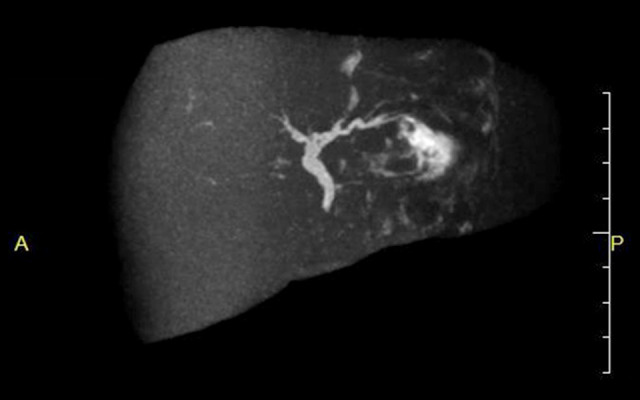
BACKGROUND Methimazole embryopathy is caused by maternal methimazole intake during early pregnancy. It causes fetal malformations such as choanal atresia, esophageal atresia, aplasia cutis, omphalomesenteric duct remnants, urachal remnants, and omphalocele. Gallbladder agenesis is sometimes complicated with other malformations, but there have been no reports of gallbladder agenesis due to methimazole or concomitant methimazole embryopathy with gallbladder agenesis. CASE REPORT The mother of a male neonate had taken methimazole for hyperthyroidism until pregnancy was recognized at 7 weeks of gestation. Ultrasonography at 12 weeks and 4 days of gestation showed the fetus had a cystic lesion in the umbilical region. The child was born at the gestational age of 38 weeks and 5 days. At birth there was omphalocele, omphalomesenteric fistula, and a scalp defect, and the child was diagnosed with methimazole embryopathy. Ultrasonography could not identify the gallbladder. Emergency surgery was performed for omphalocele with omphalomesenteric fistula on day 0. The intestine, including the omphalomesenteric fistula, was resected. Postoperative blood testing revealed hypothyroidism, so the patient was administered levothyroxine. Although MRI did not detect the gallbladder, it showed dilatation of the biliary duct. Hypothyroidism was well controlled by levothyroxine, so the patient was discharged with outpatient follow-up for the gallbladder agenesis. Six months later, the patient is asymptomatic. CONCLUSIONS Concomitant gallbladder agenesis with methimazole embryopathy has not been previously reported. In the case of a dilated common bile duct, even when asymptomatic in the neonatal period, gallbladder agenesis demands long-term follow-up because of possible manifestation of choledocholithiasis or biliary malignant tumors.
Conflict of interest statement
Figures


References
-
- Andersen SL, Olsen J, Wu CS, Laurberg P. Birth defects after early pregnancy use of antithyroid drugs: A Danish nationwide study. J Clin Endocrinol Metab. 2013;98(11):4373–81. - PubMed
-
- Yoshihara A, Noh J, Yamaguchi T, et al. Treatment of Graves’ disease with antithyroid drug in the first trimester of pregnancy and the prevalence of congenital malformation. J CIin Endocrinol Metab. 2012;97(7):2396–403. - PubMed
-
- Clementi M, Di Gianantonio E, Pelo E, et al. Methimazole embryopathy: Delineation of the phenotype. Am J Med Genet. 1999;83(1):43–46. - PubMed
-
- Martinovici D, Ransy V, Vanden Eijnden S, et al. Neonatal hemochromatosis and Martinez-Frias syndrome of intestinal atresia and diabetes mellitus in a consanguineous newborn. Eur J Med Genet. 2010;53(1):25–28. - PubMed
Publication types
MeSH terms
Substances
LinkOut - more resources
Full Text Sources
Medical
