. 2020 Sep 8;14(9):e0008365.
doi: 10.1371/journal.pntd.0008365. eCollection 2020 Sep.

Biodistribution of degradable polyanhydride particles in Aedes aegypti tissues

Affiliations

Biodistribution of degradable polyanhydride particles in Aedes aegypti tissues

Edmund J Norris et al. PLoS Negl Trop Dis. .

Abstract

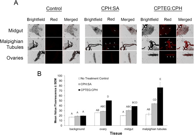
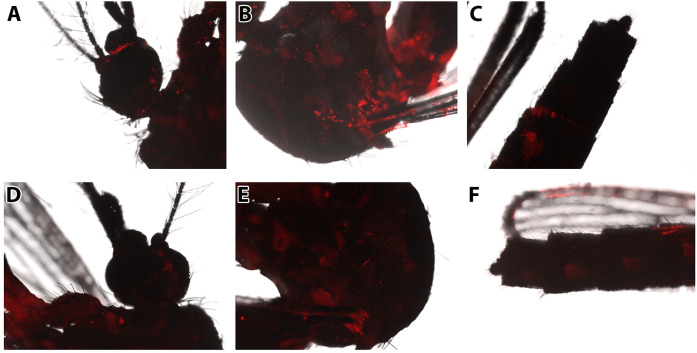
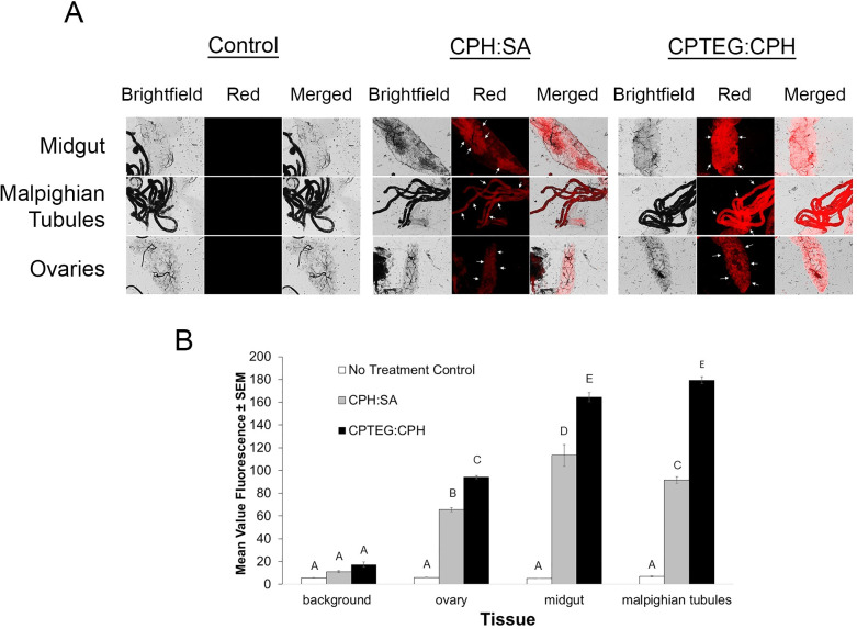
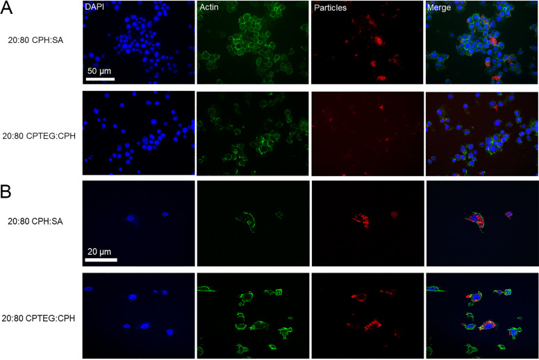
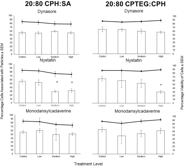
Insecticide resistance poses a significant threat to the control of arthropods that transmit disease agents. Nanoparticle carriers offer exciting opportunities to expand the armamentarium of insecticides available for public health and other pests. Most chemical insecticides are delivered by contact or feeding, and from there must penetrate various biological membranes to reach target organs and kill the pest organism. Nanoparticles have been shown to improve bioactive compound navigation of such barriers in vertebrates, but have not been well-explored in arthropods. In this study, we explored the potential of polyanhydride micro- and nanoparticles (250 nm- 3 μm), labeled with rhodamine B to associate with and/or transit across insect biological barriers, including the cuticle, epithelium, midgut and ovaries, in female Ae. aeygpti mosquitoes. Mosquitoes were exposed using conditions to mimic surface contact with a residual spray or paint, topical exposure to mimic contact with aerosolized insecticide, or per os in a sugar meal. In surface contact experiments, microparticles were sometimes observed in association with the exterior of the insect cuticle. Nanoparticles were more uniformly distributed across exterior tissues and present at higher concentrations. Furthermore, by surface contact, topical exposure, or per os, particles were detected in internal organs. In every experiment, amphiphilic polyanhydride nanoparticles associated with internal tissues to a higher degree than hydrophobic nanoparticles. In vitro, nanoparticles associated with Aedes aegypti Aag2 cells within two hours of exposure, and particles were evident in the cytoplasm. Further studies demonstrated that particle uptake is dependent on caveolae-mediated endocytosis. The propensity of these nanoparticles to cross biological barriers including the cuticle, to localize in target tissue sites of interest, and to reach the cytoplasm of cells, provides great promise for targeted delivery of insecticidal candidates that cannot otherwise reach these cellular and subcellular locations.


Conflict of interest statement

The authors have declared that no competing interests exist.

Figures

Fig 1
Fig 1. End group functionalization of polyanhydride copolymers with Rho.
(a) Chemical synthesis scheme of Rho functionalization by melt condensation. (b) 1H NMR spectra of Rho-functionalized polymers and precursors. (c) NP-HPLC chromatograms of Rho-functionalized polymers for quantifying attached vs unattached Rho. Attached Rho elutes at 3.7 min and unattached Rho elutes at 5 min. Rho-functionalized polymers appear to contain significant amounts of unattached Rho.
Fig 2
Fig 2. Scanning electron micrographs of Rho-functionalized particles.
Size distributions are provided in Table 2.
Fig 3
Fig 3. Mass of particles associated with adult female Aedes aegypti legs or whole bodies (without legs) after a 48-h exposure to nanoparticles via contact with a treated surface.
Mosquitoes were collected in groups of ten and legs were removed. Legs or bodies (without legs) were then homogenized in phosphate buffered saline for 2 h, before RhodamineB content was measured for each treatment group. Standard curves were used to calculate the initial mass of particles associated with mosquitoes. CPH:SA (C:S) particles were observed in association legs than with bodies with legs removed. The opposite trend was observed for CPTEG:CPH (C:C) particles. BD = below detectability.
Fig 4
Fig 4. Representative images of Aedes aegypti external structures associated with Rho-labeled CPH:SA and CPTEG:CPH nanoparticles following surface exposure.
Mosquitoes were exposed to particles by contact with a particle-loaded filter paper in a WHO insecticide bioassay arena. Rhodamine labeled CPH:SA nanoparticles were detected in association with the A) femur, B) tibiae, C) tarsi, D) ventral proboscis, and E) ventral abdomen of exposed mosquitoes. Rhodamine labeled CPTEG:CPH nanoparticles were detected in association with tibiae (F) and tarsi (G).
Fig 5
Fig 5. Internal tissues labeled with both Rho-functionalized CPH:SA and CPTEG:CPH nanoparticles via treated-surface contact.
A) Rho labeling was apparent throughout various explored tissues by both nanoparticle chemistries. Arrows represent tissues with high levels of labeling compared to the control. B) Mean value fluorescence of tissue samples from mosquitoes exposed to a particle-treated surface. Letters A-D represent statistically significant differences between fluorescence intensity among various tissues and particle exposures according to an ANOVA with a Bonferroni post-hoc analysis (α = 0.05) to assess differences among treatment groups and tissues. Malpighian tubules were labeled most intensely as compared to other tissues, and CPTEG:CPH was more often observed in internal tissues, in general, compared with CPH:SA particles.
Fig 6
Fig 6. Evidence of association of Rho-functionalized CPH:SA and CPTEG:CPH nanoparticles with the cuticle of female Aedes aegypti after topical exposure.
Particles were deposited in 0.2 μl volumes on the pronotum of the thorax and mosquitoes were returned to rearing conditions for 48h prior to imaging. Rho from CPH:SA nanoparticles was broadly distributed from the point of initial contact, throughout the A) head, B) thorax, and C) abdomen and was most apparent at intersegmental membranes between sclerites of the exposed insect. External labeling of mosquitoes after topical application with a suspension containing CPTEG:CPH nanoparticles was also observed in the D) head, E) thorax, and F) abdomen of mosquitoes. External Rho labeling was less intense in CPTEG:CPH trials compared with labeling observed in the CPH:SA trials. Particle labeling was diffuse throughout external tissues and even apparent on the wings of treated mosquitoes for both nanoparticle chemistries.
Fig 7
Fig 7. Nanoparticle internalization of CPHSA: or CPTEG:CPH Rho labeled particle suspensions in female Aedes aegypti exposed via topical application.
A) Control and experimental group mosquitoes were dissected to observe midgut, Malphighian tubule and ovary tissues. Representative images are shown for each group and tissue type as they appear using bright field and fluorescence microscopy. B) Mean fluorescence intensity (MFI) was quantified for each tissue and both particle types.
Fig 8
Fig 8. Evidence of association of Rho-functionalized CPH:SA and CPTEG:CPH nanoparticles with the internal organs of female Aedes aegypti after per os exposure for 48, 120 or 240 hours.
Control and experimental group mosquitoes were dissected to observe midgut, Malphighian tubule and ovary tissues. Representative images are shown for each group and tissue type as they appear using bright field and fluorescence microscopy.
Fig 9
Fig 9. Evidence of uptake of Rho-functionalized CPH:SA and CPTEG:CPH nanoparticles in Aedes aegypti Aag2 cells in culture.
DAPI was used to visualize the nucleus and phalloidin-Alexafluor 488 (green) was used to visualize the actin of the cells. Rho-labeled particles appear as red. Visualization via these methods (A: epifluorescent microscopy and B: confocal microscopy) illustrates the ability of particles to be internalized into the cytoplasm of exposed cells. Particles were not observed in association with the nuclei of exposed cells.
Fig 10
Fig 10. Evidence of mechanism of endocytosis of Rho-functionalized CPH:SA and CPTEG:CPH nanoparticles in Aedes aegypti Aag2 using pharmacological inhibitors of clathrin-dependent endocytosis (dynasore) receptor-mediated endocytosis (monodansylcadaverine), and caveolae-mediated endocytosis (nystatin).
Cells were exposed to inhibitors for 3 h, then to particles for 2 h, and were fixed an imaged to quantify particle uptake. The percentage of Aag2 cells associated with particles ± SEM is shown with bars and the viability of Aag2 cells ± SEM is shown with lines. Dynasore and monodansylcadaverine did not produce a statistically significant effect on the association of Aag2 cells with particles. Nystatin did significantly decrease the percentage of cells that associated with both CHP:SA and CPTEG:CPH particles.

References

    1. Webb JE, Green RA. 1945. On the penetration of insecticides through the insect cuticle. J Exp Biol.22: 8–20. - PubMed
    1. Hurst H. Permeability of insect cuticle. 1940. Nature. 45: 462–463.
    1. Rockstein M. 1973. The Physiology of Insecta. 2nd ed. Elsevier Science Ltd;
    1. Chapman RF. 2012. The Insects: Structure and Function. 5th ed. Cambridge University Press.
    1. Halstead NT, Civitello DJ, Rohr JR. 2015. Comparative toxicities of organophosphate and pyrethroid insecticides to aquatic macroarthropods. Chemosphere. 35: 265–271. - PubMed

Publication types