. 2020 Sep 8;16(9):e1008842.
doi: 10.1371/journal.ppat.1008842. eCollection 2020 Sep.

The p150 Isoform of ADAR1 Blocks Sustained RLR signaling and Apoptosis during Influenza Virus Infection

Affiliations

The p150 Isoform of ADAR1 Blocks Sustained RLR signaling and Apoptosis during Influenza Virus Infection

Olivia A Vogel et al. PLoS Pathog. .

Abstract

Signaling through retinoic acid inducible gene I (RIG-I) like receptors (RLRs) is tightly regulated, with activation occurring upon sensing of viral nucleic acids, and suppression mediated by negative regulators. Under homeostatic conditions aberrant activation of melanoma differentiation-associated protein-5 (MDA5) is prevented through editing of endogenous dsRNA by RNA editing enzyme Adenosine Deaminase Acting on RNA (ADAR1). In addition, ADAR1 is postulated to play pro-viral and antiviral roles during viral infections that are dependent or independent of RNA editing activity. Here, we investigated the importance of ADAR1 isoforms in modulating influenza A virus (IAV) replication and revealed the opposing roles for ADAR1 isoforms, with the nuclear p110 isoform restricting versus the cytoplasmic p150 isoform promoting IAV replication. Importantly, we demonstrate that p150 is critical for preventing sustained RIG-I signaling, as p150 deficient cells showed increased IFN-β expression and apoptosis during IAV infection, independent of RNA editing activity. Taken together, the p150 isoform of ADAR1 is important for preventing sustained RIG-I induced IFN-β expression and apoptosis during viral infection.


Conflict of interest statement

The authors have declared that no competing interests exist.

Figures

Fig 1
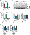
Fig 1. ADAR1 is an important host factor for IAV replication.
(A) Schematic representation of the p110 and p150 isoforms of ADAR1, including the functional domains in each isoform. The red arrow upstream of the third dsRNA binding domain indicates the region targeted by the sgRNA. (B) Western blot analysis of ADAR1 expression in ADAR1 KO and control cells. ADAR1 KO, CTRL, and WT A549s were mock treated or treated with IFN-I for 24 hours and expression of ADAR1 was examined by western blot. Expression of Ku is shown as a loading control. (C) IAV replication in ADAR1 KOs. ADAR1 KOs and CTRL A549s were infected with H5N1 (MOI = 0.001) or VSV (MOI = 0.001) and viral titers were measured at 48 hours. Data are represented as mean titer of triplicate samples ± SD. * denotes p-value ≤ 0.5. ** denotes p-value ≤ 0.01. *** denotes p-value ≤ 0.001. NS denotes p-value ≥ 0.05. Data are representative of at least three independent experiments. See also S1 Fig.
Fig 2
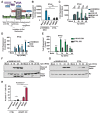
Fig 2. The p150 isoform of ADAR1 is critical for IAV replication.
(A) Schematic representation of the promoters for the p110 and p150 isoforms of ADAR1. Exons are shown as boxes with labels. The promoter for p110 is upstream of exon 1B and the start methionine for p110 is within exon 2. The promoter for p150 is upstream of exon 1A and the start methionine for p150 is within exon 1A. The green arrows depict the sgRNA target sites for p110 KO and the blue arrows depict the sgRNA target sites for p150 KO. (B) Western blot analysis of ADAR1 expression in isoform specific KO A549s. ADAR1 KO, p110 KOs, p150 KOs, and CTRL A549s were mock treated or treated with IFN for 24 hours and ADAR1 expression was examined by western blot. Expression of Ku is shown as a loading control. (C) IAV replication in isoform specific KO A549s. ADAR1 KOs, p110 KOs, p150 KOs, and CTRL A549s were infected with H5N1 (MOI = 0.001) or VSV (MOI = 0.001) and viral titers were measured at 48 hours. For easy comparison across different KOs, viral titers in this panel are shown as percentage of CTRL cell viral titers. (D) Western blot analysis of V5 tagged p150 in complemented p150 KO A549s. Two clones of p150 complements p150 KO (p150KO/p150 #1, #2), p150 KO/Vector and CTRL A549s were analyzed for expression of ADAR1 and V5 by western blot. Expression of Ku is shown as a loading control. (E) IAV replication in p150 KO/p150#1. p150 KO/p150#2, p150 KO/Vector, and CTRL A549s were infected with H5N1 (MOI = 0.001) and VSV (MOI = 0.001) and viral titers were measured at 48 hours. Data are represented as mean titer of triplicate samples ± SD. * denotes p-value ≤ 0.5. ** denotes p-value ≤ 0.01. *** denotes p-value ≤ 0.001. NS denotes p-value ≥ 0.05. Data are representative of at least three independent experiments. See also S2 Fig.
Fig 3
Fig 3. p150 supports IAV replication independent of MDA5 suppression.
(A) qRT-PCR analysis of basal IFN-β expression in ADAR1 KOs, isoform specific KOs, and CTRL A549s. Total RNA was extracted from various KOs and IFN-β mRNA levels were determined by qRT-PCR assay. Data are represented as fold expression relative to vector control A549s. (B) Western blot analysis of MDA5 and ADAR1 isoforms in various KOs. p150/MDA5 DKOs, p150 KOs, MDA5 KOs, and CTRL A549s were mock treated or treated with IFN-I for 24 hours. ADAR1 and MDA5 expression were examined by western blot. NS denotes non-specifc band. (C) qRT-PCR analysis of basal IFN-β expression in p150/MDA5 DKOs. Total RNA was extracted from various KOs and IFN-β mRNA levels were determined by qRT-PCR assay. Data are represented as fold expression relative to CTRL A549s. (D) IAV replication in p150/MDA5 DKOs. p150/MDA5 DKOs, p150 KOs, MDA5 KOs, and CTRL A549s were infected with H5N1 (MOI = 0.001) and VSV (MOI = 0.001) and viral titers were measured at 48 hours. Data are represented as mean titer of triplicate samples ± SD. (E) IAV replication in p150/MDA5 DKOs expressing wild-type V5-tagged p150. Two clones of p150/MDA5 DKOs complemented with wildtype p150 were infected with H1N1 (MOI = 0.01) and H7N7 (MOI = 0.01). p150/MDA5 DKOs that have been transduced with empty vector and CTRL A549s were also infected. Viral titers were measured at 72 hours. Data are represented as mean titer of triplicate samples ± SD. * denotes p-value ≤ 0.5. ** denotes p-value ≤ 0.01. *** denotes p-value ≤ 0.001. NS denotes p-value ≥ 0.05. Data are representative of at least three independent experiments. See also S3 Fig.
Fig 4
Fig 4. p150/MDA5 DKO A549s show elevated IFN-β expression and increased apoptosis upon RLR stimulation.
(A) Schematic representation of the RLR-Induced IRF3-mediated Pathway of Apoptosis (RIPA) and IRF3 mediated transcriptional upregulation of antiviral genes. (B) qRT-PCR analysis of IFN-β expression in p150/MDA5 DKOs after poly I:C stimulation. p150/MDA5 DKOs and CTRL A549s were transfected with LWM and HMW pI:C for 24 hours and IFN-b expression was analyzed by qRT-PCR analysis. Data are represented as fold expression relative to mock transfected CTRL A549s. (C) qRT-PCR analysis of IFN-β expression in p150/MDA5 DKOs following Sendai virus (SeV). p150/MDA5 DKOs, p150 KOs, MDA5 KOs, and CTRL A549s were infected with SeV and mRNA levels were measured at the indicated hours post infection. Data are represented as fold expression relative to mock vector control. (D) qRT-PCR analysis of IFN-β expression in p150/MDA5 DKOs following H1N1 vRNA transfection. p150/MDA5 DKOs and CTRL A549s were transfected with H1N1 vRNA and IFN-β mRNA levels were measured by qRT-PCR at the indicated hours post transfection (hpt). (E) qRT-PCR analysis of IFN-β expression in p150 KO and vector control 293s. p150 KO and vector control 293s were infected with SeV and IFN-β mRNA levels were measured by qRT-PCR at the indicated hours post infection (hpi). Data are represented as fold expression relative to mock vector control 293s. (F-G) Western blot analysis of PARP cleavage in p150/MDA5 DKOs. (F) p150/MDA5 DKOs and CTRL A549s were transfected with H1N1 vRNA and cell lysates were collected at the indicated time points post transfection. (G) p150/MDA5 DKOs and CTRL A549s were infected with H1N1 (MOI = 1) and cell lysates were collected at the indicated time points post infection. (H) Flow cytometric analysis of Annexin V+ cells following H1N1 vRNA transfection. ADAR1 KOs and CTRL A549s were transfected with H1N1 vRNA and stained with Annexin V-PE at 40 hours post transfection. The levels of Annexin V were analyzed by flow cytometry. * denotes p-value ≤ 0.5. ** denotes p-value ≤ 0.01. *** denotes p-value ≤ 0.001. NS denotes p-value ≥ 0.05. Data are representative of at least three independent experiments. See also S4 Fig.
Fig 5
Fig 5. p150 suppresses RIG-I-signaling mediated induction of IFN-β and apoptosis.
(A) Western blot analysis of PARP cleavage in p150/MDA5 DKOs following siRNA knockdown of RIG-I or IRF3 expression. RIG-I and IRF3 were knocked down in p150/MDA5 DKOs via siRNA transfection. Control siRNA were also transfected into p150/MDA5 DKOs and CTRL A549s. After 24h post siRNA transfection, cells were transfected with H1N1 vRNA for additional 24 hours. The cell lysates were analyzed for expression of RIG-I, IRF3, cleaved PARP, and Ku by western blot. (B) qRT-PCR analysis of IFN-β expression in p150/MDA5/RIG-I and p150/MDA5/MAVS TKOs after SeV infection. p150/MDA5/RIG-I TKOs, p150/MDA5/MAVS TKOs, p150/MDA5 DKOs, and CTRL A549s were infected with SeV and IFN-β mRNA levels were measured by qRT-PCR assay at 24h post-infection. Data are represented as fold expression relative to mock vector control A549s. (C) Western blot analysis of PARP cleavage TKOs following H1N1 infection. p150/MDA5/RIG-I TKOs, p150/MDA5/MAVS TKOs, p150/MDA5 DKOs, and CTRL A549s were infected with H1N1 (MO1 = 1) and lysates were collected after 40 hours. The levels of cleaved PARP and Ku were determined by western blot. (D) IAV replication in TKOs. p150/MDA5/RIG-I TKOs, p150/MDA5/MAVS TKOs, p150/MDA5 DKOs, and CTRL A549s were infected with H5N1 (MOI = 0.001) and viral titers were measured at the indicated time points post infection. Data are represented as mean titer of triplicate samples ± SD. * denotes p-value ≤ 0.5. ** denotes p-value ≤ 0.01. *** denotes p-value ≤ 0.001. NS denotes p-value ≥ 0.05. Data are representative of at least three independent experiments.
Fig 6
Fig 6. Increased type I IFN signaling and apoptosis reduce IAV replication in cells lacking p150.
(A) Western blot analysis of PARP Cleavage in p150/Bax DKOs following vRNA transfection. Two clones of p150/Bax DKOs, p150 KOs, and CTRL A549s were transfected with IAV vRNA for 24 hours. PARP cleavage and Ku expression were analyzed by western blot. (B) qRT-PCR analysis of IFN-β expression in p150/Bax DKOs following IAV vRNA transfection. Two clones of p150/Bax DKOs, p150 KOs, and CTRL A549s were transfected with H1N1 vRNA and IFN-β mRNA levels were determined 24 hours post transfection. Data are represented as fold expression relative to mock vector control. (C) IAV replication in p150/Bax DKOs. Two clones of p150/Bax DKOs, p150 KOs, and CTRL A549s were infected with H5N1 (MOI = 0.001) and viral titers were measured at the indicated time points post infection. Data are represented as mean titer of triplicate samples ± SD. (D) IAV replication in p150/Bax DKOs following inhibition of Janus kinases. p150/Bax DKOs, p150 KOs, and CTRL A549s were treated with 0.2 μM Ruxolitinib for 24 hours prior to infection. The following day cells were infected with H5N1(MOI = 0.001) in the presence of Ruxolitinib and viral titers were measured at 48 hours. Data are represented as mean titer of triplicate samples ± SD. * denotes p-value ≤ 0.5. ** denotes p-value ≤ 0.01. *** denotes p-value ≤ 0.001. NS denotes p-value ≥ 0.05. Data are representative of at least three independent experiments.
Fig 7
Fig 7. RNA binding activity of p150 is required for suppression of RIG-I signaling.
(A) A schematic representation of different p150 mutants. (B) Analysis of IFN-β-Firefly luciferase reporter activity. A firefly luciferase reporter under the control of the IFN-β promoter was transfected along with wild-type and mutant p150 constructs into 293s. At 24h post-transfection, cells were infected with SeV and luciferase activity was measured 48 hours post-transfection. Data are represented as percent luciferase activity relative to GFP+SeV. (C) Western blot analysis of V5-tagged p150 expression in complemented cells. Lysates from p150/MDA5 DKOs stably expressing wild-type and mutant p150 constructs were analyzed by western blot using an anti-V5 antibody. (D) qRT-PCR analysis of IFN-β in V5-p150 complemented p150/MDA5 DKOs. p150/MDA5 DKOs complemented with different V5-p150 mutants were transfected with H1N1 vRNA and IFN-β mRNA levels were measured 24 hours post transfection. Data are represented as fold expression relative to mock CTRL A549s. (E) Western blot analysis of PARP cleavage in complemented p150/MDA5 DKOs. p150/MDA5 DKOs complemented with p150 mutants were infected with H1N1 (MOI = 1) and cell lysates were collected at 40 hours post infection for western blot analysis of cleaved PARP. (F) IAV replication in complemented p150/MDA5 DKOs. Complemented p150/MDA5 DKOs were infected with H5N1 (MOI = 0.001) and viral titers were measured at 48 hours. Data are represented as mean titer of triplicate samples ± SD. * denotes p-value ≤ 0.5. ** denotes p-value ≤ 0.01. *** denotes p-value ≤ 0.001. NS denotes p-value ≥ 0.05. Data are representative of at least three independent experiments.

Similar articles

Cited by

References

    1. Pichlmair A, Schulz O, Tan CP, Naslund TI, Liljestrom P, Weber F, et al. RIG-I-mediated antiviral responses to single-stranded RNA bearing 5'-phosphates. Science. 2006;314(5801):997–1001. - PubMed
    1. Hornung V, Ellegast J, Kim S, Brzozka K, Jung A, Kato H, et al. 5'-Triphosphate RNA is the ligand for RIG-I. Science. 2006;314(5801):994–7. - PubMed
    1. Schlee M, Roth A, Hornung V, Hagmann CA, Wimmenauer V, Barchet W, et al. Recognition of 5' triphosphate by RIG-I helicase requires short blunt double-stranded RNA as contained in panhandle of negative-strand virus. Immunity. 2009;31(1):25–34. - PMC - PubMed
    1. Schmidt A, Schwerd T, Hamm W, Hellmuth JC, Cui S, Wenzel M, et al. 5'-triphosphate RNA requires base-paired structures to activate antiviral signaling via RIG-I. Proc Natl Acad Sci U S A. 2009;106(29):12067–72. - PMC - PubMed
    1. Kato H, Takeuchi O, Mikamo-Satoh E, Hirai R, Kawai T, Matsushita K, et al. Length-dependent recognition of double-stranded ribonucleic acids by retinoic acid-inducible gene-I and melanoma differentiation-associated gene 5. J Exp Med. 2008;205(7):1601–10. - PMC - PubMed

Publication types