. 2020 Sep 8;15(9):e0238862.
doi: 10.1371/journal.pone.0238862. eCollection 2020.

Outgrowth of erlotinib-resistant subpopulations recapitulated in patient-derived lung tumor spheroids and organoids

Affiliations

Outgrowth of erlotinib-resistant subpopulations recapitulated in patient-derived lung tumor spheroids and organoids

Malathi Banda et al. PLoS One. .

Abstract

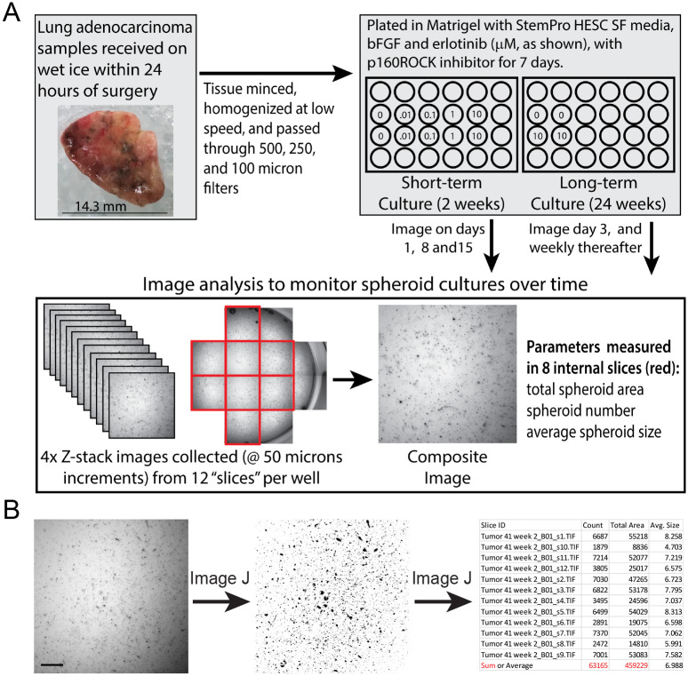
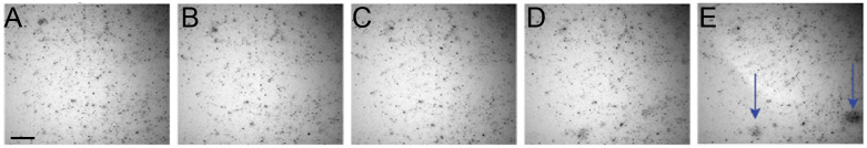
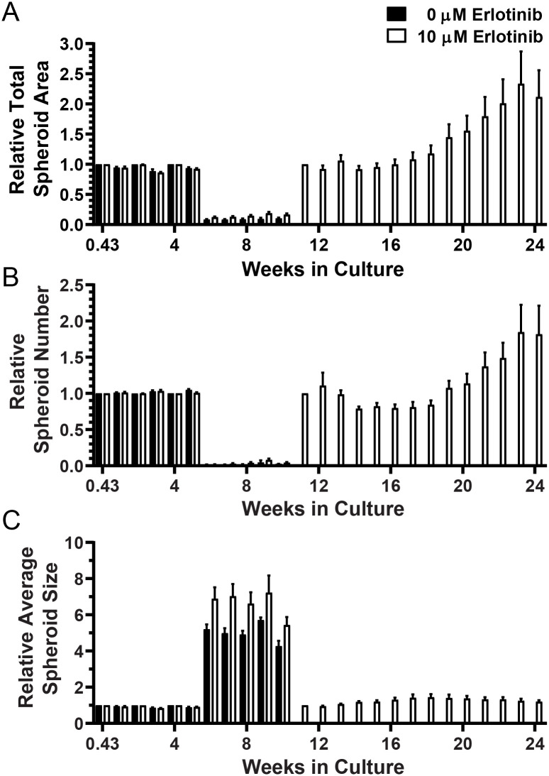
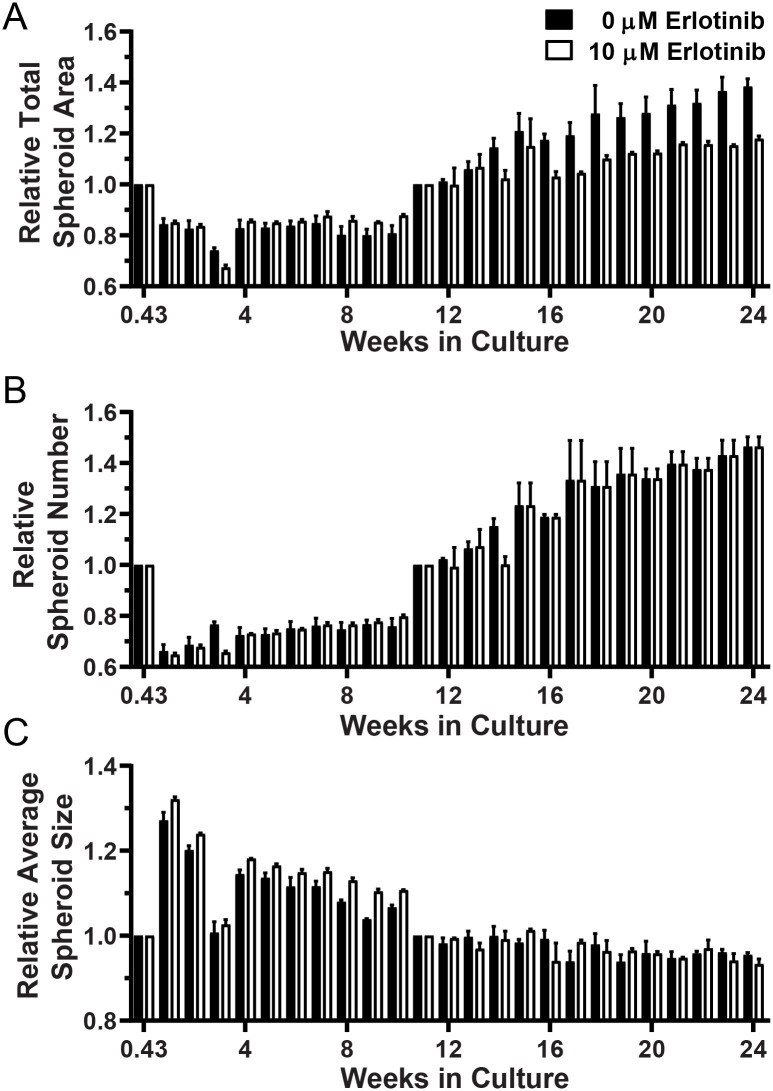
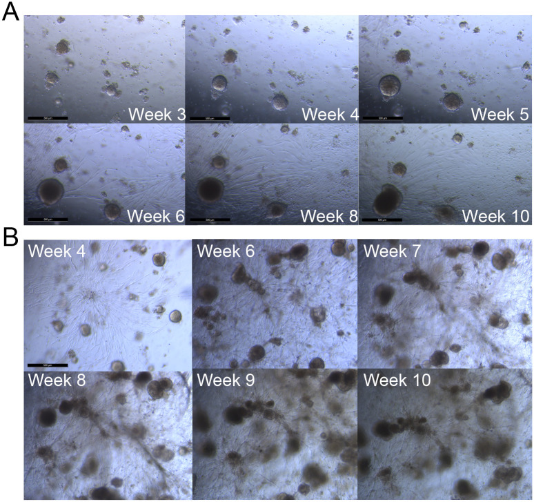
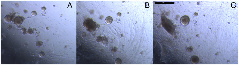
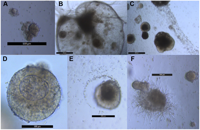
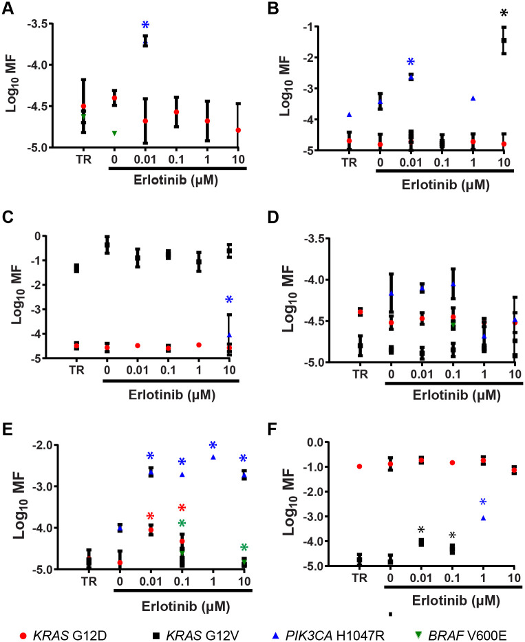
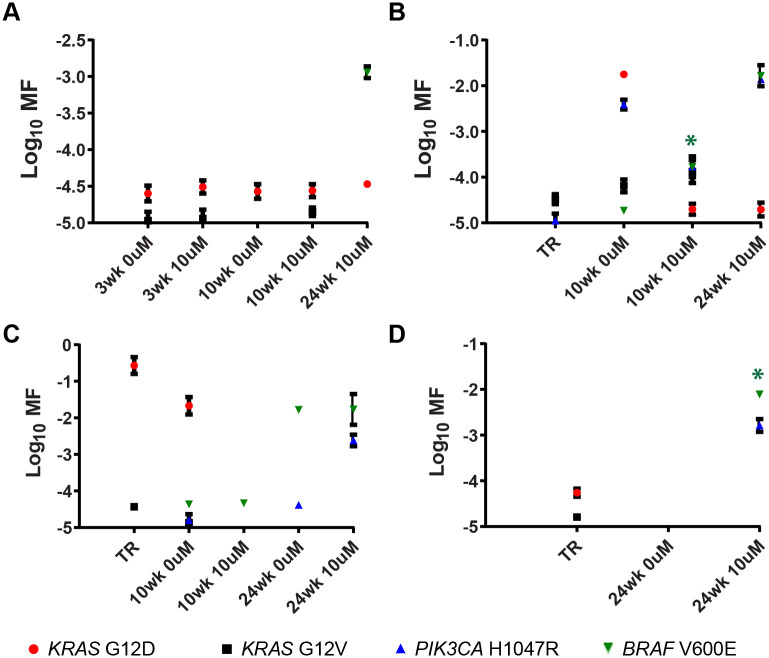
A model that recapitulates development of acquired therapeutic resistance is needed to improve oncology drug development and patient outcomes. To achieve this end, we established methods for the preparation and growth of spheroids from primary human lung adenocarcinomas, including methods to culture, passage, monitor growth, and evaluate changes in mutational profile over time. Primary lung tumor spheroids were cultured in Matrigel® with varying concentrations of erlotinib, a small molecule kinase inhibitor of epidermal growth factor receptor (EGFR) that is ineffective against KRAS mutant cells. Subtle changes in spheroid size and number were observed within the first two weeks of culture. Spheroids were cultured for up to 24 weeks, during which time interactions between different cell types, movement, and assembly into heterogeneous organoid structures were documented. Allele-specific competitive blocker PCR (ACB-PCR) was used to quantify low frequency BRAF V600E, KRAS G12D, KRAS G12V, and PIK3CA H1047R mutant subpopulations in tumor tissue residue (TR) samples and cultured spheroids. Mutant subpopulations, including multiple mutant subpopulations, were quite prevalent. Twelve examples of mutant enrichment were found in eight of the 14 tumors analyzed, based on the criteria that a statistically-significant increase in mutant fraction was observed relative to both the TR and the no-erlotinib control. Of the mutants quantified in erlotinib-treated cultures, PIK3CA H1047 mutant subpopulations increased most often (5/14 tumors), which is consistent with clinical observations. Thus, this ex vivo lung tumor spheroid model replicates the cellular and mutational tumor heterogeneity of human lung adenocarcinomas and can be used to assess the outgrowth of mutant subpopulations. Spheroid cultures with characterized mutant subpopulations could be used to investigate the efficacy of lung cancer combination therapies.


Conflict of interest statement

The authors have declared that no competing interests exist.

Figures

Fig 1
Fig 1. Processing, culture and image analysis of primary lung tumor spheroids.
Short- and long-term cultures were plated in Matrigel, and Z-stack images were collected approximately weekly as shown (A). Eight composite images from each well were analyzed using an Image J analyze particle macro to quantify total spheroid area, spheroid number, and average spheroid size (B). The scale bar in (B) is 500 μm.
Fig 2
Fig 2. Composite images show only a fraction of spheroids growing over time.
Composite images of the same well slice of Tumor 11 collected at (A) Day 3, (B) Week 2, (C) Week 4, (D) Week 6, and (E) Week 8. The scale bar in (B) is 500 μm.
Fig 3
Fig 3. Long-term culture and growth parameters for Tumor 11.
(A) Total spheroid area (measured in Px), (B) spheroid number, and (C) average spheroid size measured at Day 3 (0.43 weeks) and after passage at Weeks 3 and 10 were set to 1 for each well and relative repeat measures over time are plotted.
Fig 4
Fig 4. Long-term culture and growth parameters for Tumor 14.
(A) Total spheroid area (measured in Px), (B) spheroid number, and (C) average spheroid size measured at Day 3 (0.43 weeks) and after passage at Week 10 were set to 1 for each well and relative repeat measures over time are plotted.
Fig 5
Fig 5. Bright field images of Tumor 11 showing growth of spheroids and fibroblast-like cells over time in 10 μM erlotinib.
Sets of images were collected from the same fields of view for two different wells (A and B) between Weeks 3–10 in culture. The scale bars are 500 μm in length.
Fig 6
Fig 6. Bright field images of Tumor 11 shows multiple cell types and movement over time.
Images collected at (A) Week 4, (B) Week 5, and (C) Week 6. The scale bar (C) is 500 μm.
Fig 7
Fig 7. Large and heterogeneous organoids develop in long-term spheroid cultures.
The scale bars in panels A-E are 500 μm. The scale bar in panel F is 100 μm.
Fig 8
Fig 8. Quantification of mutant subpopulations in DNA harvested from short-term spheroid cultures.
Six of the cultures derived from the ten tumors are shown: (A) Tumor 2, (B) Tumor 3, (C) Tumor 4, (D) Tumor 5, (E) Tumor 7, and (F) Tumor 9. Asterisks denote MF measurements significantly greater than that of both TR and the corresponding 0 μM erlotinib culture (one-sided Mann Whitney test, P = 0.0500).
Fig 9
Fig 9. Quantification of mutant subpopulations in DNA harvested from long-term spheroid cultures.
Mutant quantification in cultures derived from: (A) Tumor 11, (B) Tumor 12, (C) Tumor 13, (D) and Tumor 14 are shown. Asterisks denote MF measurements significantly greater than that of both TR and its corresponding 0 μM erlotinib culture (one-sided Mann Whitney test, P = 0.0500).

References

    1. Jin M-Z, Han R-R, Qiu G-Z, Ju X-C, Lou G, Jin W-L. Organoids: An intermediate modeling platform in precision oncology Cancer Letters. 2018;414:174–80. 10.1016/j.canlet.2017.11.021 - DOI - PubMed
    1. Saglam-Metiner P, Gulce-Iz S, Biray-Avci C. Bioengineering-inspired three-dimensional culture systems: Organoids to create tumor microenvironment. Gene. 2019;686:203–12. 10.1016/j.gene.2018.11.058 - DOI - PubMed
    1. Vlachogiannis G, Hedayat S, Vatsiou A, Jamin Y, Fernández-Mateos J, Khan K, et al. Patient-derived organoids model treatment response of metastatic gastrointestinal cancers. Science. 2018;359(6378):920 10.1126/science.aao2774 - DOI - PMC - PubMed
    1. Cantrell MA, Kuo CJ. Organoid modeling for cancer precision medicine. Genome Medicine. 2015;7(1):32 10.1186/s13073-015-0158-y - DOI - PMC - PubMed
    1. Clevers H. Modeling Development and Disease with Organoids. Cell. 2016;165(7):1586–97. 10.1016/j.cell.2016.05.082 - DOI - PubMed

Publication types

MeSH terms