. 2020 Aug;10(4):e139.
doi: 10.1002/ctm2.139.

Transfer of metastatic traits via miR-200c in extracellular vesicles derived from colorectal cancer stem cells is inhibited by atractylenolide I

Affiliations

Transfer of metastatic traits via miR-200c in extracellular vesicles derived from colorectal cancer stem cells is inhibited by atractylenolide I

Dongxin Tang et al. Clin Transl Med. 2020 Aug.

Abstract

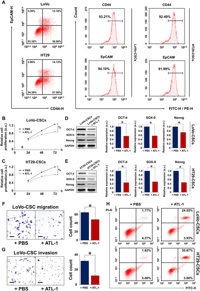
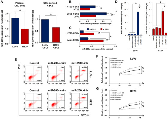
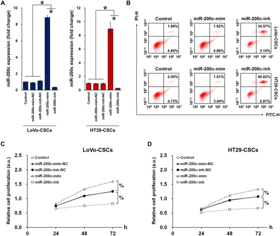
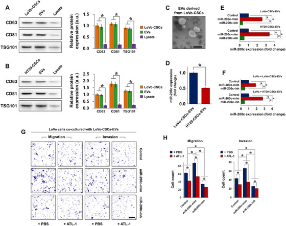
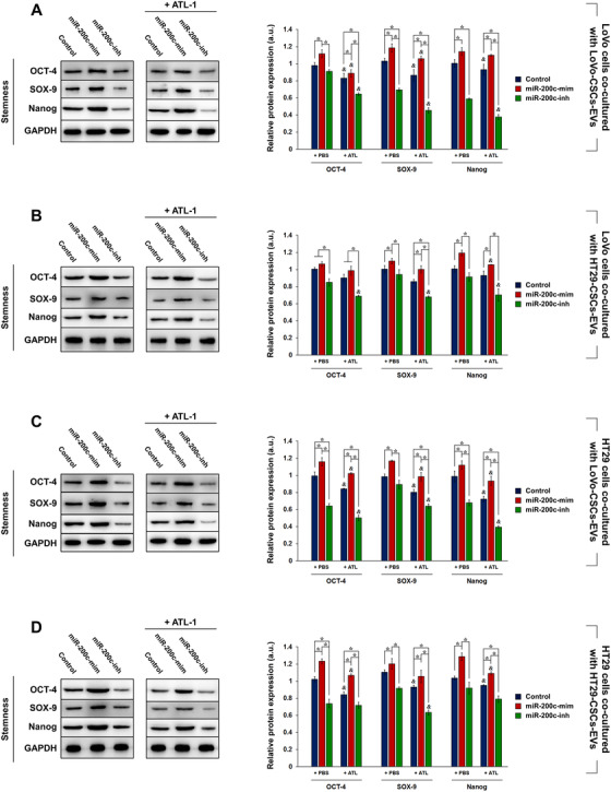
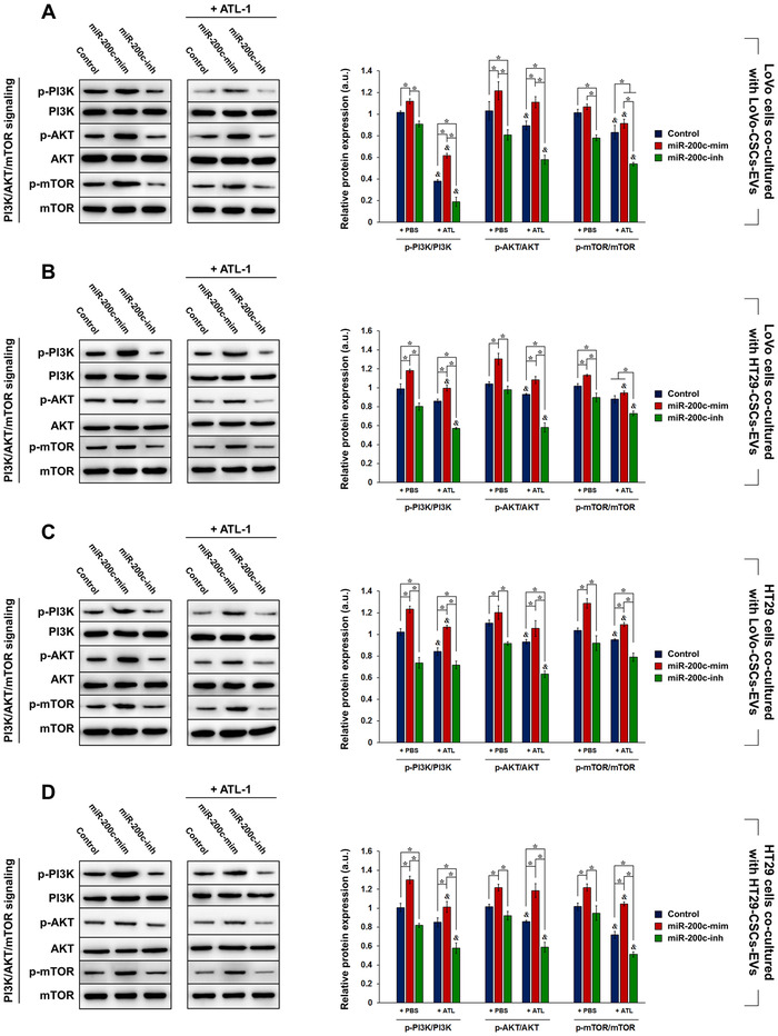
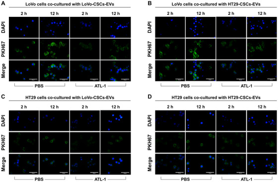
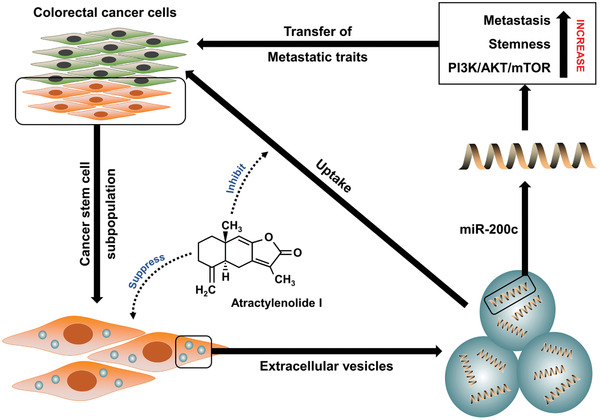
Cancer stem cells (CSCs) are important factors contributing to tumorigenesis. We examined whether CSCs isolated from colorectal cancer (CRC) cells possess metastatic properties that can be transferred to non-CSCs via the delivery of miR-200c enclosed in extracellular vesicles (EVs). The inhibitory effect of atractylenolide I (ATL-1), a traditional Chinese medicinal compound, on miR-200c activity and metastatic transfer was investigated. EVs were isolated from colorectal CSCs. The expression of miR-200c was evaluated in CSCs and CSC-derived EVs, and horizontal transfer of metastatic properties via EVs to non-CSCs was investigated in terms of cell behavior and phosphatidylinositol-4,5-bisphosphate 3-kinase (PI3K)/protein kinase B (Akt)/mammalian target of rapamycin (mTOR) signaling. CSCs isolated from metastatic CRC cells exhibited higher levels of miR-200c than those in nonmetastatic CRC cells. Overexpression of miR-200c in CSCs enhanced metastatic potential by promoting proliferation and inhibiting apoptosis, in turn leading to the release of EVs carrying an excess of miR-200c. Non-CSCs co-cultured with miR-200c-containing EVs exhibited enhanced invasion and stemness maintenance associated with PI3K/Akt/mTOR activation, demonstrating successful metastatic transfer via EV delivery. Furthermore, ATL-1 impaired the EV-mediated transfer of metastatic properties by suppressing miR-200c activity and disrupting EV uptake by non-CSCs. EVs are critical signal transducers that facilitate intercellular communication and exchange of metastatic properties, which can be controlled by ATL-1. The findings are useful in the development of microRNA-based anticancer strategies by targeting EV-mediated activity, especially using natural compounds.

Keywords: PI3K/Akt/mTOR; atractylenolide I; extracellular vesicles; metastasis; stemness.


Conflict of interest statement

The authors declare no conflict of interest.

Figures

FIGURE 1
FIGURE 1
Characterization of colorectal CSCs isolated from LoVo and HT29 cells. A, Flow cytometric sorting of CSCs using CD44 and EpCAM as markers. The percentage of parental LoVo and HT29 cells expressing both CD44 and EpCAM was approximately 15%, representing the proportion of CSCs. CSCs isolated from parental LoVo and HT29 cells (denoted as LoVo‐CSCs and HT29‐CSCs, respectively) exhibited high expression of both markers (>90%). Relative proliferation of (B) LoVo‐CSCs and (C) HT29‐CSCs was inhibited by ATL‐1 at 200 μM for up to 72 h, compared to that of control (+PBS) CSCs. The protein expression of stemness markers OCT‐4, SOX‐9, and Nanog, relative to GAPDH, in (D) LoVo‐CSCs and (E) HT29‐CSCs was downregulated by ATL‐1 at 200 μM after 48 h of culture, compared to that of control (+PBS) CSCs. Transwell assay of the (F) migration and (G) invasion of LoVo‐CSCs demonstrated decreased cell counts in both cases when ATL‐1 was administered at 200 μM, compared to those of control (+PBS) CSCs. H, The percentage of apoptotic LoVo‐CSCs and HT29‐CSCs was elevated in the presence of ATL‐1 at 200 μM, compared to that of control (+PBS) CSCs. The data represent the mean ± SD of three independent technical replicates (t‐test); * P < .05; % P < .05 at 72 h
FIGURE 2
FIGURE 2
miR‐200c expression in parental CRC cells and colorectal CSCs. A, Relative basal expression of miR‐200c in LoVo cells (high metastatic potential) and HT29 cells (low metastatic potential), as well as isolated CSCs (LoVo‐CSCs and HT29‐CSCs), demonstrates a possible relationship between miR‐200c and metastatic property. B, miR‐200c expression in LoVo and HT29 cells relative to that of their corresponding CSCs. The same number of colorectal CSCs clearly exhibited higher miR‐200c expression than did CRC cells. C, miR‐200c expression was attenuated by ATL‐1 at 200 μM in both LoVo‐CSCs and HT29‐CSCs. D, Transfection efficiency of miR‐200c mimics (miR‐200c‐mim), inhibitors (miR‐200c‐inh), and their corresponding negative controls (miR‐200c‐mim‐NC and miR‐200c‐inh‐NC) in LoVo and HT29 cells. miR‐200c‐mim and miR‐200c‐inh, respectively, induced significant upregulation and downregulation of miR‐200c in LoVo and HT29 cells. E, The percentage of apoptotic LoVo and HT29 cells was increased by miR‐200c‐inh but remained unchanged in the case of miR‐200c‐mim. Relative proliferation of (F) LoVo and (G) HT29 cells subjected to transfection of miR‐200c mimics or inhibitors (or their corresponding NC). miR‐200c‐mim and miR‐200c‐inh, respectively, enhanced and inhibited the proliferation of both types of parental CRC cells. The data represent the mean ± SD of three independent technical replicates (t‐test or ANOVA); * P < .05; % P < .05 at 72 h
FIGURE 3
FIGURE 3
Transfection of miR‐200c mimics/inhibitors in colorectal CSCs. A, Transfection efficiency of miR‐200c mimics (miR‐200c‐mim), inhibitors (miR‐200c‐inh), and their corresponding negative controls (miR‐200c‐mim‐NC and miR‐200c‐inh‐NC) in LoVo‐CSCs and HT29‐CSCs. miR‐200c‐mim and miR‐200c‐inh, respectively, induced significant upregulation and downregulation of miR‐200c in both LoVo‐CSCs and HT29‐CSCs. B, The percentage of apoptotic LoVo‐CSCs and HT29‐CSCs was increased by miR‐200c‐inh but remained unchanged in the case of miR‐200c‐mim. Relative proliferation of (C) LoVo‐CSCs and (D) HT29‐CSCs subjected to transfection of miR‐200c mimics or inhibitors (or their corresponding NC). miR‐200c‐mim and miR‐200c‐inh, respectively, enhanced and inhibited the proliferation of both types of colorectal CSCs. The data represent the mean ± SD of three independent technical replicates (ANOVA); * P < .05; % P < .05 at 72 h
FIGURE 4
FIGURE 4
Isolation and characterization of CSC‐derived EVs as a carrier of miR‐200c. A, Expression of exosomal markers CD63, CD81, and TSG101 was detected in LoVo‐CSCs, EVs isolated from LoVo‐CSCs, and cell lysates after isolation. B, Expression of exosomal markers CD63, CD81, and TSG101 was detected in HT29‐CSCs, EVs isolated from HT29‐CSCs, and cell lysates after isolation. In both cases, the lysates of the CSCs showed negligible expression of these markers compared to that in CSCs and CSC‐derived EVs. C, Transmission electron microscopy of EVs derived from LoVo‐CSCs (scale bar = 200 nm). D, Comparison of the relative basal expression of miR‐200c in EVs derived from nontransfected LoVo‐CSCs and HT29‐CSCs. LoVo‐CSCs‐EVs expressed higher levels of miR‐200c than did HT29‐CSCs‐EVs. E, Expression of miR‐200c in EVs derived from LoVo‐CSCs or HT29‐CSCs that were first transfected with miR‐200c mimics (miR‐200cmim) or inhibitors (miR‐200c‐inh). EVs derived from nontransfected CSCs are denoted as Control. miR‐200c‐mim and miR‐200c‐inh, respectively, induced significant upregulation and downregulation of miR‐200c in both LoVo‐CSCs‐EVs and HT29‐CSCs‐EVs relative to Control levels. F, Expression of miR‐200c in LoVo cells co‐cultured with LoVo‐CSCs‐EVs and HT29‐CSCs‐EVs (CSCs were subjected to various transfections). Transfection of CSCs with miR‐200c‐mim and miR‐200c‐inh resulted in the secretion of EVs that, respectively, induced significant upregulation and downregulation of miR‐200c in LoVo cells relative to Control levels. G, Transwell assay of the migration and invasion of LoVo cells co‐cultured with LoVo‐CSCs‐EVs (CSCs were subjected to various transfections), with or without ATL‐1 administration at 200 μM. Relative to Control levels, migration and invasion were enhanced by EVs derived from miR‐200c‐mim‐transfected CSCs, but suppressed by those derived from miR‐200c‐inh‐transfected CSCs. In all cases, ATL‐1 reduced the degree of migration and invasion. Scale bar = 50 μm. H, Quantification of the data in (F) by cell count. The data represent the mean ± SD of three independent technical replicates (t‐test or ANOVA) * P < .05
FIGURE 5
FIGURE 5
Effect of EV co‐culture on stemness maintenance in LoVo and HT29 cells. LoVo‐CSCs and HT29‐CSCs were first transfected with miR‐200c mimics (miR‐200c‐mim) or inhibitors (miR‐200c‐inh). LoVo cells were co‐cultured for 48 h with EVs isolated from nontransfected (Control) or transfected (A) LoVo‐CSCs or (B) HT29‐CSCs, with or without ATL‐1 administration at 200 μM. Similarly, HT29 cells were co‐cultured for 48 h with EVs isolated from nontransfected (Control) or transfected (C) LoVo‐CSCs or (D) HT29‐CSCs, with or without ATL‐1 administration at 200 μM. Western blot and quantification of stemness maintenance markers OCT‐4, SOX‐9, and Nanog relative to GAPDH were carried out. Overexpression of miR‐200c in EVs enhanced the stem‐like properties of LoVo and HT29 cells via horizontal transfer, whereas miR‐200c interference suppressed these traits. The data represent the mean ± SD of three independent technical replicates (ANOVA); * P < .05; P < .05 versus the same group with PBS only (+PBS)
FIGURE 6
FIGURE 6
Effect of EV co‐culture on PI3K/AKT/mTOR signaling in LoVo and HT29 cells. LoVo‐CSCs and HT29‐CSCs were first transfected with miR‐200c mimics (miR‐200c‐mim) or inhibitors (miR‐200c‐inh). LoVo cells were co‐cultured for 48 h with EVs isolated from nontransfected (Control) or transfected (A) LoVo‐CSCs or (B) HT29‐CSCs, with or without ATL‐1 administration at 200 μM. Similarly, HT29 cells were co‐cultured for 48 h with EVs isolated from nontransfected (Control) or transfected (C) LoVo‐CSCs or (D) HT29‐CSCs, with or without ATL‐1 administration at 200 μM. Western blot and quantification of the phosphorylation levels of PI3K, AKT, and mTOR relative to the total level of the corresponding protein were carried out. Overexpression of miR‐200c in EVs enhanced the activation of the PI3K/AKT/mTOR signaling pathway in LoVo and HT29 cells via horizontal transfer, whereas miR‐200c interference suppressed these traits. The data represent the mean ± SD of three independent technical replicates (ANOVA); * P < .05; P < .05 versus the same group with PBS only (+PBS)
FIGURE 7
FIGURE 7
EV uptake in parental CRC cells in the presence or absence of ATL‐1. EV uptake by parental CRC cells (non‐CSCs) was evaluated by PKH67 staining. Positive green fluorescence indicates PKH67 signal, representing successful EV uptake. Blue staining represents DAPI (nuclei). EVs were labeled with PKH67 and imaging was performed after 2 and 12 h in LoVo cells co‐cultured with (A) LoVo‐CSCs‐EVs or (B) HT29‐CSCs‐EVs, or in HT29 cells co‐cultured with (C) LoVo‐CSCs‐EVs or (D) HT29‐CSCs‐EVs, in the presence or absence (PBS) of ATL‐1 at 200 μM. The CSCs from which EVs were derived were not transfected. In all cases, the stronger PKH67 fluorescence signal at 12 h compared to that at 2 h indicated the continuous uptake and accumulation of EVs by CRC cells. Relative to PBS, ATL‐1 induced lower EV uptake in all groups at both time points, as demonstrated by the weaker green fluorescence intensity. EV uptake was generally more efficient in LoVo cells than that in HT29 cells. Scale bar = 50 μm
FIGURE 8
FIGURE 8
Schematic outline of the mechanism revealed in this study. A key factor that contributes to colorectal tumor metastasis is the production of a large number of CSCs in the tumor microenvironment. These CSCs secrete EVs that act as carrier vehicles to transport miR‐200c, which regulates stemness maintenance in CRC cells and promotes CRC metastasis. Horizontal transfer of miR‐200c‐mediated properties is facilitated via the co‐culture of CRC cells and miR‐200c‐containing EVs, and the uptake of EVs by CRC cells results in increased metastatic potential, enhanced stem‐like traits, and activation of PI3K/AKT/mTOR signaling. ATL‐1 suppresses the activity of CSCs while inhibiting the uptake of EVs by CRC cells to prevent metastasis

Similar articles

Cited by

References

    1. Siegel RL, Miller KD, Jemal A. Cancer statistics, 2018. CA Cancer J Clin. 2018;68:7‐30. - PubMed
    1. Hur K, Toiyama Y, Schetter AJ, et al. Identification of a metastasis‐specific MicroRNA signature in human colorectal cancer. J Natl Cancer Inst. 2015;107:dju492. - PMC - PubMed
    1. Cheng CJ, Bahal R, Babar IA, et al. MicroRNA silencing for cancer therapy targeted to the tumour microenvironment. Nature. 2015;518:107‐110. - PMC - PubMed
    1. Wiggins JF, Ruffino L, Kelnar K, et al. Development of a lung cancer therapeutic based on the tumor suppressor microRNA‐34. Cancer Res. 2010;70:5923‐5930. - PMC - PubMed
    1. Chen J, Wang W, Zhang Y, Hu T, Chen Y. The roles of miR‐200c in colon cancer and associated molecular mechanisms. Tumour Biol. 2014;35:6475‐6483. - PubMed