. 2020 Nov 1;467(1-2):108-117.
doi: 10.1016/j.ydbio.2020.08.008. Epub 2020 Sep 6.

A systematic, label-free method for identifying RNA-associated proteins in vivo provides insights into vertebrate ciliary beating machinery

Affiliations

A systematic, label-free method for identifying RNA-associated proteins in vivo provides insights into vertebrate ciliary beating machinery

Kevin Drew et al. Dev Biol. .

Abstract

Cell-type specific RNA-associated proteins are essential for development and homeostasis in animals. Despite a massive recent effort to systematically identify RNA-associated proteins, we currently have few comprehensive rosters of cell-type specific RNA-associated proteins in vertebrate tissues. Here, we demonstrate the feasibility of determining the RNA-associated proteome of a defined vertebrate embryonic tissue using DIF-FRAC, a systematic and universal (i.e., label-free) method. Application of DIF-FRAC to cultured tissue explants of Xenopus mucociliary epithelium identified dozens of known RNA-associated proteins as expected, but also several novel RNA-associated proteins, including proteins related to assembly of the mitotic spindle and regulation of ciliary beating. In particular, we show that the inner dynein arm tether Cfap44 is an RNA-associated protein that localizes not only to axonemes, but also to liquid-like organelles in the cytoplasm called DynAPs. This result led us to discover that DynAPs are generally enriched for RNA. Together, these data provide a useful resource for a deeper understanding of mucociliary epithelia and demonstrate that DIF-FRAC will be broadly applicable for systematic identification of RNA-associated proteins from embryonic tissues.

Keywords: Cilia; DIF-FRAC; Proteomics; RNA; Xenopus.


Figures

Figure 1:
Figure 1:. Differential Fractionation (DIF-FRAC) identifies RNA-associated proteins in a mucociliary epithelium.
(A) Experimental workflow of the RNAse DIF-FRAC experiment on Xenopus animal cap explants. (B) Venn diagram displaying overlap of RNA-associated proteins in Xenopus animal caps with previously published data from HEK293T cells (Mallam et al., 2019). The p-value represents the probability of overlap based on chance using the hypergeometric test. (C) Venn diagram of high confidence hits identified in replicate experiments and previously annotated RNA associated proteins. The set of previously annotated RNA associated proteins was constructed by including those with >10 peptide spectral matches in either of the replicates. (D-E) Gene ontology molecular function enrichment analysis of high confidence DIF-FRAC hits from replicate 1 (D) and replicate 2 (E).
Figure 2:
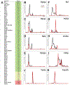
Figure 2:. Individual DIF-FRAC elution profiles show distinct changes consistent with RNAse sensitivity.
(A) Table of DIF-FRAC calculated Z-scores for ribosomal proteins and selected others. Values highlighted in green and yellow are considered high confidence. Values in red are of borderline confidence and should be evaluated with prior knowledge. (B-D) Individual profiles for ribosomal subunits. X-axis represents SEC fractions from larger molecular weight to smaller. Y-axis represents observed abundance in MS by unique peptide spectral matches normalized to the highest value for that protein. (E) Ribosomal subunit, Rpl35a, had a Z-score below the cutoff (A), but its elution profile shows consistent behavior with other ribosomal subunits. (F) Known RNA-binding protein, Nucleolin, shows shift in molecular weight. (G) Known RNA-binding protein, Puf60, shows increased observed abundance. (H-I) Profiles of RNA-binding proteins with known roles in Xenopus development. (J-K) Elution profiles of negative controls do not change.
Figure 3:
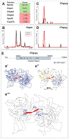
Figure 3:. DIF-FRAC identifies a ciliopathy protein as RNA associated.
(A) Table of DIF-FRAC calculated Z-scores for selected motile cilia-related proteins; Rps3A, Vps35 and Cops7b serve as positive and negative controls. (B) Elution profile of Hspe1 shows loss of observed abundance. (C) Elution profile of the inner arm dynein tethering protein Cfap44 shows a gain of observed abundance. (D) Elution profile of Cfap43 shows similar behavior to Cfap44. (E) Graphic of modeled region of Cfap44 showing identified WD40 repeat segments. (e’) Homology model of Cfap44 WD-40 domains (blue) with an RNA molecule (red) is modeled from Gemin5 crystal structure (PDB ID: 5GXH). (e’’) Homology model of Cfap44 colored to show amino acid conservation where blue is highly conserved and yellow is variable. (e’’’) Homology model of Cfap44 highlighting highly conserved residues in proximity (<5.0 Å) to modeled RNA molecule.
Figure 4:
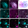
Figure 4:. Cfap44 and RNA are present in DynAPs:
(A, a’) Cfap44-GFP localizes to axonemes in Xenopus motile cilia, as indicated by co-labelling with the membrane-RFP. (B) Overlap of Cfapp+ and Ktu+ cytosolic foci in MCCs. (C, c’, c’’) Cfap44-GFP labels cytosolic foci, some of which partially co-localize with DynAPs as indicated by co-labelling with Ktu-GFP. This partial co-localization in DynAps is reminiscent of that observed for inner or outer arm dynein subunits (Lee et al., 2020). (D, d’, d’’) Staining with SytoRNA Select highlights RNA in the nucleus and also in DynAPs, as indicated by co-labelling with Ktu-RFP.

References

    1. Aizer A, Brody Y, Ler LW, Sonenberg N, Singer RH, and Shav-Tal Y. 2008. The dynamics of mammalian P body transport, assembly, and disassembly in vivo. Mol. Biol. Cell 19:4154–4166. - PMC - PubMed
    1. Ariizumi T, Takahashi S, Chan T.c., Ito Y, Michiue T, and Asashima M. 2009. Isolation and differentiation of Xenopus animal cap cells. Current protocols in stem cell biology. 9:1D. 5.1–1D. 5.31. - PubMed
    1. Artiles KL, Fire AZ, and Frøkær-Jensen C. 2019. Assessment and maintenance of unigametic germline inheritance for C. elegans. Dev. Cell 48:827–839. e829. - PMC - PubMed
    1. Ashkenazy H, Abadi S, Martz E, Chay O, Mayrose I, Pupko T, and Ben-Tal N. 2016. ConSurf 2016: an improved methodology to estimate and visualize evolutionary conservation in macromolecules. Nucleic Acids Res. 44:W344–350. - PMC - PubMed
    1. Baltz AG, Munschauer M, Schwanhausser B, Vasile A, Murakawa Y, Schueler M, Youngs N, Penfold-Brown D, Drew K, Milek M, Wyler E, Bonneau R, Selbach M, Dieterich C, and Landthaler M. 2012. The mRNA-bound proteome and its global occupancy profile on protein-coding transcripts. Mol. Cell 46:674–690. - PubMed

Publication types