. 2020 Dec:223:117349.
doi: 10.1016/j.neuroimage.2020.117349. Epub 2020 Sep 6.

Ultra-high field (10.5 T) resting state fMRI in the macaque

Affiliations

Ultra-high field (10.5 T) resting state fMRI in the macaque

Essa Yacoub et al. Neuroimage. 2020 Dec.

Abstract

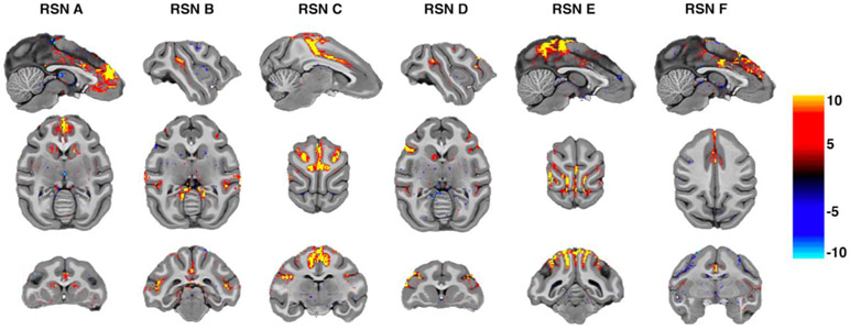
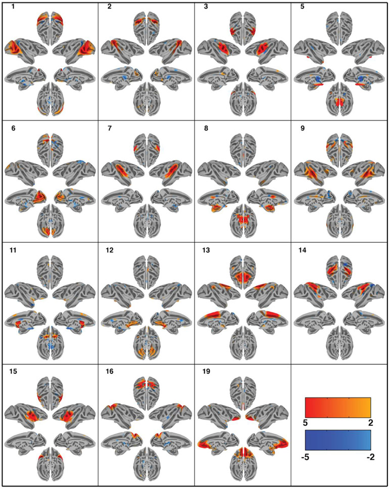
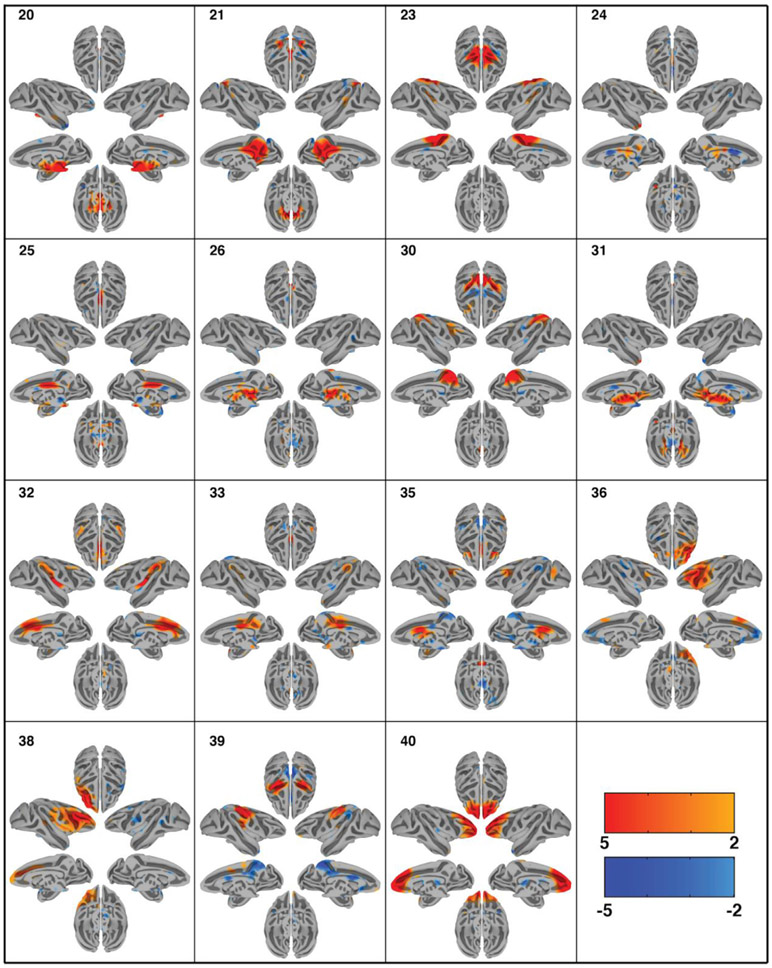
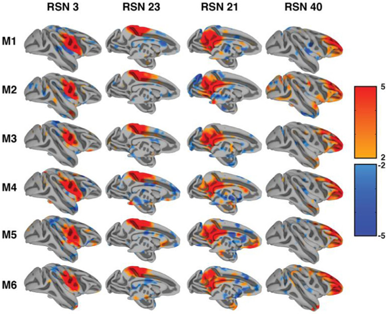
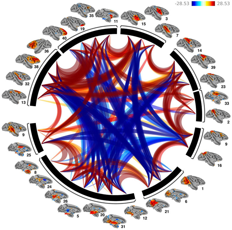
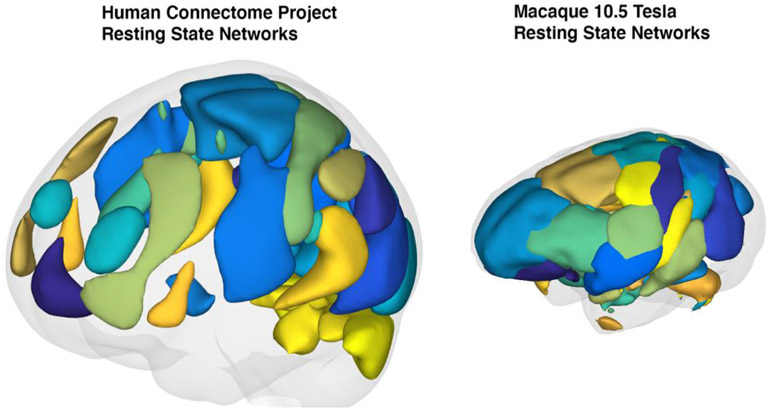
Resting state functional connectivity refers to the temporal correlations between spontaneous hemodynamic signals obtained using functional magnetic resonance imaging. This technique has demonstrated that the structure and dynamics of identifiable networks are altered in psychiatric and neurological disease states. Thus, resting state network organizations can be used as a diagnostic, or prognostic recovery indicator. However, much about the physiological basis of this technique is unknown. Thus, providing a translational bridge to an optimal animal model, the macaque, in which invasive circuit manipulations are possible, is of utmost importance. Current approaches to resting state measurements in macaques face unique challenges associated with signal-to-noise, the need for contrast agents limiting translatability, and within-subject designs. These limitations can, in principle, be overcome through ultra-high magnetic fields. However, imaging at magnetic fields above 7T has yet to be adapted for fMRI in macaques. Here, we demonstrate that the combination of high channel count transmitter and receiver arrays, optimized pulse sequences, and careful anesthesia regimens, allows for detailed single-subject resting state analysis at high resolutions using a 10.5 Tesla scanner. In this study, we uncover thirty spatially detailed resting state components that are highly robust across individual macaques and closely resemble the quality and findings of connectomes from large human datasets. This detailed map of the rsfMRI 'macaque connectome' will be the basis for future neurobiological circuit manipulation work, providing valuable biological insights into human connectomics.

Keywords: Functional MRI (fMRI); Functional connectivity; Resting-state; Rhesus macaque; Spontaneous activity.


Figures

Fig. 1.
Fig. 1.
Independent components data in individual subject space. Displayed are 6 RSNs in one unsmoothed macaque dataset processed without spatial smoothing. For this visualization thresholds were set to 4,−4. The results show robust results in the unsmoothed volumes at native resolution demonstrating our high SNR regime. Networks adhere to the cortical ribbon topography without any registration performed.
Fig. 2.
Fig. 2.
Cortical surface representation of the first 15 resting state networks (RSNs) identified using group independent component analysis (GICA) in 6 macaque monkeys. Overlayed color maps represent thresholded z-scores. Individual subjects were normalized to the NMT template. Each component shows the medial and lateral view of each hemisphere independently as well as the dorsal and ventral view of the hemispheres combined.
Fig. 3.
Fig. 3.
Cortical surface representation of the second 15 resting state networks (RSNs) identified using group independent component analysis (GICA) in 6 macaque monkeys. Overlayed color maps represent thresholded z-scores. Individual subjects were normalized to the NMT template. Each component shows the medial and lateral view of each hemisphere independently as well as the dorsal and ventral view of the hemispheres combined.
Fig. 4.
Fig. 4.
Consistency of single subject resting state networks. The figure demonstrates the same four resting state components in all six monkeys. Thresholds were kept consistent with group analysis results.
Fig. 5.
Fig. 5.
Example resting state networks (RSNs) of one macaque following single-subject independent component analysis (ICA). Images are normalized to the NMT space and overlaid onto the NMT-template. Overlaid color maps represent thresholded z-scores. Arbitrary slices were selected to demonstrate the diversity of components from subcortical to cortical components as well as the detailed anatomical correspondence.
Fig. 6.
Fig. 6.
Functional Network Connectivity of the 30 extracted Independent components. Lines portray significant connections between the networks and color indicates positive (red) and negative (blue) functional connectivity. Networks were clustered using hierarchical clustering. Results demonstrate strong positive within cluster network connections and primarily negative between cluster connections.
Fig. 7.
Fig. 7.
Qualitative comparison between resting state networks identified from the Human Connectome Project database (supplied by the CONN toolbox and Connectome Workbench) and our six subject macaque group resting state networks. Individual regions of interest were smoothed using a 10 mm spherical kernel interpolated in 3D. Gray shading represents a reconstruction of the estimated pial surface.

References

    1. Abou-Elseoud A, Starck T, Remes J, Nikkinen J, Tervonen O, Kiviniemi V, 2010. The effect of model order selection in group PICA. Hum. Brain Mapp 31. doi: 10.1002/hbm.20929, NA-NA. - DOI - PMC - PubMed
    1. Allen EA, Damaraju E, Plis SM, Erhardt EB, Eichele T, Calhoun VD, 2014. Tracking whole-brain connectivity dynamics in the resting state. Cereb. Cortex 24, 663–676. doi: 10.1093/cercor/bhs352. - DOI - PMC - PubMed
    1. Autio JA, Glasser MF, Ose T, Donahue CJ, Bastiani M, Ohno M, Kawabata Y, Urushibata Y, Murata K, Nishigori K, Yamaguchi M, Hori Y, Yoshida A, Go Y, Coalson TS, Jbabdi S, Sotiropoulos SN, Kennedy H, Smith S, Essen DCV, Hayashi T, 2020. Towards HCP-Style macaque connectomes: 24-Channel 3T multi-array coil, MRI sequences and preprocessing. Neuroimage 215, 116800. doi: 10.1016/j.neuroimage.2020.116800. - DOI - PMC - PubMed
    1. Avants BB, Tustison NJ, Stauffer M, Song G, Wu B, Gee JC, 2014. The Insight ToolKit image registration framework. Front. Neuroinform 8 (44). doi: 10.3389/fninf.2014.00044. - DOI - PMC - PubMed
    1. Avants BB, Tustison NJ, Wu J, Cook PA, Gee JC, 2011. An Open Source Multivariate Framework for n-Tissue Segmentation with Evaluation on Public Data. Neuroinformatics 9, 381–400. doi: 10.1007/s12021-011-9109-y. - DOI - PMC - PubMed

Publication types