Review
. 2020 Sep 3;25(17):4017.
doi: 10.3390/molecules25174017.

PET Radiotracers for CNS-Adrenergic Receptors: Developments and Perspectives

Affiliations
Review

PET Radiotracers for CNS-Adrenergic Receptors: Developments and Perspectives

Santosh Reddy Alluri et al. Molecules. .

Abstract

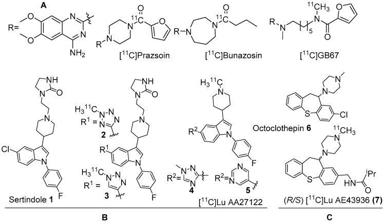
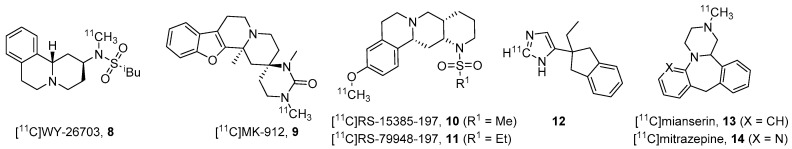
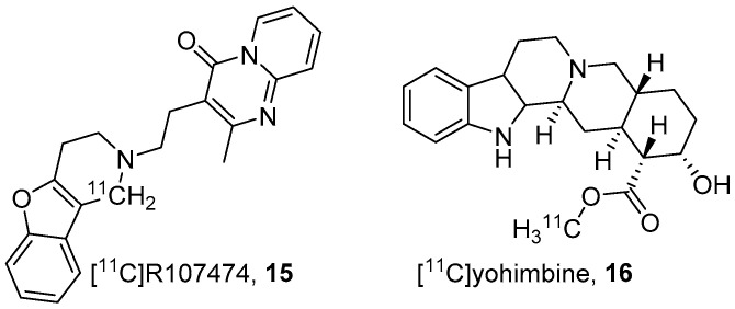
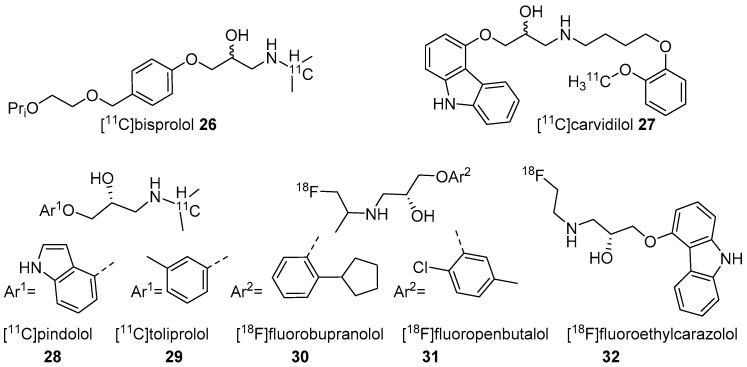
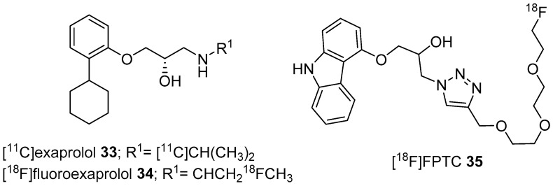
Epinephrine (E) and norepinephrine (NE) play diverse roles in our body's physiology. In addition to their role in the peripheral nervous system (PNS), E/NE systems including their receptors are critical to the central nervous system (CNS) and to mental health. Various antipsychotics, antidepressants, and psychostimulants exert their influence partially through different subtypes of adrenergic receptors (ARs). Despite the potential of pharmacological applications and long history of research related to E/NE systems, research efforts to identify the roles of ARs in the human brain taking advantage of imaging have been limited by the lack of subtype specific ligands for ARs and brain penetrability issues. This review provides an overview of the development of positron emission tomography (PET) radiotracers for in vivo imaging of AR system in the brain.

Keywords: adrenergic receptor; positron emission tomography; radiotracer.


Conflict of interest statement

The authors declare no conflict of interest.

Figures

Figure 1
Figure 1
(A) Earlier PET radiotracers, [11C]Prazosin, [11C]Bunazosin, and [11C]GB67 for cardiac α1-AR imaging. (B) Antagonist PET radiotracers based on sertindole. (C) Antagonist PET radiotracers based on octoclothepin for brain α1-AR imaging.
Figure 2
Figure 2
Various classes of α2-ARs antagonist radiotracers.
Figure 3
Figure 3
Anti-depressive & antihypertensive based α2-AR PET radiotracers.
Figure 4
Figure 4
Parametric maps of 16 in living porcine brain. (A) Baseline study using 16 showed regional differences in its distribution. (B) Blocking experiment (yohimbine at 0.07 mg/kg) reduced the scale of distribution volume (Vd) to ~2 mL g−1 in all the α2-AR bound regions. (C) Increased dose of yohimbine (1.6 mg/kg) had no further significant effect in comparison to the low dose (n = 3) Maps are superimposed on the MR image. Adapted from JNM publication by Jacobsen S, Pedersen, K.; Smith, D.F.; Jensen, S.B.; Munk, O.L.; Cumming P [97]. Permission obtained from SNMMI.
Figure 5
Figure 5
α2A-antagonist (17) and agonist (18) PET radiotracers.
Figure 6
Figure 6
PET radiotracers for α2C-ARs.
Figure 7
Figure 7
PET/CT images and time-activity curves of 21 for striatum and cerebellar cortex of (A) α2A KO (B) α2AC KO and (C) WT mice. Brain uptake of 21 in α2AC KO is negligible and is similar in α2A KO and WT mice with 7.8–8.1% ID/g at 1 min and 1.2% ID/g at 30 min after 21 injection. The striatum to cerebellar cortex radioactivity ratios (at 5–15 min) for α2AC KO mice did not differ and for α2A KO and WT mice are alike. Adapted from JNM publication by Arponen E.; Helin, S.; Marjamäki, P.; Grönroos, T.; Holm, P.; Löyttyniemi, E.; Någren, K.; Scheinin, M.; Haaparanta-Solin, M.; Sallinen, J.; [36]. Permission obtained from SNMMI.
Figure 8
Figure 8
Early PET radiotracers for cerebral β-ARs.
Figure 9
Figure 9
Radiotracers based on various β-AR blockers.
Figure 10
Figure 10
Another set of latest β-AR PET radiotracers.

Similar articles

Cited by

References

    1. Scott J.A. Positron Emission Tomography: Basic Science and Clinical Practice. Am. J. Roentgenol. 2004;182:418. doi: 10.2214/ajr.182.2.1820418. - DOI
    1. Shukla A., Kumar U. Positron emission tomography: An overview. J. Med. Phys. 2006;31:13–21. doi: 10.4103/0971-6203.25665. - DOI - PMC - PubMed
    1. McCluskey S.P., Plisson C., Rabiner E.A., Howes O. Advances in CNS PET: The state-of-the-art for new imaging targets for pathophysiology and drug development. Eur. J. Nucl. Med. Mol. Imaging. 2020;47:451–489. doi: 10.1007/s00259-019-04488-0. - DOI - PMC - PubMed
    1. Rankovic Z. CNS Drug Design: Balancing Physicochemical Properties for Optimal Brain Exposure. J. Med. Chem. 2015;58:2584–2608. doi: 10.1021/jm501535r. - DOI - PubMed
    1. Adenot M., Lahana R. Blood-Brain Barrier Permeation Models: Discriminating between Potential CNS and Non-CNS Drugs Including P-Glycoprotein Substrates. J. Chem. Inf. Comput. Sci. 2004;44:239–248. doi: 10.1021/ci034205d. - DOI - PubMed

MeSH terms