. 2020 Sep 3;21(17):6403.
doi: 10.3390/ijms21176403.

Cellular and Subcellular Localisation of Kv4-Associated KChIP Proteins in the Rat Cerebellum

Affiliations

Cellular and Subcellular Localisation of Kv4-Associated KChIP Proteins in the Rat Cerebellum

Rocío Alfaro-Ruíz et al. Int J Mol Sci. .

Abstract

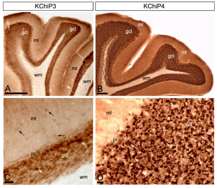
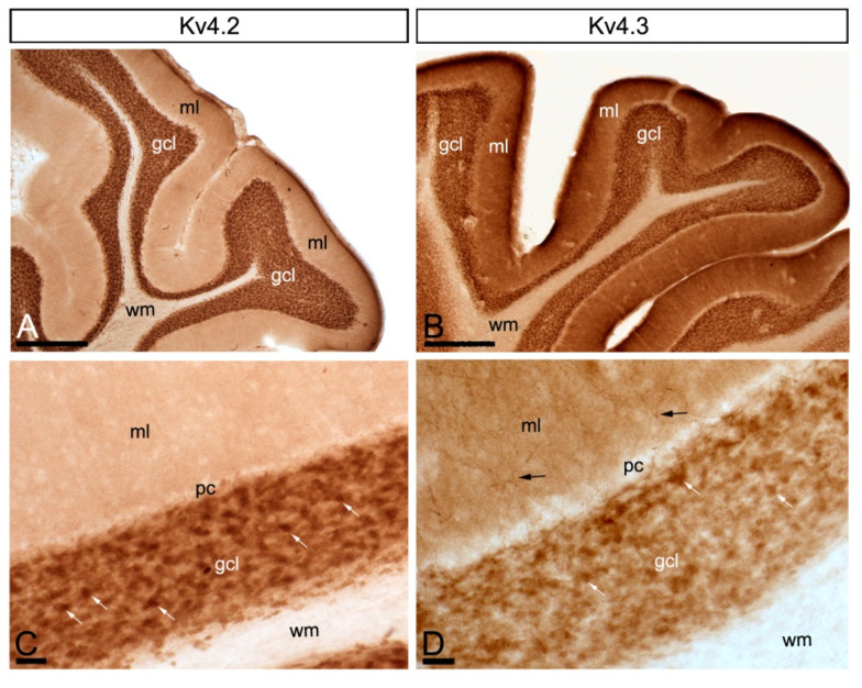
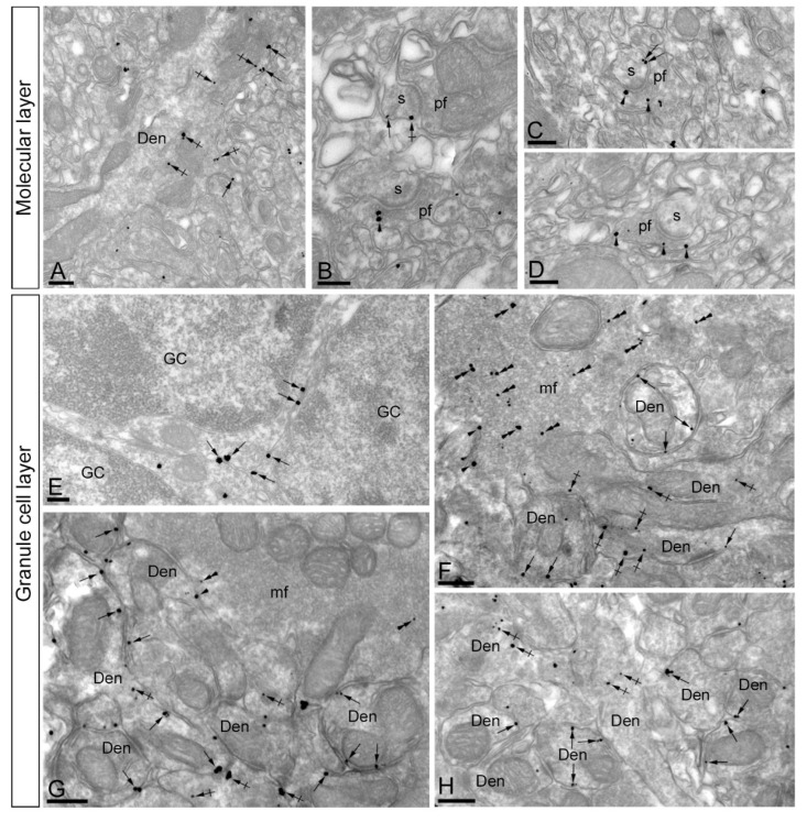
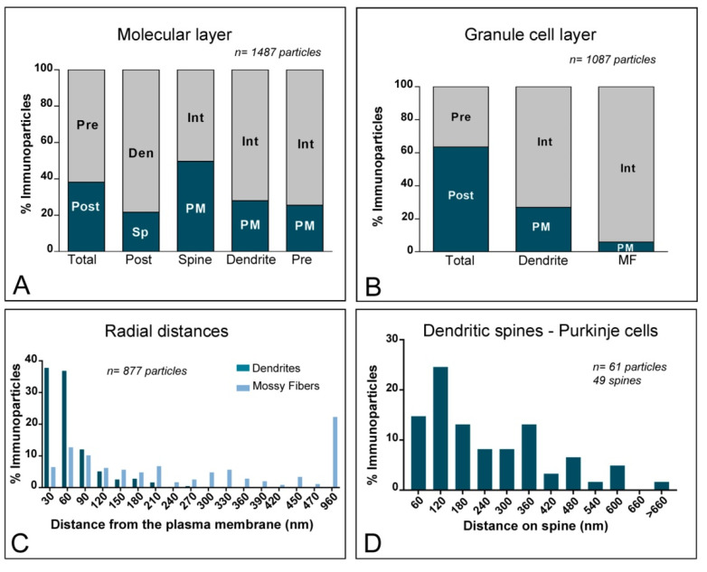
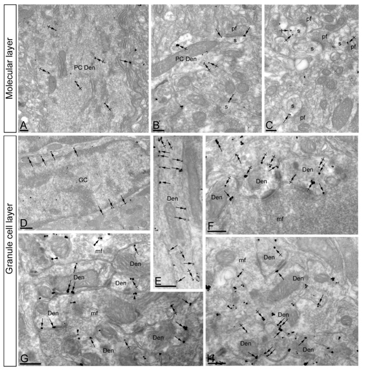
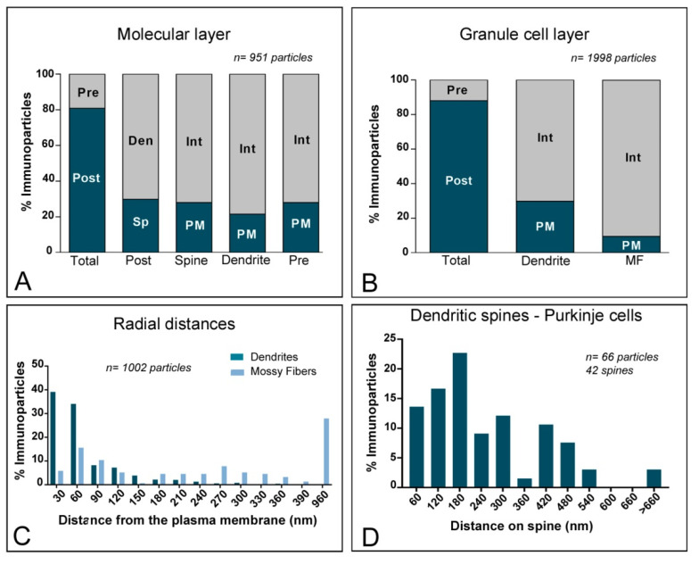
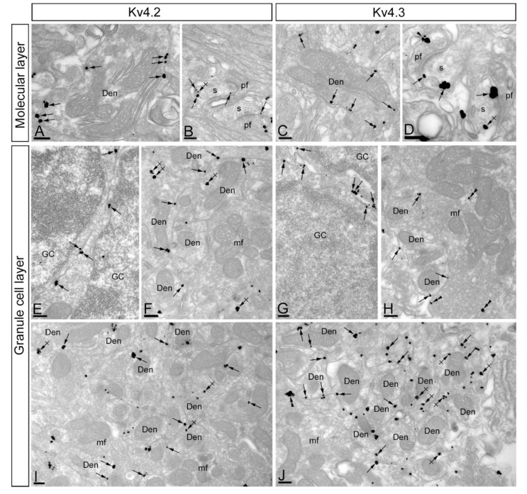
The K+ channel interacting proteins (KChIPs) are a family of cytosolic proteins that interact with Kv4 channels, leading to higher current density, modulation of channel inactivation and faster recovery from inactivation. Using immunohistochemical techniques at the light and electron microscopic level combined with quantitative analysis, we investigated the cellular and subcellular localisation of KChIP3 and KChIP4 to compare their distribution patterns with those for Kv4.2 and Kv4.3 in the cerebellar cortex. Immunohistochemistry at the light microscopic level demonstrated that KChIP3, KChIP4, Kv4.2 and Kv4.3 proteins were widely expressed in the cerebellum, with mostly overlapping patterns. Immunoelectron microscopic techniques showed that KChIP3, KChIP4, Kv4.2 and Kv4.3 shared virtually the same somato-dendritic domains of Purkinje cells and granule cells. Application of quantitative approaches showed that KChIP3 and KChIP4 were mainly membrane-associated, but also present at cytoplasmic sites close to the plasma membrane, in dendritic spines and shafts of Purkinje cells (PCs) and dendrites of granule cells (GCs). Similarly, immunoparticles for Kv4.2 and Kv4.3 were observed along the plasma membrane and at intracellular sites in the same neuron populations. In addition to the preferential postsynaptic distribution, KChIPs and Kv4 were also distributed presynaptically in parallel fibres and mossy fibres. Immunoparticles for KChIP3, KChIP4 and Kv4.3 were detected in parallel fibres, and KChIP3, KChIP4, Kv4.2 and Kv4.3 were found in parallel fibres, indicating that composition of KChIP and Kv4 seems to be input-dependent. Together, our findings unravelled previously uncharacterised KChIP and Kv4 subcellular localisation patterns in neurons, revealed that KChIP have additional Kv4-unrelated functions in the cerebellum and support the formation of macromolecular complexes between KChIP3 and KChIP4 with heterotetrameric Kv4.2/Kv4.3 channels.

Keywords: KChIP proteins; cerebellum; electron microscopy; immunohistochemistry; potassium channel.


Conflict of interest statement

The authors of this manuscript declare that they have no competing interest.

Figures

Figure 1
Figure 1
Regional and cellular distribution of Kv channel interacting proteins (KChIPs) subunits KChIP3 and KChIP4 in the cerebellum. (AD) Immunoreactivity for KChIP3 and KChIP4 in the cerebellar cortex using a pre-embedding immunoperoxidase method at the light microscopic level. Parasagittal photomicrographs of the cerebellar cortex. Immunoreactivity for both KChIP3 and KChIP4 was widely distributed in the cerebellar cortex with mostly overlapping labelling patterns. Although with some differences in intensity of labelling, strong immunoreactivity for KChIP3 and KChIP4 was found in the granule cell layer (gcl) and weaker in the molecular layer (ml) and the white matter (wm) was devoid of any staining. In the molecular layer, KChIP3 and KChIP4 was mostly neuropilar and KChIP3 labelling was also detected in cell bodies and dendrites of stellate and basket cells (black arrows). In the granule cell layer, KChIP3 and KChIP4 particularly concentrated in glomeruli (white arrows) and surrounding GCs. Scale bars: (A), 50 µm; (B), 100 µm; (C,D), 25 µm.
Figure 2
Figure 2
Regional and cellular distribution of voltage-gated potassium (Kv) channel subunits Kv4.2 and Kv4.3 in the cerebellum. (AD) Immunoreactivity for Kv4.2 and Kv4.3 in the rat cerebellar cortex using a pre-embedding immunoperoxidase method at the light microscopic level. Parasagittal photomicrographs of the cerebellar cortex. The strongest immunoreactivity for Kv4.2 and Kv4.3 was found in the granule cell layer (gcl). Strong immunoreactivity for Kv4.3 was also observed in the molecular layer (ml), but weaker for Kv4.2. The white matter (wm) was always devoid of any immunolabelling. Immunoreactivity for Kv4.2 and Kv4.3 in the molecular layer was mostly neuropilar, but Kv4.3 labelling was also detected in cell bodies and dendrites of basket cells (black arrows). In the granule cell layer, Kv4.2 and Kv4.3 immunolabelling particularly concentrated in glomeruli (white arrows) and surrounding GCs. Scale bars: (A,B), 50 µm; (C,D), 25 µm.
Figure 3
Figure 3
Subcellular localisation of Kv channel interacting protein (KChIP) subunit KChIP3 in the cerebellum. Electron micrographs showing immunoparticles for KChIP3 in the cerebellar cortex, as detected using the pre-embedding immunogold technique. (AD) In dendritic shafts (Den) and spines (s) of PCs, located in the molecular layer, KChIP3 immunoparticles were observed postsynaptically along the extrasynaptic plasma membrane (arrows). Less frequently, KChIP3 immunoparticles were observed intracellularly (crossed arrows). Presynaptically, KChIP3 immunoparticles were detected along the extrasynaptic membrane and at intracellular sites (arrowheads) of parallel fibres (pf). (EH) In the granule cell layer, KChIP3 immunoparticles were observed along the somatic plasma membrane (arrows) and dendrites (Den, arrows) and intracellular sites (crossed arrows) of granule cells (GC). Presynaptically, KChIP3 immunoparticles were observed along the plasma membrane (arrowheads) of mossy fibre axon terminals (mf), but most of them were localised intracellularly (double arrowheads) associated with cytoplasmic membranes. Scale bars: (AC), 200 nm; (DH), 250 nm.
Figure 4
Figure 4
Compartmentalisation of Kv channel interacting protein (KChIP) subunit KChIP3 in cerebellar cells. (A) Bar graphs showing the percentage of immunoparticles for KChIP3 at post- and pre-synaptic compartments in the molecular layer. A total of 1487 immunoparticles in the molecular layer were analysed, of which 38.1% were postsynaptic and 61.9% were presynaptic. Postsynaptically, immunoparticles were detected in dendritic spines (21.7%) and in dendritic shafts (78.35%), distributed along the plasma membrane (49.6% in spines; 27.9% in dendrites) and at cytoplasmic sites (50.4% in spines; 72.1% in dendrites). Presynaptically, immunoparticles were detected in parallel fibres of ML (61.9%) distributed along the plasma membrane (25.5%) and at cytoplasmatic sites (74.5%). (B) Bar graphs showing the percentage of KChIP3 immunoparticles at post- and pre-synaptic compartments, and along the plasma membrane and intracellular sites in dendritic shafts of granule cells and mossy fibres in the granule cell layer. A total of 1087 immunoparticles in the granular cell layer were analysed, of which 63.5% were postsynaptic and 36.5% were presynaptic. Postsynaptically, immunoparticles were detected in dendrites of GCs (63.5%), distributed along the plasma membrane (27%) and at cytoplasmic sites (73%). Presynaptically, immunoparticles were detected in mossy fibre terminals (36.5%), distributed mostly at cytoplasmic sites (94%) and very few along the plasma membrane (6%). (C) Histogram showing the radial distribution of KChIP3 immunoparticles from the plasma membrane towards cytoplasmic sites in dendrites of GCs and mossy fibres. In dendrites, immunoparticles for KChIP3 showed a skewed frequency distribution in the plasma membrane direction, but in mossy fibres they were more equally distributed across the axoplasm. (D) Histogram showing the distribution of immunoreactive KChIP3 in relation to glutamate release sites in dendritic spines PCs. The data show the proportion of KChIP3 immunoparticles at a given distance from the edge of the postsynaptic density. About 68.9% of immunolabelled KChIP3 are located in a 60–300 nm wide band, and then the density decreased markedly further in the spine membrane.
Figure 5
Figure 5
Subcellular localisation of Kv channel interacting protein (KChIP) subunit KChIP4 in the cerebellum. Electron micrographs showing immunoparticles for KChIP4 in the cerebellar cortex, as detected using the pre-embedding immunogold technique. (AC) In the molecular layer, KChIP4 immunoparticles were detected in Purkinje cells, distributed along the extrasynaptic plasma membrane (arrows) of dendritic shafts (Den) and dendritic spines (s), as well as at intracellular sites (crossed arrows). (DH) In the granule cell layer, immunoparticles for KChIP4 were mostly distributed along the somatic plasma membrane (arrows) and dendrites (Den) of granule cells (GC), as well as at intracellular sites (crossed arrows). At presynaptic sites, immunoparticles for KChIP4 were found along the plasma membrane (arrowheads) and intracellular sites (double arrowheads) of mossy fibre axon terminals (mf). Scale bars: (AC,EH), 250 nm; (D), 500 nm.
Figure 6
Figure 6
Compartmentalisation of Kv channel interacting protein (KChIP) subunit KChIP4 in cerebellar cells. (A) Bar graphs showing the percentage of immunoparticles for KChIP4 at post- and pre-synaptic compartments in the molecular layer. A total of 951 immunoparticles in the molecular layer were analysed, of which 80.9% were postsynaptic and 19.1% were presynaptic. Postsynaptically, immunoparticles were detected in dendritic spines (29.8%) and in dendritic shafts (70.2%), distributed along the plasma membrane (27.9% in spines; 21.5% in dendrites) and at cytoplasmic sites (72.1% in spines; 78.5% in dendrites). (B) Bar graphs showing the percentage of KChIP4 immunoparticles at post- and pre-synaptic compartments, and along the plasma membrane and intracellular sites in dendritic shafts of granule cells and mossy fibres in the granule cell layer. A total of 1998 immunoparticles in the granular cell layer were analysed, of which 87.9% were postsynaptic and 12.1% were presynaptic. Postsynaptically, immunoparticles were detected in dendrites of GCs (87.9%), distributed along the plasma membrane (29.9%) and at cytoplasmic sites (70.1%). Presynaptically, immunoparticles were detected in mossy fibre terminals (12.1%), distributed mostly at cytoplasmic sites (90.4%) and very few along the plasma membrane (9.6%). (C) Histogram showing the radial distribution of KChIP4 immunoparticles from the plasma membrane towards cytoplasmic sites in dendrites of GCs and mossy fibres. In dendrites, immunoparticles for KChIP4 showed a skewed frequency distribution in the plasma membrane direction, but in mossy fibres, they were more equally distributed across the axoplasm. (D) Histogram showing the distribution of immunoreactive KChIP4 in relation to glutamate release sites in dendritic spines PCs. The data show the proportion of KChIP4 immunoparticles at a given distance from the edge of the postsynaptic density. About 74.2% of immunolabelled KChIP4 are located in a 60–300 nm wide band, and then the density decreased markedly further in the spine membrane.
Figure 7
Figure 7
Subcellular distribution of voltage-gated potassium (Kv) channel subunits Kv4.2 and Kv4.3 channels. Immunoreactivity for Kv4.2 and Kv4.3 in the cerebellar cortex as demonstrated by pre-embedding immunogold labelling. (AD) Immunoparticles for both Kv4.2 and Kv4.3 in the molecular layer were found in the plasma membrane (arrows) and intracellular sites (crossed arrows) of PC dendrites (Den) and spines (s) establishing synapses with parallel fibres (pf). Although, at low frequency, Kv4.3 immunoparticles were also found in the plasma membrane (arrowheads) of parallel fibres (pf). (EJ) Immunoparticles for both Kv4.2 and Kv4.3 in the granule cell layer were found in the plasma membrane (arrows) of GC somata and GC dendrites (Den) in cerebellar glomeruli. In addition, immunolabelling was found intracellularly (crossed arrows). Few immunoparticles were also observed presynaptically along the plasma membrane (arrowheads) and intracellular sites (double arrowheads) in mossy fibre (mf) axon terminals. Scale bars: (AJ), 200 nm.
Figure 8
Figure 8
Compartmentalisation of voltage-gated potassium (Kv) channel subunits Kv4.2 and Kv4.3 in cerebellar cells. (A,B) Bar graphs showing the percentage of immunoparticles for Kv4.2 and Kv4.3 in neuronal compartment in the molecular layer. Immunoparticles for Kv4.2 were mostly localised at the postsynaptic compartment (99% of all particles), while Kv4.3 was distributed at postsynaptic (88.4%) and presynaptic (11.6%) compartments. (C,D) Bar graphs showing the percentage of immunoparticles for Kv4.2 and Kv4.3 in the neuronal compartment in the granule cell layer. A total of 667 immunoparticles for Kv4.2 and 771 for Kv4.3 were analysed. Postsynaptically, immunoparticles were detected in dendrites of GCs (91.6% for Kv4.2; 82.2% for Kv4.3), distributed along the plasma membrane (46.3% for Kv4.2; 62.3% for Kv4.3) and at cytoplasmic sites (53.7% for Kv4.2; 37.7% for Kv4.3). Presynaptically, immunoparticles were detected in mossy fibre terminals (8.4% for Kv4.2; 17.8% for Kv4.3), distributed at cytoplasmic sites (87.5% for Kv4.2; 53.3% for Kv4.3) and along the plasma membrane (12.5% for Kv4.2; 46.7% for Kv4.3). (E,F) Histogram showing the distribution of immunoparticles for Kv4.2 and Kv4.3 in relation to glutamate release sites in dendritic spines of PCs. About 46% of immunolabelled Kv4.2 and 52% of immunolabelled Kv4.3 were located in a 60–300 nm wide band. These data show that Kv4.2 immunoparticles were more equally distributed along PC spines, while immunoparticles Kv4.3 were skewed toward the PSD of PC spines.

References

    1. Sotelo C. Cellular and genetic regulation of the development of the cerebellar system. Prog. Neurobiol. 2004;72:295–339. doi: 10.1016/j.pneurobio.2004.03.004. - DOI - PubMed
    1. Ito M. Cerebellar Long-Term Depression: Characterization, Signal Transduction, and Functional Roles. Physiol. Rev. 2001;81:1143–1195. doi: 10.1152/physrev.2001.81.3.1143. - DOI - PubMed
    1. Ito M. Cerebellar circuitry as a neuronal machine. Prog. Neurobiol. 2006;78:272–303. doi: 10.1016/j.pneurobio.2006.02.006. - DOI - PubMed
    1. Sillitoe R.V., Joyner A.L. Morphology, molecular codes, and circuitry produce the three-dimensional complexity of the cerebellum. Annu. Rev. Cell Dev. Biol. 2007;23:549–577. doi: 10.1146/annurev.cellbio.23.090506.123237. - DOI - PubMed
    1. Trimmer J.S. Subcellular localization of K+ channels in mammalian brain neurons: Remarkable precision in the midst of extraordinary complexity. Neuron. 2015;85:238–256. doi: 10.1016/j.neuron.2014.12.042. - DOI - PMC - PubMed

Substances

LinkOut - more resources