Review
. 2020 Sep 3;11(9):1041.
doi: 10.3390/genes11091041.

Gut Microbiota between Environment and Genetic Background in Familial Mediterranean Fever (FMF)

Affiliations
Review

Gut Microbiota between Environment and Genetic Background in Familial Mediterranean Fever (FMF)

Agostino Di Ciaula et al. Genes (Basel). .

Abstract

The gastrointestinal tract hosts the natural reservoir of microbiota since birth. The microbiota includes various bacteria that establish a progressively mutual relationship with the host. Of note, the composition of gut microbiota is rather individual-specific and, normally, depends on both the host genotype and environmental factors. The study of the bacterial profile in the gut demonstrates that dominant and minor phyla are present in the gastrointestinal tract with bacterial density gradually increasing in oro-aboral direction. The cross-talk between bacteria and host within the gut strongly contributes to the host metabolism, to structural and protective functions. Dysbiosis can develop following aging, diseases, inflammatory status, and antibiotic therapy. Growing evidences show a possible link between the microbiota and Familial Mediterranean Fever (FMF), through a shift of the relative abundance in microbial species. To which extent such perturbations of the microbiota are relevant in driving the phenotypic manifestations of FMF with respect to genetic background, remains to be further investigated.

Keywords: MEFV; amyloidosis; colchicine; inflammasome; interleukin-1b.


Conflict of interest statement

The authors declare no conflict of interest.

Figures

Figure 1
Figure 1
Schematic structure of MEFV gene and encoded pyrin (marenostrin) protein. The MEFV gene encodes for the pyrin protein (781 amino acids). The most common mutations in Familial Mediterranean Fever (FMF) are in exon 10 encoding the B30.2 domain. The most important interaction partners appear below the pyrin structure. ASC and Pro-Caspase 1 are also drawn. Pyrin structure includes five different domains, each one responsible for protein-protein interaction, and each domain has a role in the regulation of innate response. From left to right, PYRIN (PYD) domain (residues 1–92) interacts with ASC (apoptosis-associated speck-like protein containing a CARD—caspase-recruitment domain). bZIP transcription factor basic domain (residues 266–280) interacts with the p65 subunit (transcription factor p65) of NF-κB, and IκB-α. The B-box zinc finger domain (residues 375–412) and α-helical (CC, coiled-coil) domain (residues 420–440) likely influence the oligomerization of pyrin, and interact with the PAPA protein (also named PSTPIP1, proline serine threonine phosphatase-interacting protein, also known as CD2BP1 involved in the organization of the cytoskeleton) and the regulation of IL-1β secretion. The B30.2 domain (PRYSPRY) (residues 597–776) is the most important, and interacts with caspase-1 and the proapoptotic protein Siva. Further pyrin interactions include binding to microtubules (starting from the N-terminal to bZIP), interaction with 14.3.3 (14-3-3 protein), and with the PKN1/2 (serine-threonine kinases PKN1 and PKN2) at the three serine residues 208, 209, 242 between PYD and bZIP. The position of Asp330 between bZIP and the B-box indicates the caspase-1 cleavage site. Mutations in the B30.2 domain tend to be transmitted in an autosomal-recessive fashion. Mutations in exons 2, 3 and 5 generally exhibit autosomal-dominant pattern of inheritance [24,34,35,36,41,43,48,49,50,51,52,53,54,55,56,57,58].
Figure 2
Figure 2
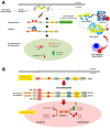
Mechanisms underlying the assembly of the pyrin inflammasome. (A) With a normally functioning pyrin (i.e., when pyrin is non-mutated), the mevalonate kinase pathway provides geranyl-geranyl phosphate and, together with release of GEF-H1 (increased by colchicine acting as the inhibitor of the microtubule polymerization), activates RhoA. PKN1 and PKN2 are effector kinases of RhoA mediating the phosphorylation of pyrin and binding to the inhibitory proteins 14-3-3. Inhibition of pyrin can increase with agents activating PKN1/2 or following the release of GEF-H1 (i.e., colchicine). (B) If the pyrin phosphorylation decreases, (i.e., in FMF patients lacking the control by pyrin/marenostrin caused by pathogenic MEFV variants), with low GEF-H1 or defective function of the MVK-pathway, the activation of PKN1/2 also decreases. This step results in pyrin inflammasome activation and release of mature IL-1β and IL-18. The plasma membrane pore-forming N-terminal fragment of gasdermin D facilitates IL-1β and IL-18 release. Appropriate stimuli can also lead to the assembly of the inflammasome. The first step is the PYD-PYD homotypic interaction of ASC resulting in oligomerization into ASC specks. Pro-caspase-1 is recruited because of CARD-CARD interaction with ASC. This step anticipates the auto-cleavage of pro-caspase-1 into active caspase-1 tetramers (p10/p20) governing the transformation of pro-IL-1/18 into mature IL-1/18. The pyroptosis mediated by Gasdermin D also contributes to cytoplasmic enrichment with IL-1/18, and further reinforces the inflammatory pathway. Colchicine inhibits the polymerization of intracellular β-tubulin by forming colchicine-tubulin complexes via contact of A and C rings with the C domain of the tubulin β-subunit. These supramolecular interactions block the dockage of tubulin into the (+) ends of microtubules (cytoskeleton), thus preventing inflammasome activation in neutrophils and monocytes. The colchicine-dependent inhibition of tubulin also efficiently affects the migration and degranulation of white blood cells [43,50].
Figure 3
Figure 3
Schematic appearance of time-dependent changes of body temperature in Familial Mediterranean Fever (FMF) before starting the treatment with colchicine. The profile refers to a typical case belonging to a cluster of families identified in the region of Apulia, Italy [24]. (A) Frequency of febrile attacks in a year. The dotted horizontal line is placed at 37 °C (cut-off value). In between attacks, the temperature has been conventionally set at 36.5 °C. This patient reported a total of 14 attacks in a year. The black arrow indicates the febrile attack described in panel B. (B) The single febrile attack lasts about 48 h and is associated with a major auto-inflammatory status and symptoms. Adapted from Portincasa et al. Familial Mediterranean fever: a fascinating model of inherited auto inflammatory disorder. Eur J Clin Invest 43, 1314-1327 (2013) with permission from John Wiley & Sons Ltd. [24].
Figure 4
Figure 4
Therapeutic agents effective in Familial Mediterranean Fever (FMF). (A) Colchicine: chemical structure, IUPAC names, chemical formula, molecular weight and three-dimensional (3D) structures. (B) Canakinumab: 3D structure, chemical formula and molecular weight.
Figure 5
Figure 5
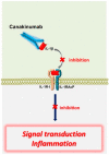
Mechanism of action of biologic agent canakinumab in FMF. Canakinumab is a fully human selective anti-IL-1β monoclonal antibody and binds human IL-1β. Subsequent binding of IL-1β to the IL-1R is inhibited with prevention of intracellular signal transduction and further proinflammatory events. Abbreviations: IL, interleukin; IL-1R-I, interleukin-1 receptor, type1; IL-1RAcP, IL-1 receptor accessory protein. Adapted from [49,50,51].
Figure 6
Figure 6
Distribution and function of microbiota in human intestine during health or disease. The bacterial community is the core component of microbiota in the gastrointestinal tract and there are eukaryotic microbes. In healthy subjects, microbial density and types vary across the gastrointestinal tract, with density increasing dramatically in oro-aboral direction. Microbial composition also changes. The crosstalk between bacteria and host contribute to maintain physiological metabolic, protective, and structural functions. In disease, shift of relative abundance in microbial species or quantitative variations of microbiota occur, generating chronic low-grade inflammation, atypical gases and fecal levels of organic acids, impaired metabolism of proteins and carbohydrates and immunological responses [2,102,103,104,105,106].

Similar articles

Cited by

References

    1. Kuramitsu H.K., He X., Lux R., Anderson M.H., Shi W. Interspecies interactions within oral microbial communities. Microbiol. Mol. Biol. Rev. 2007;71:653–670. doi: 10.1128/MMBR.00024-07. - DOI - PMC - PubMed
    1. Bonfrate L., Tack J., Grattagliano I., Cuomo R., Portincasa P. Microbiota in health and irritable bowel syndrome: Current knowledge, perspectives and therapeutic options. Scand. J. Gastroenterol. 2013;48:995–1009. doi: 10.3109/00365521.2013.799220. - DOI - PubMed
    1. Gerritsen J., Smidt H., Rijkers G.T., de Vos W.M. Intestinal microbiota in human health and disease: The impact of probiotics. Genes Nutr. 2011;6:209–240. doi: 10.1007/s12263-011-0229-7. - DOI - PMC - PubMed
    1. Young V.B. The intestinal microbiota in health and disease. Curr. Opin. Gastroenterol. 2012;28:63. doi: 10.1097/MOG.0b013e32834d61e9. - DOI - PMC - PubMed
    1. Penders J., Thijs C., Vink C., Stelma F.F., Snijders B., Kummeling I., van den Brandt P.A., Stobberingh E.E. Factors influencing the composition of the intestinal microbiota in early infancy. Pediatrics. 2006;118:511–521. doi: 10.1542/peds.2005-2824. - DOI - PubMed

Publication types

MeSH terms

LinkOut - more resources