. 2020 Sep 4;10(9):1750.
doi: 10.3390/nano10091750.

Physical Properties and Biofunctionalities of Bioactive Root Canal Sealers In Vitro

Affiliations

Physical Properties and Biofunctionalities of Bioactive Root Canal Sealers In Vitro

Seung Bin Jo et al. Nanomaterials (Basel). .

Abstract

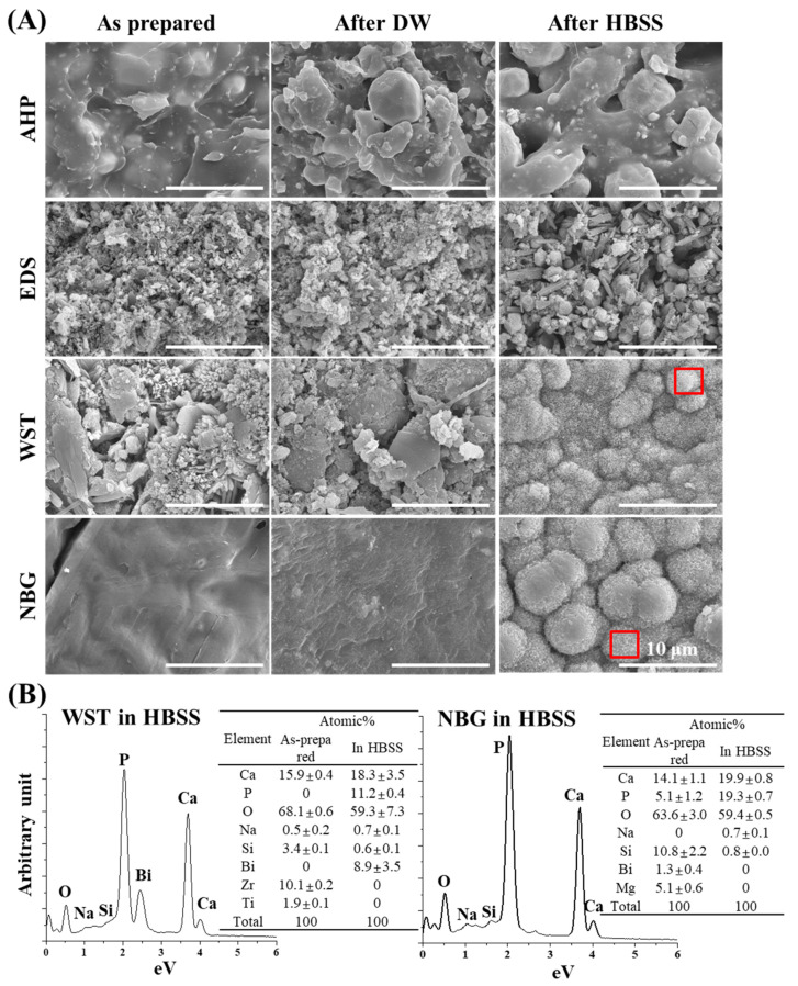
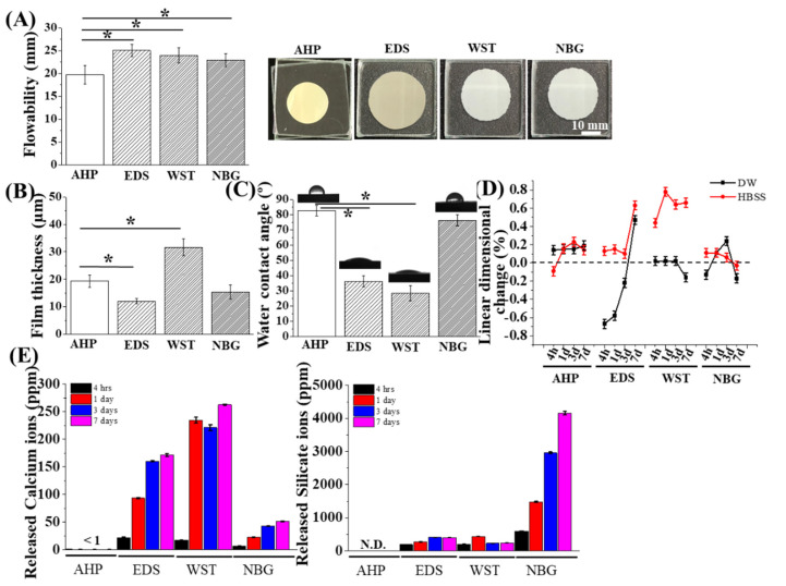
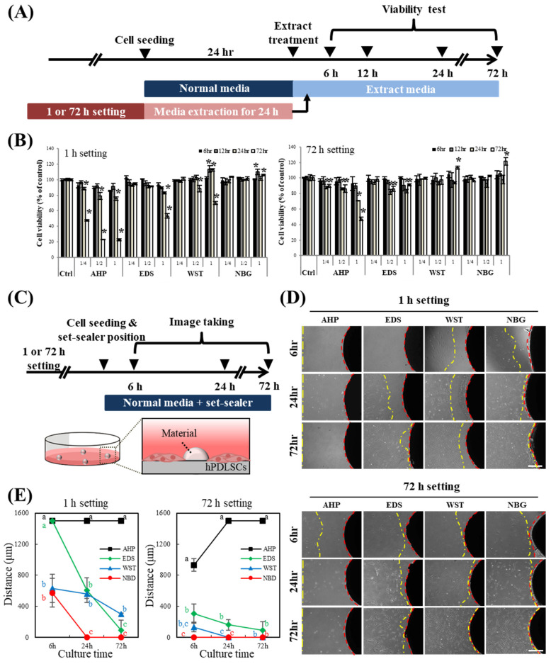
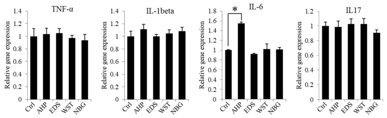
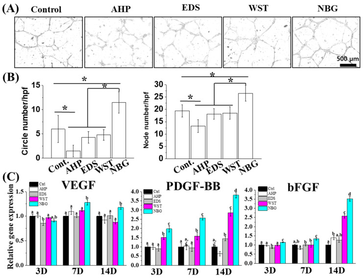
Calcium silicate-based bioactive glass has received significant attention for use in various biomedical applications due to its excellent bioactivity and biocompatibility. However, the bioactivity of calcium silicate nanoparticle-incorporated bioactive dental sealer is not much explored. Herein, three commercially available bioactive root canal sealers (Endoseal MTA (EDS), Well-Root ST (WST), and Nishika Canal Sealer BG (NBG)) were compared with a resin-based control sealer (AH Plus (AHP)) in terms of physical, chemical, and biological properties. EDS and NBG showed 200 to 400 nm and 100 to 200 nm nanoparticle incorporation in the SEM image, respectively, and WST and NBG showed mineral deposition in Hank's balanced salt solution after 28 days. The flowability and film thickness of all products met the ISO 3107 standard. Water contact angle, linear dimensional changes, and calcium and silicate ion release were significantly different among groups. All bioactive root canal sealers released calcium ions, while NBG released ~10 times more silicon ions than the other bioactive root canal sealers. Under the cytocompatible extraction range, NBG showed prominent cytocompatibility, osteogenecity, and angiogenecity compared to other sealers in vitro. These results indicate that calcium silicate nanoparticle incorporation in dental sealers could be a potential strategy for dental periapical tissue regeneration.

Keywords: angiogenic; bioactive; osteogenic induction; root canal sealers; silicate ions.


Conflict of interest statement

The authors declare no conflict of interest.

Figures

Figure 1
Figure 1
SEM images of the bioactive set sealers before and after DW or HBSS immersion. (A) SEM images revealing surface changes of each set sealer between the as-prepared and immersed specimens. Mineralization was observed on the surfaces of HBSS-immersed WST and NBG samples. The red boxes indicate the area analysed with EDS to evaluate the composition. (B) The EDS results represent high amounts of Ca and P after HBSS immersion from WST and NBG, indicating calcium phosphate mineralization. Other specific components (Na, Si, and Bi for WST and Na and Si for NBG) were detected in each sealer set.
Figure 2
Figure 2
Physical and chemical properties of the bioactive set sealers. (A) Flowability of each material. EDS, WST, and NBG showed greater flowability than AHP, while all materials passed the ISO flowability standard (17 mm, n = 3). (B) The current sealer materials presented film thicknesses under 50 μm, which meet the ISO standard, while EDS showed the thinnest film thickness (n = 3). (C) The water contact angle of each sealer set presented different values (30–80 degrees) among groups (n = 4). (D) Linear dimensional change results between DW and HBSS solution over time (n = 3). EDS and WST revealed significant differences between DW and HBSS. (E) Ion release for 7 days (n = 3). EDS and WST exhibited excellent calcium ion release, while silicate ion release was predominant in NBG with time. The asterisks indicate significant differences at the 0.05 level between groups.
Figure 3
Figure 3
Biological characteristics of the bioactive set sealers. (A) Schematic timeline of the cell viability assay with the sealer extract in media. (B) Cell viability assay of hPDLSCs with set sealer extract or its dilute for up to 72 h. The extract media of 1- or 72-h-set-AHP showed severe cytotoxicity to hPDLSCs both at the original extract concentration and at 50% dilution, while the other extract media showed acceptable viability (~90%) under 50% dilution. Asterisks indicate significant differences compared to each control (n = 5, p < 0.05). Increased cell viability was observed for WST and NBG. (C) Schematic timeline of the direct contact assay with the set sealers. hPDLSCs were cultured directly with the set sealers. (D,E) Cells migrated towards the 1- or 72-h sealers over time. hPDLSCs cultured with bioactive root canal sealers (EDS, WST and NBG) migrated and made contact with the materials, while cells rarely migrated toward the set-AHP. Cells cultured with NBG displayed the fastest migration among the groups. Different letters indicate significant differences among the groups in each time point (n = 4, p < 0.05).
Figure 4
Figure 4
Inflammatory and anti-inflammatory gene expression of hPDLSCs in set sealer extracts. hPDLSCs showed no change in inflammatory (TNF-α, IL-1β and IL-6) or anti-inflammatory (IL-17) gene expression in 50% diluted extract from EDS, WST and NBG after 1 hr of setting, while AHP from 72 h of setting showed significant upregulation of inflammatory gene expression (IL-6). The asterisk indicates a significant difference between groups (n = 3, p < 0.05).
Figure 5
Figure 5
Osteogenic differentiation effects of the bioactive set sealers. (A,B) hPDLSCs cultured with basal media (negative control), osteogenic media (positive control), or sealer extract media (50%) revealed that both alkaline phosphatase (ALP, violet colour) activity and Alizarin red S (ARS, red colour) staining were highest in the NBG group at early (days 3 and 7) and late (day 21) differentiation stages. (C) Osteogenic gene expression (DMP1, RUNX2, and osterix) from extract (50% dilution) was upregulated in the bioactive root canal sealers, while NBG showed the highest expression among groups at days 3 and 7. Gene expression data from AHP were excluded due to low quality of RNA. Different letters indicate significant differences among the groups (n = 3, p < 0.05).
Figure 6
Figure 6
Angiogenic effects of the bioactive set sealers. (A,B) HUVECs cultured with basal media or sealer extract (25% dilution) revealed that both circle and node numbers were highest in the NBG group compared with the other groups. Asterisks indicate significant differences among the groups (n = 3, p < 0.05). (C) Angiogenic gene expression of hPDLSCs over 14 days from extract after 50% dilution revealing that angiogenic gene (VEGF, PDGF-BB and bFGF) expression was the most upregulated in the NBG group. Different letters indicate significant differences among the groups (n = 3, p < 0.05).

References

    1. Jung C., Kim S., Sun T., Cho Y.-B., Song M. Pulp-dentin regeneration: Current approaches and challenges. J. Tissue Eng. 2019;10:2041731418819263. doi: 10.1177/2041731418819263. - DOI - PMC - PubMed
    1. Hargreaves K.M., Berman L.H. Cohen’s Pathways of the Pulp Expert Consult. Elsevier Health Sciences; Amsterdam, The Netherlands: 2015.
    1. ISO . ISO 6876:2012 Dentistry—Root Canal Sealing Materials. International Organization for Standardization; Geneva, Switzerland: 2012.
    1. Grossman I., Oliet S., Del Rio E. Endodontic Practice. 11th ed. Lea and Fabringer; Philadelphia, PA, USA: 1988. pp. 145–155.
    1. Carvalho-Junior J.R., Correr-Sobrinho L., Correr A.B., Sinhoreti M.A., Consani S., Sousa-Neto M.D. Solubility and dimensional change after setting of root canal sealers: A proposal for smaller dimensions of test samples. J. Endod. 2007;33:1110–1116. doi: 10.1016/j.joen.2007.06.004. - DOI - PubMed

LinkOut - more resources