

Advances in Continuous Microfluidics-Based Technologies for the Study of HIV Infection
- PMID: 32899657
- PMCID: PMC7552050
- DOI: 10.3390/v12090982
Advances in Continuous Microfluidics-Based Technologies for the Study of HIV Infection
Abstract
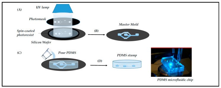
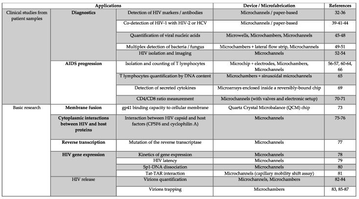
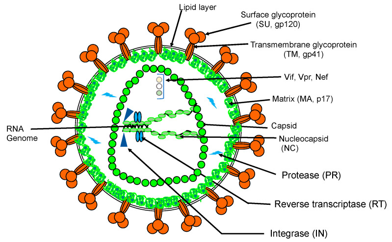
HIV-1 is the causative agent of acquired immunodeficiency syndrome (AIDS). It affects millions of people worldwide and the pandemic persists despite the implementation of highly active antiretroviral therapy. A wide spectrum of techniques has been implemented in order to diagnose and monitor AIDS progression over the years. Besides the conventional approaches, microfluidics has provided useful methods for monitoring HIV-1 infection. In this review, we introduce continuous microfluidics as well as the fabrication and handling of microfluidic chips. We provide a review of the different applications of continuous microfluidics in AIDS diagnosis and progression and in the basic study of the HIV-1 life cycle.
Keywords: HIV; diagnosis; infection; life cycle; microfluidics; replication; retrovirus.
Conflict of interest statement
The authors declare no conflict of interest.
Figures
Similar articles
-
Miniaturized devices for point of care molecular detection of HIV.Lab Chip. 2017 Jan 31;17(3):382-394. doi: 10.1039/c6lc01239f. Lab Chip. 2017. PMID: 28092381 Free PMC article. Review.
-
HIV heterogeneity and disease progression in AIDS: a model of continuous virus adaptation.AIDS. 1998;12 Suppl A:S43-52. AIDS. 1998. PMID: 9632983 Review. No abstract available.
-
Implementation and new insights in molecular diagnostics for HIV infection.Expert Rev Mol Diagn. 2018 May;18(5):433-441. doi: 10.1080/14737159.2018.1464393. Epub 2018 Apr 19. Expert Rev Mol Diagn. 2018. PMID: 29641941 Review.
-
Molecular and cellular interactions of HIV-1/HTLV coinfection and impact on AIDS progression.AIDS Rev. 2007 Jul-Sep;9(3):140-9. AIDS Rev. 2007. PMID: 17982939 Review.
-
How to win the HIV-1 drug resistance hurdle race: running faster or jumping higher?Biochem J. 2017 Apr 26;474(10):1559-1577. doi: 10.1042/BCJ20160772. Biochem J. 2017. PMID: 28446620 Review.
Cited by
-
Microfluidic Devices for HIV Diagnosis and Monitoring at Point-of-Care (POC) Settings.Biosensors (Basel). 2022 Nov 1;12(11):949. doi: 10.3390/bios12110949. Biosensors (Basel). 2022. PMID: 36354458 Free PMC article. Review.
-
Current Advancements and Future Road Map to Develop ASSURED Microfluidic Biosensors for Infectious and Non-Infectious Diseases.Biosensors (Basel). 2022 May 20;12(5):357. doi: 10.3390/bios12050357. Biosensors (Basel). 2022. PMID: 35624657 Free PMC article. Review.
-
Viro-fluidics: Real-time analysis of virus production kinetics at the single-cell level.Biophys Rep (N Y). 2022 Aug 11;2(3):100068. doi: 10.1016/j.bpr.2022.100068. eCollection 2022 Sep 14. Biophys Rep (N Y). 2022. PMID: 36425325 Free PMC article.
-
Special Issue "Function and Structure of Viral Ribonucleoproteins Complexes".Viruses. 2020 Nov 26;12(12):1355. doi: 10.3390/v12121355. Viruses. 2020. PMID: 33256140 Free PMC article.
References
-
- Agarwal A. Digital microfluidics: Techniques, their applications and advantages. J. Bioeng. Biomed. Sci. 2013;3 doi: 10.4172/2155-9538.S8-001. - DOI
-
- Chacon O.L.A., Baret J.C. Rapid stabilization of droplets by particles in microfluidics: Role of droplet formation. Chem. Syst. Chem. 2019;1:16–24. doi: 10.1002/syst.201900007. - DOI
-
- Mashaghi S., Abbaspourrad A., Weitz D.A., van Oijen A.M. Droplet microfluidics: A tool for biology, chemistry and nanotechnology. TrAC Trends Anal. Chem. 2016;82:118–125. doi: 10.1016/j.trac.2016.05.019. - DOI
Publication types
MeSH terms
LinkOut - more resources
Full Text Sources
Other Literature Sources
Medical
