Review
. 2020 Sep 4;12(9):984.
doi: 10.3390/v12090984.

Dance with the Devil: Stress Granules and Signaling in Antiviral Responses

Affiliations
Review

Dance with the Devil: Stress Granules and Signaling in Antiviral Responses

Nina Eiermann et al. Viruses. .

Abstract

Cells have evolved highly specialized sentinels that detect viral infection and elicit an antiviral response. Among these, the stress-sensing protein kinase R, which is activated by double-stranded RNA, mediates suppression of the host translation machinery as a strategy to limit viral replication. Non-translating mRNAs rapidly condensate by phase separation into cytosolic stress granules, together with numerous RNA-binding proteins and components of signal transduction pathways. Growing evidence suggests that the integrated stress response, and stress granules in particular, contribute to antiviral defense. This review summarizes the current understanding of how stress and innate immune signaling act in concert to mount an effective response against virus infection, with a particular focus on the potential role of stress granules in the coordination of antiviral signaling cascades.

Keywords: G3BP1; PKR; antiviral signaling; innate immune response; stress granules; stress response; virus.


Conflict of interest statement

The authors declare no conflict of interest. The funders had no role in the design of the study; in the collection, analyses, or interpretation of data; in the writing of the manuscript, or in the decision to publish the results.

Figures
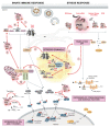
Figure 1
Figure 1
PKR at the crossroads of virus sensing, innate immune signaling, and stress response pathways. Cells evolved several cytosolic sentinels that detect virus infection and initiate defense mechanisms, including innate immune and stress responses. Cytosolic nucleic acid sensors RIG-I, MDA5, and cGAS signal via the mitochondrial adapter protein MAVS and the ER-adapter protein STING, triggering downstream signaling cascades via the IKK complex (IKKγ, IKKα, IKKβ) and IKKε/IKKγ/TBK1. The IKK complex phosphorylates the NF-κB (p50, p65) inhibitor IκB, whose degradation enables the nuclear translocation of NF-κB and transcriptional activation of IFN-β and pro-inflammatory cytokines. TBK1, on the other hand, phosphorylates IRF3/7, whose nuclear translocation mediates the transcriptional activation of IFN-α/β (left side). Cytosolic dsRNA that accumulates during viral replication is sensed by the stress kinase PKR. As a consequence, phosphorylation of eIF2α strongly represses the translation of most cellular mRNAs, while the translation of factors related to the stress response (ATF4, CHOP, and GADD34) or cell survival (XIAP and Bcl-2) is selectively favored (right side). The other eIF2α-kinases, GCN2, PERK, and HRI, contribute to translation suppression by detecting virus-induced changes in cellular homeostasis such as nutrient deprivation and accumulation of reactive oxygen species (ROS) or unfolded proteins in the ER. In the cytosol, untranslated mRNAs condense together with numerous RBPs and form SGs (1). Upon infection with certain viruses, innate immune sensors, PKR, regulators of stress and immune sensors, and interferon (IFN)-induced effectors localize in stress granules (SGs) together with viral RNA or DNA, forming a signaling platform (2) that coordinates and potentiates the antiviral response (3,4,5). PKR is at the crossroads of stress and innate immune signaling pathways: PKR interacts with RIG-I, promotes its activation, and amplifies the downstream signaling cascade (6). A similar interaction exists with cGAS (7). PKR promotes MDA5 filament formation, is activated by MDA5 to enhance downstream MAVS signaling (8), and, in turn, can also be activated by MAVS (9). PKR affects pro-inflammatory responses by interacting with TRAFs to activate the NF-κB signaling and potentially the JNK/p38 MAPK pathway leading to AP-1 activation (10), or directly regulates NF-κB activity via phosphorylation of IκB and IKK (11). Finally, PKR is involved in controlling the stability and translation of IFN mRNAs (12). Black arrows indicate signaling pathways; dashed red arrows indicate crossroads between stress response pathways, innate immune signaling, and SGs.
Figure 2
Figure 2
SGs as immune and stress signaling platforms. SGs function as immune and signaling platforms by phase-separating and concentrating regulatory proteins involved in apoptosis induction and other intracellular signaling pathways (A) and IFN signaling. (B). Certain RBPs and cellular mRNAs (e.g., cytokine mRNAs) preferentially localize in SGs (C). Viral components and ITAFs that control viral gene expression are also detected in SGs (D). SG composition is highly stress- and cell type-specific. Notably, many components have been detected in SGs under metabolic or environmental stress conditions, while an investigation in the context of viral infection is still missing. The localization and function of some SG components is dependent on the interaction with specific SG core proteins, indicated in turquoise.
Figure 3
Figure 3
SG localization in close proximity to mitochondria. Shown are live-cell microscopy images of HCV-induced dynamic assembly and disassembly of SGs. (A) human hepatocarcinoma Huh7 cells, stably expressing YFP-TIA1 (in yellow) and mTurquoise2-mito [339], a mitochondrial targeting peptide (in cyan), were infected with an HCV-mCherry reporter virus (in red). (B) Cropped section. White arrows indicate examples of SGs that are localized in close proximity to mitochondria (scale bar 10 µM).

References

    1. Mir M.A., Panganiban A.T. A protein that replaces the entire cellular eIF4F complex. EMBO J. 2008;27:3129–3139. doi: 10.1038/emboj.2008.228. - DOI - PMC - PubMed
    1. Schulz F., Yutin N., Ivanova N.N., Ortega D.R., Lee T.K., Vierheilig J., Daims H., Horn M., Wagner M., Jensen G.J., et al. Giant viruses with an expanded complement of translation system components. Science. 2017;356:82–85. doi: 10.1126/science.aal4657. - DOI - PubMed
    1. Colson P., De Lamballerie X., Yutin N., Asgari S., Bigot Y., Bideshi D.K., Cheng X.W., Federici B.A., Van Etten J.L., Koonin E.V., et al. “Megavirales”, a proposed new order for eukaryotic nucleocytoplasmic large DNA viruses. Arch. Virol. 2013;158:2517–2521. doi: 10.1007/s00705-013-1768-6. - DOI - PMC - PubMed
    1. Sullivan M.B., Huang K.H., Ignacio-Espinoza J.C., Berlin A.M., Kelly L., Weigele P.R., DeFrancesco A.S., Kern S.E., Thompson L.R., Young S., et al. Genomic analysis of oceanic cyanobacterial myoviruses compared with T4-like myoviruses from diverse hosts and environments. Environ. Microbiol. 2010;12:3035–3056. doi: 10.1111/j.1462-2920.2010.02280.x. - DOI - PMC - PubMed
    1. Sencilo A., Jacobs-Sera D., Russell D.A., Ko C.C., Bowman C.A., Atanasova N.S., Osterlund E., Oksanen H.M., Bamford D.H., Hatfull G.F., et al. Snapshot of haloarchaeal tailed virus genomes. RNA Biol. 2013;10:803–816. doi: 10.4161/rna.24045. - DOI - PMC - PubMed

Publication types