. 2020 Sep 4;21(18):6476.
doi: 10.3390/ijms21186476.

P2 × 7 Receptor Inhibits Astroglial Autophagy via Regulating FAK- and PHLPP1/2-Mediated AKT-S473 Phosphorylation Following Kainic Acid-Induced Seizures

Affiliations

P2 × 7 Receptor Inhibits Astroglial Autophagy via Regulating FAK- and PHLPP1/2-Mediated AKT-S473 Phosphorylation Following Kainic Acid-Induced Seizures

Duk-Shin Lee et al. Int J Mol Sci. .

Abstract

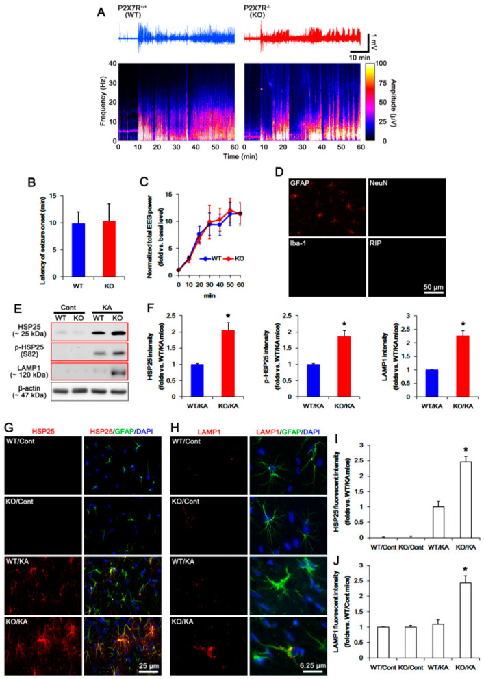
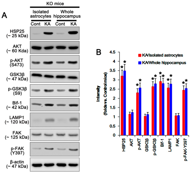
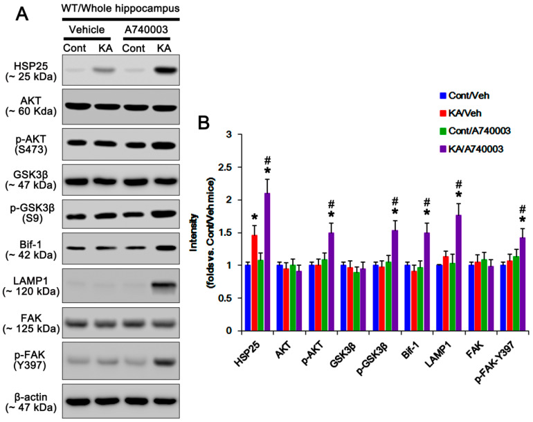
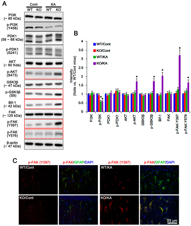
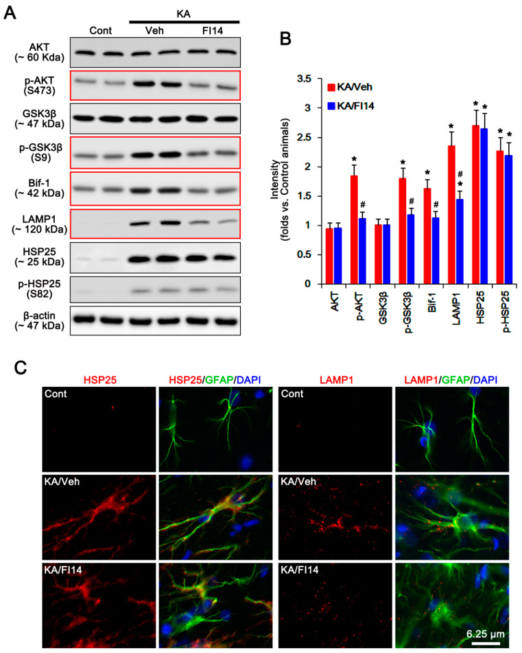
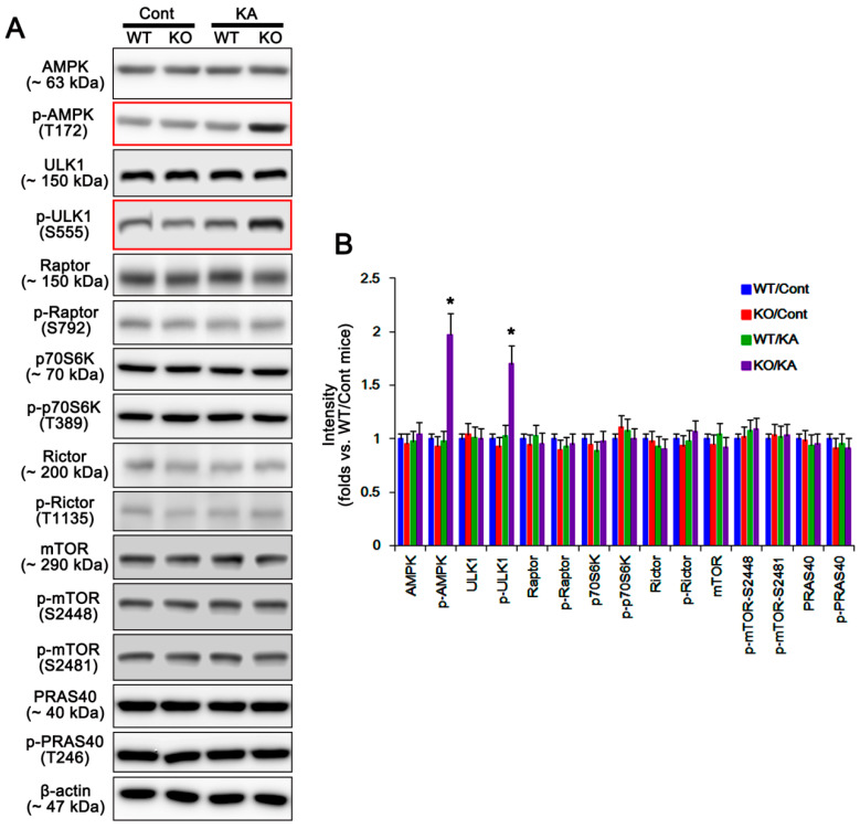
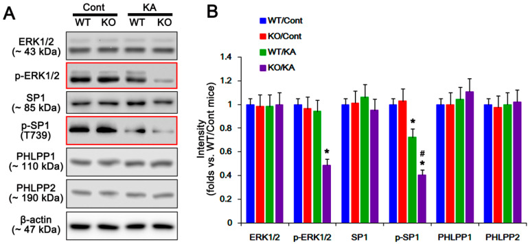
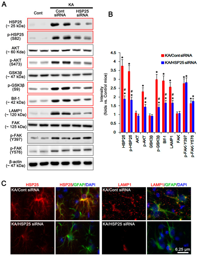
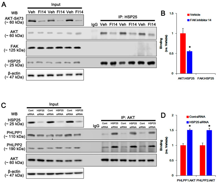
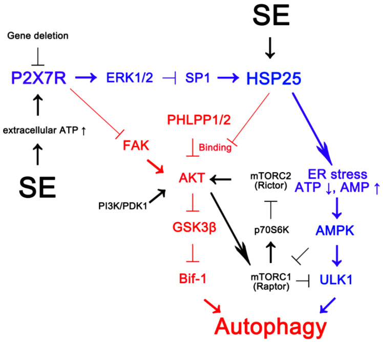
Recently, we have reported that blockade/deletion of P2X7 receptor (P2X7R), an ATP-gated ion channel, exacerbates heat shock protein 25 (HSP25)-mediated astroglial autophagy (clasmatodendrosis) following kainic acid (KA) injection. In P2X7R knockout (KO) mice, prolonged astroglial HSP25 induction exerts 5' adenosine monophosphate-activated protein kinase/unc-51 like autophagy activating kinase 1-mediated autophagic pathway independent of mammalian target of rapamycin (mTOR) activity following KA injection. Sustained HSP25 expression also enhances AKT-serine (S) 473 phosphorylation leading to astroglial autophagy via glycogen synthase kinase-3β/bax interacting factor 1 signaling pathway. However, it is unanswered how P2X7R deletion induces AKT-S473 hyperphosphorylation during autophagic process in astrocytes. In the present study, we found that AKT-S473 phosphorylation was increased by enhancing activity of focal adhesion kinase (FAK), independent of mTOR complex (mTORC) 1 and 2 activities in isolated astrocytes of P2X7R knockout (KO) mice following KA injection. In addition, HSP25 overexpression in P2X7R KO mice acted as a chaperone of AKT, which retained AKT-S473 phosphorylation by inhibiting the pleckstrin homology domain and leucine-rich repeat protein phosphatase (PHLPP) 1- and 2-binding to AKT. Therefore, our findings suggest that P2X7R may be a fine-tuner of AKT-S473 activity during astroglial autophagy by regulating FAK phosphorylation and HSP25-mediated inhibition of PHLPP1/2-AKT binding following KA treatment.

Keywords: Bif-1; FAK inhibitor 14; LAMP1; PRAS40; Raptor; Rictor; p70S6K; siRNA.


Conflict of interest statement

The authors have declared that no conflict of interest exist.

Figures

Figure 1
Figure 1
Effects of P2X7 receptor (P2X7R) deletion on seizure activity, heat shock protein 25 (HSP25) expression, and astroglial autophagy in response to kainic acid (KA). (AC) Effect of P2X7R deletion on seizure susceptibility in response to KA. P2X7R deletion does not affect the seizure susceptibility in response to KA. (A) Representative EEG traces and frequency-power spectral temporal maps. (B,C) Quantification of the latency of seizure onset (B) and total EEG power (C) in response to KA. (mean ± SEM; n = 7, respectively). (D) Identification of isolated cells using glial fibrillary acidic protein (GFAP, an astroglial marker), neuronal nuclear antigen (NeuN, a neuronal marker), ionized calcium binding protein-1 (Iba-1, a microglial marker), and RIP (an oligodendroglial marker) antibodies. (E,F) Effect of P2X7R deletion on KA-induced HSP25 and LAMP1 expressions in isolated astrocytes. As compared to wild-type (WT) astrocytes, both HSP25 and LAMP1 expression levels are higher in P2X7R knockout (KO) astrocytes 7 days after KA injection (red box). (E) Representative Western blot of HSP25 and lysosome-associated membrane protein 1 (LAMP1). Cont, control animals; KA, KA-injected animals. (F) Quantifications of HSP25 and LAMP1 expressions and HSP phosphorylation following KA injection (mean ± SEM; * p < 0.05 vs. KA-treated WT astrocytes; n = 7, respectively). (G,H) Effect of P2X7R deletion on HSP25 and LAMP1 protein expressions in the hippocampal astrocytes. As compared to WT mice, both HSP25 (G) and LAMP1 (H) expression levels are higher in P2X7R KO mice 7 days after KA injection. (I,J) Quantifications of HSP25 (I) and LAMP1 (J) fluorescent intensity following KA injection (mean ± SEM; * p < 0.05 vs. KA-treated WT mice; n = 7, respectively).
Figure 2
Figure 2
Effects of KA on expressions and phosphorylations of HSP25, AKT (also known as protein kinase B), glycogen synthase kinase-3β (GSK3β), bax interacting factor 1 (Bif-1), LAMP1, and focal adhesion kinase (FAK) in isolated astrocytes and the whole hippocampus obtained from P2X7R mice. (A) Representative Western blot of expressions and phosphorylations of HSP25, AKT, GSK3β, Bif-1, LAMP1, and FAK. Cont, control animals; KA, KA-injected animals. (B) Quantifications of expressions and phosphorylations of HSP25, AKT, GSK3β, Bif-1, LAMP1, and FAK following KA injection (mean ± SEM; * p < 0.05 vs. control mice; n = 7, respectively).
Figure 3
Figure 3
Effects of A740003 on expressions and phosphorylations of HSP25, AKT, GSK3β, Bif-1, LAMP1, and FAK in the whole hippocampus obtained from WT mice following KA injection. (A) Representative Western blot of expressions and phosphorylations of HSP25, AKT, GSK3β, Bif-1, LAMP1, and FAK. Cont, control animals; KA, KA-injected animals. (B) Quantifications of expressions and phosphorylations of HSP25, AKT, GSK3β, Bif-1, LAMP1, and FAK in KO astrocytes following KA injection (mean ± SEM; *,# p < 0.05 vs. control and vehicle-treated mice, respectively; n = 7, respectively).
Figure 4
Figure 4
Effects of P2X7R deletion on expressions and phosphorylations of phosphatidylinositol-4,5-bisphosphate 3-kinase (PI3K), phosphoinositide-dependent kinase-1 (PDK1), AKT, GSK3β, Bif-1, and FAK in isolated astrocytes following KA injection. KA increases p-AKT-S473, p-GSK3β-S9, Bif-1, p-FAK-Y397, and p-FAK-Y576 levels in KO astrocytes 7 days after KA injection (red box). (A) Representative Western blot of expressions and phosphorylations of PI3K, PDK1, AKT, GSK3β, Bif-1, and FAK. Cont, control animals; KA, KA-injected animals. (B) Quantifications of expressions and phosphorylations of PI3K, PDK1, AKT, GSK3β, Bif-1, and FAK following KA injection (mean ± SEM; * p < 0.05 vs. control WT astrocytes; n = 7, respectively). (C) Effect of P2X7R deletion on p-FAK-Y397 level in the hippocampal astrocytes. As compared to WT mice, p-FAK-Y397 level is higher in KO mice 7 days after KA injection.
Figure 5
Figure 5
Effects of FAK inhibition on expressions and phosphorylations of AKT, GSK3β, Bif-1, LAMP1, HSP25, and p-HSP25 in KO astrocytes following KA injection. As compared to vehicle, FAK inhibitor 14 (FI14) abolishes the upregulations of p-AKT-S473, p-GSK3β-S9, Bif-1, and LAMP1, but not HSP25, levels 7 days after KA injection (red box). (A) Representative Western blot of expressions and phosphorylations of AKT, GSK3β, Bif-1, LAMP1, HSP25, and p-HSP25 in KO astrocytes. Cont, control animals; KA, KA-injected animals; Veh, vehicle; FI4, FAK inhibitor 14. (B) Quantifications of expressions and phosphorylations of AKT, GSK3β, Bif-1, LAMP1, HSP25, and p-HSP25 in KO astrocytes following KA injection (mean ± SEM; *,# p < 0.05 vs. control KO astrocytes and vehicle, respectively; n = 7, respectively). (C) Effect of P2X7R deletion on HSP25 and LAMP1 expression in the hippocampal astrocytes. As compared to vehicle, FI14 abrogates LAMP1, but not HSP25, expression in KO mice 7 days after KA injection.
Figure 6
Figure 6
Effects of P2X7R deletion on expressions and phosphorylations of 5′ adenosine monophosphate-activated protein kinase (AMPK), unc-51 like autophagy activating kinase 1 (ULK1), regulatory associated protein of mammalian target of rapamycin (Raptor), p70S6 kinase (p70D6K), rapamycin-insensitive companion of mammalian target of rapamycin (Rictor), mammalian target of rapamycin (mTOR), and proline-rich AKT substrate of 40 kDa (PRAS40) in isolated astrocytes following KA injection. KA increases p-AMPK and p-ULK1 levels in KO astrocytes 7 days after KA injection (red box). (A) Representative Western blot of expressions and phosphorylations of AKT, GSK3β, Bif-1, LAMP1, HSP25, and p-HSP25 in KO astrocytes. Cont, control animals; KA, KA-injected animals. (B) Quantifications of expressions and phosphorylations of AMPK, ULK1, Raptor, p70D6K, Rictor, mTOR, and PRAS40 in isolated astrocytes following KA injection (mean ± SEM; * p < 0.05 vs. control WT astrocytes; n = 7, respectively).
Figure 7
Figure 7
Effects of P2X7R deletion on expressions and phosphorylations of extracellular regulated kinase 1/2 (ERK1/2), specificity protein 1 (SP1), pleckstrin homology domain, and leucine-rich repeat protein phosphatase 1 (PHLPP1) and PHLPP2 in isolated astrocytes following KA injection. As compared to WT astrocytes, KA reduces p-ERK1/2 and p-SP1 levels in KO astrocytes 7 days after KA injection (red box). (A) Representative Western blot of expressions and phosphorylations of ERK1/2, SP1, PHLPP1, and PHLPP2 in isolated astrocytes following KA injection. Cont, control animals; KA, KA-injected animals. (B) Quantifications of expressions and phosphorylations of ERK1/2, SP1, PHLPP1, and PHLPP2 in isolated astrocytes following KA injection (mean ± SEM; *,# p < 0.05 vs. Control WT astrocytes and KA-treated WT astrocytes, respectively; n = 7, respectively).
Figure 8
Figure 8
Effects of HSP25 knockdown on expressions and phosphorylations of HSP25, AKT, GSK3β, Bif-1, LAMP1, and FAK in KO astrocytes following KA injection. HSP25 knockdown abrogates upregulations of HSP25, p-HSP25, p-AKT-S473, p-GSK3β-S9, Bif-1, and LAMP1 induced by KA, while it does not affect FAK and its phosphorylations, 7 days after KA injection (red box). (A) Representative Western blot of expressions and phosphorylations of HSP25, AKT, GSK3β, Bif-1, LAMP1, and FAK in KO astrocytes. Cont, control animals; KA, KA-injected animals. (B) Quantifications of expressions and phosphorylations of HSP25, AKT, GSK3β, Bif-1, LAMP1, and FAK in KO astrocytes following KA injection (mean ± SEM; *,# p < 0.05 vs. control KO astrocytes and control siRNA, respectively; n = 7, respectively). (C) Effects of HSP25 knockdown on HSP25 and LAMP1 expression in the hippocampal astrocytes. As compared to control siRNA, FI14 abrogates LAMP1, but not HSP25, expression in KO mice 7 days after KA injection.
Figure 9
Figure 9
Effects of FAK inhibition and HSP25 knockdown on the bindings of AKT-HSP25, FAK-HSP25, PHLPP1-AKT, and PHLPP1-AKT in the hippocampus of KO mice following KA injection. (A,B) Effects of FI14 on the bindings of AKT-HSP25 and FAK-HSP25. FI14 attenuates AKT-HSP25 binding with reducing AKT-S473 phosphorylation, 7 days after KA injection. FAK does not bind with HSP25. (A) Representative Western blot of p-AKT-S473, AKT, FAK, and HSP25 in the KO hippocampus. Veh, vehicle; FI14, FAK inhibitor 14. (B) Quantifications of the bindings of AKT-HSP25 and FAK-HSP25 (mean ± SEM; * p < 0.05 vs. vehicle, respectively; n = 7, respectively). (C,D) Effects of HSP25 knockdown on the bindings of PHLPP1-AKT and PHLPP2-AKT. HSP25 siRNA ameliorates PHLPP1-AKT and PHLPP2-AKT bindings, 7 days after KA injection. (C) Representative Western blot of HSP25, PHLPP1, PHLPP2, and AKT in the KO hippocampus. (D) Quantifications of the bindings of PHLPP1-AKT and PHLPP2-AKT (mean ± SEM; * p < 0.05 vs. vehicle, respectively; n = 7, respectively).
Figure 10
Figure 10
Scheme of inhibitory role of P2X7R in AKT-S473 hyperphosphorylation during astroglial autophagy induced by KA injection based on the present data (red) and previous reports (blue and black) [6,14,21,22,23]. After KA injection, P2X7R activation inhibits FAK phosphorylation and HSP25 transactivation via ERK1/2-mediated SP1-T739 phosphorylation. P2X7R deletion leads to sustained HSP25 expression, which activates AMPK/ULK1-mediated astroglial autophagy (blue) [6]. In addition, P2X7R deletion increases FAK autophosphorylation [20]. Subsequently, the activated FAK phosphorylates AKT, and the prolonged HSP25 expression abrogates the binding of PHLPP1/2 to AKT, which result in AKT-S473 hyperphosphorylation. Sustained AKT-S473 phosphorylation exerts AKT/GSK3β-mediated Bif-1 induction that triggers astroglial autophagy (red), independent of PI3K/PDK1 and mTOR complex (mTORC) 1/2 activities (black). Thus, these findings suggest that P2X7R may be a fine-tuner of autophagic process in astrocytes by regulating AKT-S473 phosphorylation.

References

    1. Jäkel S., Dimou L. Glial cells and their function in the adult brain: A journey through the history of their ablation. Front. Cell Neurosci. 2017;11:24. doi: 10.3389/fncel.2017.00024. - DOI - PMC - PubMed
    1. Kang T.C., Kim D.S., Kwak S.E., Kim J.E., Won M.H., Kim D.W., Choi S.Y., Kwon O.S. Epileptogenic roles of astroglial death and regeneration in the dentate gyrus of experimental temporal lobe epilepsy. Glia. 2006;54:258–271. doi: 10.1002/glia.20380. - DOI - PubMed
    1. Sugawara T., Lewén A., Noshita N., Gasche Y., Chan P.H. Effects of global ischemia duration on neuronal, astroglial, oligodendroglial, and microglial reactions in the vulnerable hippocampal CA1 subregion in rats. J. Neurotrauma. 2002;19:85–98. doi: 10.1089/089771502753460268. - DOI - PubMed
    1. Olabarria M., Noristani H.N., Verkhratsky A., Rodríguez J.J. Concomitant astroglial atrophy and astrogliosis in a triple transgenic animal model of Alzheimer’s disease. Glia. 2010;58:831–838. doi: 10.1002/glia.20967. - DOI - PubMed
    1. Penfield W. Neuroglia and microglia—The interstitial tissue of the central nervous system. In: Cowdry E.V., editor. Special Cytology, the Form and Function of the Cell in Health and Disease. Hoeber; New York, NY, USA: 1928. pp. 1033–1068.

MeSH terms