. 2020 Sep 4;9(9):731.
doi: 10.3390/pathogens9090731.

A Novel Genotype and First Record of Trypanosoma lainsoni in Argentina

Affiliations

A Novel Genotype and First Record of Trypanosoma lainsoni in Argentina

Anahí G Díaz et al. Pathogens. .

Abstract

Trypanosomes are a group of parasitic flagellates with medical and veterinary importance. Despite many species having been described in this genus, little is known about many of them. Here, we report a genetic and morphological characterization of trypanosomatids isolated from wild mammals from the Argentine Chaco region. Parasites were morphologically and ultrastructurally characterized by light microscopy and transmission electron microscopy. Additionally, 18s rRNA and gGAPDH genes were sequenced and analyzed using maximum likelihood and Bayesian inference. Morphological characterization showed clear characteristics associated with the Trypanosoma genus. The genetic characterization demonstrates that the studied isolates have identical sequences and a pairwise identity of 99% with Trypanosoma lainsoni, which belongs to the clade of lizards and snakes/rodents and marsupials. To date, this species had only been found in the Amazon region. Our finding represents the second report of T. lainsoni and the first record for the Chaco region. Furthermore, we ultrastructurally described for the first time the species. Finally, the host range of T. lainsoni was expanded (Leopardus geoffroyi, Carenivora, Felidae; and Calomys sp., Rodentia, Cricetidae), showing a wide host range for this species.

Keywords: 18S rDNA genes; Calomys spp.; Leopardus geoffroyi; Trypanosoma lainsoni; gGAPDH genes; morphology; transmission electron microscopy.


Conflict of interest statement

The authors declare no conflict of interest. The funders had no role in the design of the study; in the collection, analyses, or interpretation of the data; in the writing of the manuscript, or in the decision to publish the results.

Figures
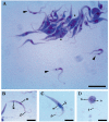
Figure 1
Figure 1
Microphotography at 1000× of parasites in cultures with May Grünwald and Giemsa. (A) Exponential phase culture: trypomastigotes (arrow) were observed among the epimastigotes; the dividing epimastigotes are denoted with an asterisk; (B) Trypomastigote; (C) Epimastigote; (D) Spheromastigote. Nucleus = n, kinetoplast = k, flagellum = f. Scale bars: (A) 20 µm and (BD) 10 µm.
Figure 2
Figure 2
Microphotography at 1000× of parasites in cultures with May Grünwald and Giemsa. (A) The difference in sizes between a long epimastigote (LE) and a simple epimastigote (E) is observed; (B) Dividing epimastigote with two nuclei, two flagella, and one kinetoplast; (C) Trypomastigote (T) smaller than epimastigote (E); (D) Transitional form (t). Nucleus = n, kinetoplast = k, flagellum = f. Scale bar (AD) 10 µm.
Figure 3
Figure 3
Electron micrograph of the exponential growth phase. (A) Epimastigote with a tapered shape, central nucleus (n), anterior kinetoplast (k) in a position close to the flagellar pocket (fp); (B) epimastigote showing the position of the nucleus (n), kinetoplast (k), cytostome (cyt) and acidocalcisomes (Ac); (C) Nucleus (n) with chromatin spots on the inner side of the nuclear membrane; (D) Rod shaped kinetoplast (k); (E) Structure of the flagellum: basal body (bb), probasal body (pb), and cross section of the flagellum; (F) Cross section cytostome (cyt); (G) Acidocalcisomes (ac). Scale bar (AB) 1 µm; (CG) 0.5 µm).
Figure 4
Figure 4
Transmission electron micrograph of the isolated parasites in the exponential phase. (A) Division cell with two nuclei (n), a kinetoplast (k), and a single flagellar pocket (fp); (B) Flagellar pocket with two basal body (bb); (C) Numerous vesicular bodies highlighting lipid bodies (l) and reservosomes (r). Scale bar (AC) 0.5 µm.
Figure 5
Figure 5
Phylogenetic analysis performed by the maximum likelihood method of the concatenated 18S rDNA and gGAPDH genes in 33 species of trypanosomatids. Species of the genera Herpetomonas and Crithidia were included as out groups. The two values at nodes represent: Bayesian analysis support/ML method bootstrap value. Trypanosoma lainsoni and Le29 are found within the LSRM clade.

References

    1. Vickman K. The Diversity of the kinetoplastid flagellates. In: Lumsden W.H.R., Evans D.A., editors. Biology of the Kinetoplastida. Academic Press; London, UK: New York, NY, USA: San Francisco, CA, USA: 1976. pp. 1–34.
    1. Svobodová M., Zídková L., Cepicka I., Obornik M., Lukes J., Votrpka J. Sergeia podlipaevi gen. Nov., sp. voc. (Tryoanosomatida, Kinetoplastida), parasite of biting midges (Ceratopogonidae, Diptera) Int. J. Syst. Evol. Microbiol. 2007;57:423–432. doi: 10.1099/ijs.0.64557-0. - DOI - PubMed
    1. Jaskowska E., Mayordomo C., Preston G., Kelly S. Phytomonas: Tripanosomátidos adaptados a los ambientes vegetales. PLoS Pathog. 2015;11:e1004484. - PMC - PubMed
    1. Seward E.A., Votypka J., Kment P., Lukes J., Kelly S. Description of Phytomonas oxycareni n. sp. from the Salivary Glands of Oxycarenus lavaterae. Protist. 2017;168:71–79. doi: 10.1016/j.protis.2016.11.002. - DOI - PubMed
    1. Hamilton P.B., Gibson W.C., Stevens J.R. Patterns of co-evolution between trypanosomes and their hosts deduced from ribosomal RNA and protein-coding gene phylogenies. Mol. Phylogenetics Evol. 2007;44:15–25. doi: 10.1016/j.ympev.2007.03.023. - DOI - PubMed

LinkOut - more resources