. 2020 Sep 6;12(9):574.
doi: 10.3390/toxins12090574.

Bee Venom Melittin Protects against Cisplatin-Induced Acute Kidney Injury in Mice via the Regulation of M2 Macrophage Activation

Affiliations

Bee Venom Melittin Protects against Cisplatin-Induced Acute Kidney Injury in Mice via the Regulation of M2 Macrophage Activation

Hyunseong Kim et al. Toxins (Basel). .

Abstract

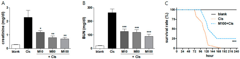
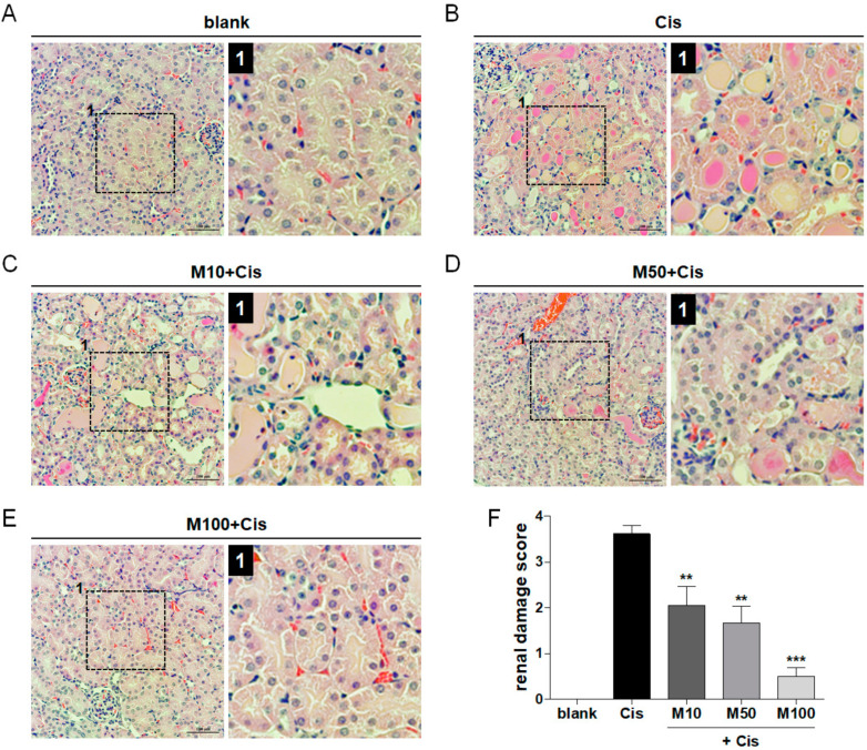
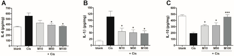
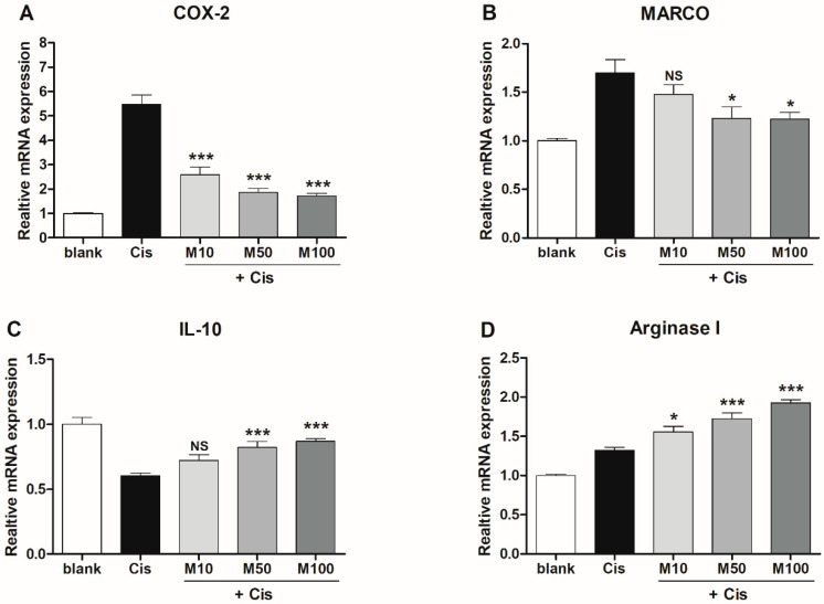
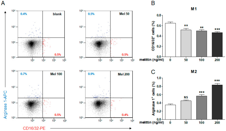
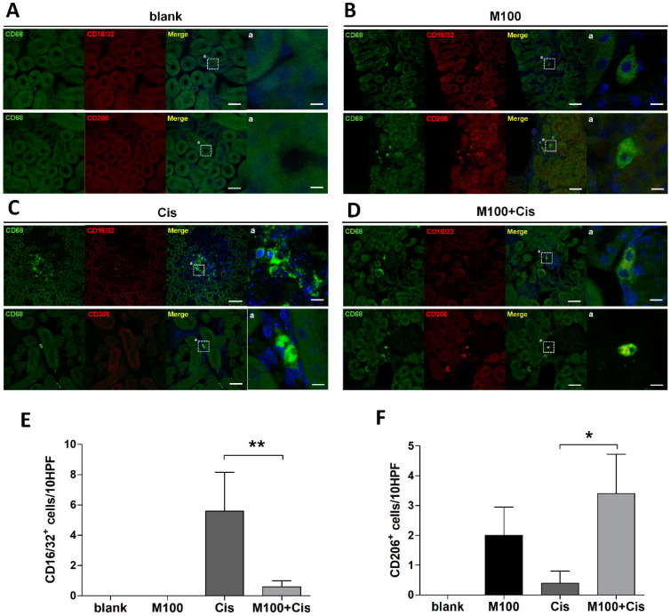
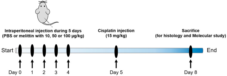
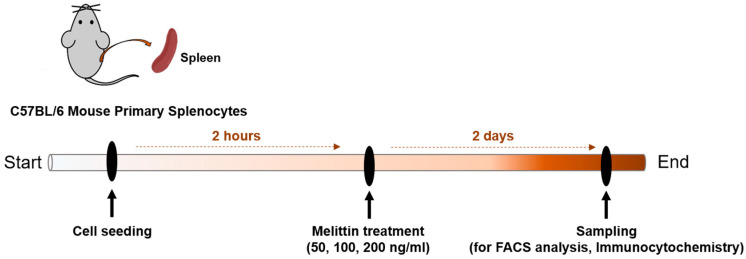
Inflammation is an essential biological response that eliminates pathogenic bacteria and repairs tissue after injury. Acute kidney injury (AKI) is associated with systemic and intrarenal inflammation as the inflammatory process decreases renal function and promotes progression to advanced chronic kidney disease. Macrophages are key mediators of the inflammatory response; their activation influences the immune system and may have various effects. Classically activated type I macrophages (M1) produce a variety of pro-inflammatory cytokines at the lesion site. However, anti-inflammatory type II macrophages (M2) are alternatively activated upon exposure to anti-inflammatory cytokines and are associated with wound healing and tissue repair following AKI. Here, we used melittin from bee venom to enhance the polarization of M2 macrophages and promote renal recovery after AKI. Melittin was administered to mice intraperitoneally for 5 days at various concentrations (10, 50, and 100 µg/kg); serum creatinine and blood urea nitrogen (BUN) levels were analyzed 72 h after cisplatin administration to confirm renal dysfunction. Melittin inhibited the cisplatin-induced increase in creatinine and BUN, an indicator of renal dysfunction. The expression of M1 markers (CD16/32) decreased significantly, whereas that of M2 markers (CD206, Arg1nase I) increased after melittin administration. Consistently, tubular necrosis was substantially reduced in melittin-treated mice. Thus, melittin alleviates cisplatin-induced AKI by regulating M2 macrophage expression.

Keywords: M2 macrophage; acute kidney injury; cisplatin; melittin.


Conflict of interest statement

The authors declare no conflict of interest.

Figures

Figure 1
Figure 1
Protective effects of melittin on cisplatin-induced acute kidney injury. (A) Serum creatinine and (B) blood urea nitrogen (BUN) levels were measured 72 h after cisplatin injection in each group (n = 8). Mice injected with melittin presented a dose-dependent significant decrease in BUN and creatinine values at 72 h after cisplatin injection compared with the Cis group. (C) Survival curves of mice injected with 100 µg/kg melittin, starting 72 h after cisplatin administration (15 mg/kg, n = 12). Data are expressed as the means ± standard error of the mean (SEM). Significant differences indicated as * p < 0.05, ** p < 0.01, and *** p < 0.001 vs. the cisplatin group were analyzed via one-way analysis of variance (ANOVA) with Tukey’s post-hoc test.
Figure 2
Figure 2
Reduction of cisplatin-induced renal tissue damage by melittin administration. (A) Blank group showing normal pattern of renal tubules and glomeruli. (B) The majority of the renal tubules were severely damaged, showing vacuolation and degeneration of renal tubules and brush border loss. (CE) Melittin dose-dependently improves recovery of renal damage in the acute kidney injury (AKI) model. Scale bar = 100 or 50 μm. Low-magnification view to show region (box outlined with black dashed line) examined at higher magnification and indicated by number 1. (F) Renal damage was quantified in each group by analyzing the renal damage score (n = 6). Data are expressed as the means ± SEM. Significant differences indicated as * p < 0.05, ** p < 0.01, and *** p < 0.001 vs. the cisplatin group were analyzed via one-way ANOVA with Tukey’s post-hoc test.
Figure 3
Figure 3
Effects of melittin on pro- and anti-inflammatory cytokine production in mice with AKI. The expression of (A) IL-6 and (B) IL-1β (pro-inflammatory), and (C) IL-10 (anti-inflammatory) was detected in 72 h after cisplatin injection via enzyme-linked immunosorbent assay (ELISA) in mice injected with PBS (blank group), cisplatin (Cis group), or melittin at various concentrations (10, 50, or 100 µg/kg) in the presence of cisplatin (n = 6). Significant differences indicated as * p < 0.05, ** p < 0.01, and *** p < 0.001 vs. the cisplatin group were analyzed via one-way ANOVA with Tukey’s post-hoc test.
Figure 4
Figure 4
Effects of melittin on the expression of M1 and M2 macrophage-associated genes in mice with AKI. mRNA expression of M1 markers (A) cyclooxygenase 2 (COX-2), (B) macrophage receptor with collagenous structure (MARCO), the M2 markers (C) IL-10 and (D) arginase 1 in the renal tissue of mice in each group, measured 72 h after cisplatin administration (n = 6). Significant differences indicated as * p < 0.05, ** p < 0.01, and *** p < 0.001 vs. the cisplatin group were analyzed via one-way ANOVA with Tukey’s post-hoc test.
Figure 5
Figure 5
Effects of melittin on M1 and M2 macrophage populations in mouse splenocytes. (A) Representative flow cytometry gating shows M1 macrophages (CD16/32) and M2 macrophage (Arg1nase1) populations in mouse splenocytes at 2 days after treatment of melittin with various concentration (50, 100, or 200 ng/mL). Quantification of the percentage of cells positive for (B) CD16/32 (M1 macrophage marker) and (C) Arg1 (M2 macrophage marker) relative to that in the blank group (n = 6). Significant differences indicated at * p < 0.05, ** p < 0.01, and *** p < 0.001 vs. the blank group were analyzed via one-way ANOVA with Tukey’s post-hoc test.
Figure 6
Figure 6
Effects of melittin on M2 macrophage infiltration into the kidney. (AD) Representative images of renal tissues stained with M1 macrophages (CD16/32 positive, double-stained with CD68) and M2 macrophage (CD206 positive, double-stained with CD68) in each group, detected 72 h after cisplatin administration. Scale bar = 50 and 10 μm. Quantification of (E) CD16/32- and (F) CD206-positive cells per 10 consecutive high-power fields (HPF) in each group (n = 6). Significant differences indicated as * p < 0.05, ** p < 0.01, and *** p < 0.001 vs. the cisplatin group were analyzed via one-way ANOVA with Tukey’s post-hoc test.
Scheme 1
Scheme 1
Schematic illustration of the experimental timeline. C57/BL6 mouse was injected with PBS or melittin with 10, 50 or 100 µg/kg into an abdominal cavity through an i.p. for 5 days. Mouse received a single injection of cisplatin (15 mg/kg i.p.) with or without melittin pretreatment. Mouse was sacrificed three days later.
Scheme 2
Scheme 2
Schematic illustration of the experimental timeline. Mouse primary splenocytes was isolated from spleen of C57BL/6 mouse. Cells were seeded at a density of 4 × 10⁵ cells/well in 24 well plates. After 2 h, splenocytes were treated with different concentration (50, 100, 200 ng/mL) of melittin for 2 days. We also evaluated the effect of melittin on macrophage polarization in mouse splenocytes using FACS analysis and immunocytochemistry.

References

    1. Dasari S., Tchounwou P.B. Cisplatin in cancer therapy: Molecular mechanisms of action. Eur. J. Pharmacol. 2014;740:364–378. doi: 10.1016/j.ejphar.2014.07.025. - DOI - PMC - PubMed
    1. Oh G.S., Kim H.J., Shen A., Bin Lee S., Khadka D., Pandit A., So H.S. Cisplatin-induced Kidney Dysfunction and Perspectives on Improving Treatment Strategies. Electrolytes Blood Press. 2014;12:55–65. doi: 10.5049/EBP.2014.12.2.55. - DOI - PMC - PubMed
    1. Tbahriti H.F., Meknassi D., Moussaoui R., Messaoudi A., Zemour L., Kaddous A., Bouchenak M., Mekki K. Inflammatory status in chronic renal failure: The role of homocysteinemia and pro-inflammatory cytokines. World J. Nephrol. 2013;2:31–37. doi: 10.5527/wjn.v2.i2.31. - DOI - PMC - PubMed
    1. Rabb H., Griffin M.D., McKay D.B., Swaminathan S., Pickkers P., Rosner M.H., Kellum J.A., Ronco C. Inflammation in AKI: Current Understanding, Key Questions, and Knowledge Gaps. J. Am. Soc. Nephrol. 2015;27:371–379. doi: 10.1681/ASN.2015030261. - DOI - PMC - PubMed
    1. Domitrović R., Cvijanovic O., Pernjak-Pugel E., Zagorac G.B., Mahmutefendic H., Škoda M. Luteolin ameliorates cisplatin-induced nephrotoxicity in mice through inhibition of platinum accumulation, inflammation and apoptosis in the kidney. Toxicology. 2013;310:115–123. doi: 10.1016/j.tox.2013.05.015. - DOI - PubMed

Publication types

MeSH terms

LinkOut - more resources