. 2020 Sep 6;9(9):2038.
doi: 10.3390/cells9092038.

Nuclear Heparanase Regulates Chromatin Remodeling, Gene Expression and PTEN Tumor Suppressor Function

Affiliations

Nuclear Heparanase Regulates Chromatin Remodeling, Gene Expression and PTEN Tumor Suppressor Function

Rada Amin et al. Cells. .

Abstract

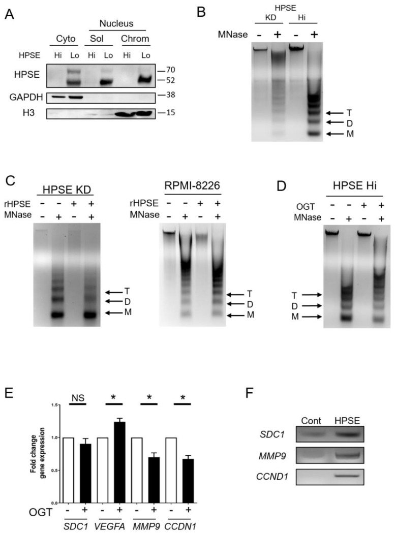
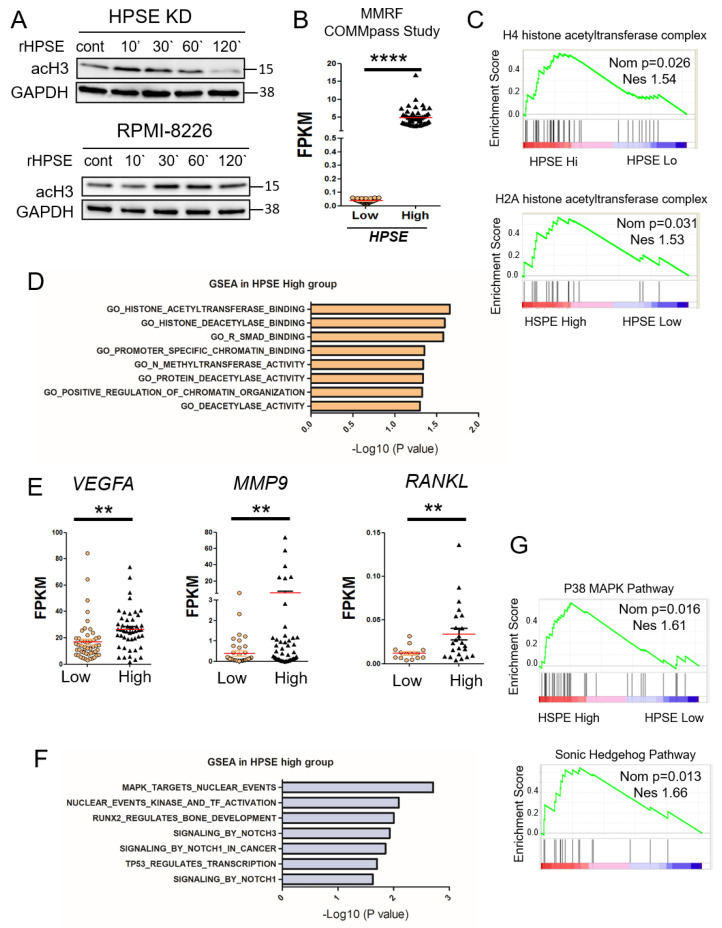
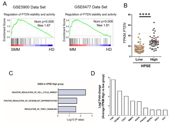
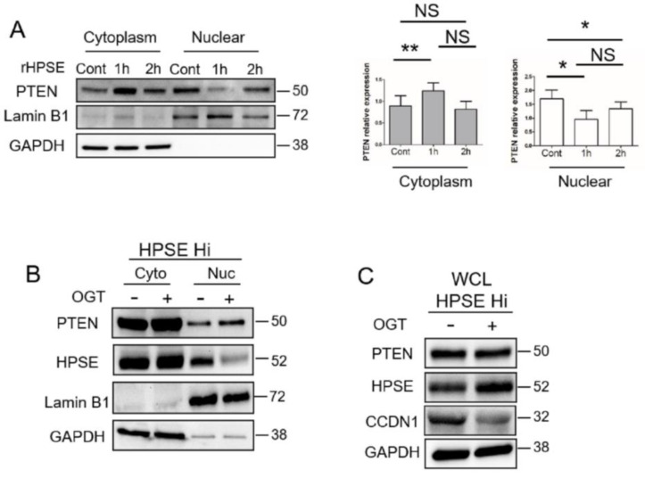
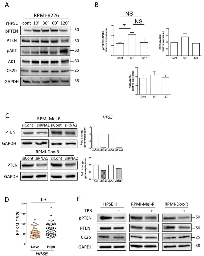
Heparanase (HPSE) is an endoglycosidase that cleaves heparan sulfate and has been shown in various cancers to promote metastasis, angiogenesis, osteolysis, and chemoresistance. Although heparanase is thought to act predominantly extracellularly or within the cytoplasm, it is also present in the nucleus, where it may function in regulating gene transcription. Using myeloma cell lines, we report here that heparanase enhances chromatin accessibility and confirm a previous report that it also upregulates the acetylation of histones. Employing the Multiple Myeloma Research Foundation CoMMpass database, we demonstrate that patients expressing high levels of heparanase display elevated expression of proteins involved in chromatin remodeling and several oncogenic factors compared to patients expressing low levels of heparanase. These signatures were consistent with the known function of heparanase in driving tumor progression. Chromatin opening and downstream target genes were abrogated by inhibition of heparanase. Enhanced levels of heparanase in myeloma cells led to a dramatic increase in phosphorylation of PTEN, an event known to stabilize PTEN, leading to its inactivity and loss of tumor suppressor function. Collectively, this study demonstrates that heparanase promotes chromatin opening and transcriptional activity, some of which likely is through its impact on diminishing PTEN tumor suppressor activity.

Keywords: PTEN; chromatin remodeling; gene transcription; heparanase; multiple myeloma.


Conflict of interest statement

The authors declare no conflict of interest.

Figures

Figure 1
Figure 1
Heparanase enhances chromatin accessibility. (A) To determine localization of heparanase, sequential cell fractionation of myeloma cells was used to isolate cytoplasmic (cyto), nucleus-soluble (sol), and nucleus-chromatin (chrom) fractions from CAG wild-type (HPSE Lo) and transfected CAG HPSE Hi cells prior to Western blot analysis. GAPDH and histone H3 (H3) were probed to assess the quality of the fractions. (B) Chromatin accessibility in CAG HPSE knockdown (KD) and HPSE Hi cells were assessed by sensitivity to micrococcal nuclease (MNase) digestion. Nuclei from KD and HPSE Hi cells were prepared and digested with 0.1 U/μL MNase and subjected to electrophoresis. Black arrows denote Tri (T), Di (D) and Mono (M)—nucleosomes. (C) Nuclei from CAG HPSE KD and RPMI-8226 myeloma cells were treated for 2 h with or without rHPSE before being subjected to Mnase digestion for chromatin accessibility. (D) CAG HPSE Hi cells were incubated with 20 µM of the HPSE inhibitor OGT2115 and chromatin accessibility was assessed following exposure of nuclei to MNase. (E) mRNA expression analysis by RT-PCR of syndecan-1 (SDC1), vascular endothelial growth factor A (VEGFA), matrix metalloproteinase-9 (MMP9) and cyclin D1 (CCDN1) in CAG HPSE Hi cells in the presence or absence of OGT2115, compared to respective control. * p < 0.05 by one-tailed, paired t-test. (F) Chromatin immunoprecipitation assay was performed on CAG HPSE Hi cells to analyze HPSE binding to the promoter regions of SDC1, MMP9 and CCND1. PCR was performed using primers probing the promoter regions of indicated genes after pulldown with an antibody control (Cont) or anti-heparanase (HPSE) and result was assessed by gel electrophoresis.
Figure 2
Figure 2
Heparanase stimulates histone acetylation and upregulation of genes that promote an aggressive tumor phenotype. (A) CAG HPSE KD and RPMI-8226 cells were incubated with 1 µg/mL of rHPSE for the indicated time and acetyl-histone 3 expression assessed by Western blot. (B) Data from the MMRF CoMMpass study were mined to assess the level of heparanase expression in a set of patients with >50% CD138+ cells. Results were plotted for patients with low heparanase (HPSE-low) expression (FPKM < 0.1; n = 50) and high heparanase (HPSE-high) expression (FPKM > 2, n = 50). The red bars represent the mean expression level of each group; **** p < 0.0001 by one-tailed, impaired t-test. (C) Gene set enrichment analysis (GSEA) was performed on the HPSE-high and HPSE-low patient groups, revealing enhanced acetyl-transferase signatures for H4 and H2A in the HPSE-high group. (D) GSEA revealed a number of genes elevated in the HPSE-high patient group that are associated with chromatin organization. (E) Expression levels in HPSE-low and HPSE-high groups of VEGFA, MMP9 and RANKL mRNA, genes known to be associated with an aggressive tumor phenotype. ** p < 0.01 by one-tailed, unpaired t-test. (F) Top significant genes from Reactome gene sets enriched in HPSE-high patient group and ranked by -log10 p-value. (G) Biocarta gene set for p38-MAPK and Sonic-Hedgehog pathway show upregulation in HPSE-high compared to HPSE-low expressing patient tumors. Nominal p-value (Nom p) and normalized enrichment score (NES) are shown for each GSEA.
Figure 3
Figure 3
PTEN stability and activity are upregulated in myeloma. (A) GSEA analysis of datasets comparing healthy donors (HD) to smoldering myeloma patients (SMM) and healthy donors to multiple myeloma patients (MM). PTEN signatures were enriched in patients compared to healthy donors (red bar tracks high enrichment, blue bar tracks low enrichment of genes in the signature). Nominal p-value (Nom p) and normalized enrichment score (NES) were displayed for every GSEA analysis (MM, multiple myeloma; SMM, smoldering multiple myeloma). (B) Expression of PTEN in patients from the HPSE-high and HPSE-low group (as shown in Figure 2B), **** p < 0.0001 by one-tailed, impaired t-test. Comparison of myeloma tumor cells from the HPSE-high patient group versus the HPSE-low group reveals elevated expression in the HPSE-high group of gene sets (C) and genes (D) known to be correlated with downregulation of PTEN activity (log2 transformation of fold change of the relative FPKM expression in (D) comparing HPSE-high group with low group).
Figure 4
Figure 4
HPSE reduces nuclear PTEN level. (A) RPMI-8226 cells were treated with PBS as a control (Cont) or rHPSE (1 µg/mL) for the designated time followed by isolation of cytoplasmic and nuclear fractions. PTEN present in each fraction was assessed by Western blotting. LaminB1 and GAPDH were used as loading control and to assess the quality of the isolated fractions. The bar graphs show the mean relative expression of PTEN normalized with GAPDH for the cytoplasmic fraction or normalized with LaminB1 for the nuclear fraction in three independent experiments. ** p < 0.01 and * p < 0.05; NS—not significant. (B) CAG HPSE Hi cells were treated with OGT2115 and the levels of PTEN and HPSE present in the cytoplasmic and nuclear fractions were evaluated by Western blot. LaminB1 and GAPDH were utilized for assessing the quality of the fractions. (C) HPSE Hi cells were incubated without or with the heparanase inhibitor OGT2115 for 16h. PTEN, CyclinD1 (CCND1) and HPSE expression were evaluated in whole cell lysates (WCL) by Western blot.
Figure 5
Figure 5
Heparanase increases PTEN phosphorylation through CK2 and promotes PTEN protein stability and inactivity. (A) RPMI-8226 control cells were treated with 1 ug/mL of recombinant heparanase (rHPSE) for the indicated time and pPTEN, PTEN, p-AKT, AKT and CK2b protein expression were investigated by Western blot. (B) Quantification of Western blot shown in panel A corresponding to the relative expression of pPTEN, PTEN and CK2b normalized with GAPDH expression, from three independent experiments. * p < 0.05 by one tailed Mann Whitney test. (C) RPMI-Mel-R and RPMI-Dox-R were transfected with siRNA control (siCont) or with two non-overlapping siRNAs to silence HPSE and PTEN level evaluated by Western blot. Right panel, HPSE silencing was evaluated by RT-PCR. (D) CK2b levels present in HPSE-high and HPSE-low patient groups. ** p < 0.01 by one-tailed, unpaired t-test. (E) CAG HPSE Hi and melphalan-resistant (Mel-R) and doxycycline-resistant (Dox-R) RMPI-8226 cell lines were incubated with CK2 inhibitor TBB for 6 h and Western blots performed to assess levels of pPTEN, PTEN and CK2b.

References

    1. Vlodavsky I., Ilan N., Sanderson R.D. Forty years of basic and translational heparanase research. Adv. Exp. Med. Biol. 2020;1221:3–59. - PMC - PubMed
    1. Thompson C.A., Purushothaman A., Ramani V.C., Vlodavsky I., Sanderson R.D. Heparanase regulates secretion, composition, and function of tumor cell-derived exosomes. J. Biol. Chem. 2013;288:10093–10099. doi: 10.1074/jbc.C112.444562. - DOI - PMC - PubMed
    1. Roucourt B., Meeussen S., Bao J., Zimmermann P., David G. Heparanase activates the syndecan-syntenin-alix exosome pathway. Cell Res. 2015;25:412–428. doi: 10.1038/cr.2015.29. - DOI - PMC - PubMed
    1. Schubert S.Y., Ilan N., Shushy M., Ben-Izhak O., Vlodavsky I., Goldshmidt O. Human heparanase nuclear localization and enzymatic activity. Lab. Investig. 2004;84:535–544. doi: 10.1038/labinvest.3700084. - DOI - PubMed
    1. Stewart M.D., Sanderson R.D. Heparan sulfate in the nucleus and its control of cellular functions. Matrix Biol. 2014;35:56–59. doi: 10.1016/j.matbio.2013.10.009. - DOI - PMC - PubMed

Publication types