. 2020 Sep 6;9(9):833.
doi: 10.3390/antiox9090833.

Antioxidative Effects of Ascorbic Acid and Astaxanthin on ARPE-19 Cells in an Oxidative Stress Model

Affiliations

Antioxidative Effects of Ascorbic Acid and Astaxanthin on ARPE-19 Cells in an Oxidative Stress Model

Sanghyeon Oh et al. Antioxidants (Basel). .

Abstract

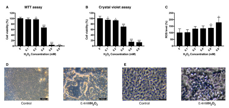
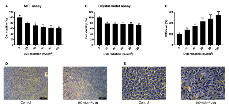
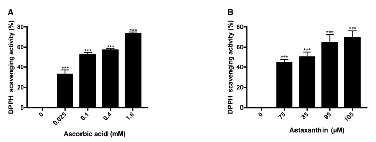
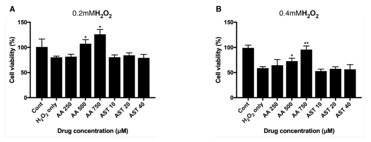
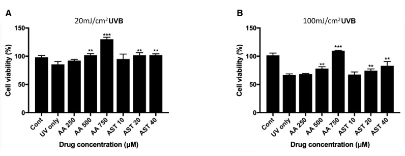
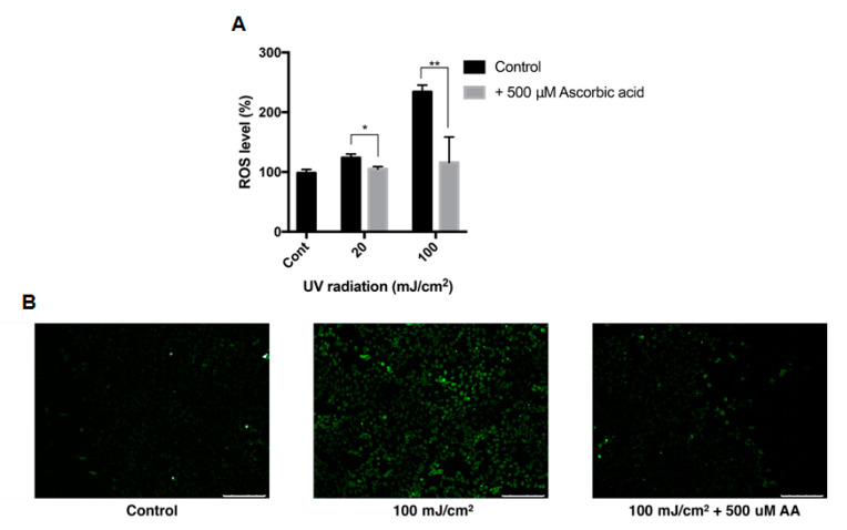
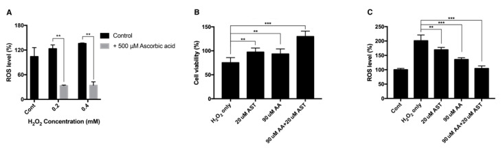
Oxidative stress has been implicated as critical pathogenic factors contributing to the etiology of diabetic retinopathy and other retinal diseases. This study investigated antioxidative effect of ascorbic acid and astaxanthin on ARPE-19 cells within an oxidative stress model induced by common biological sources of reactive oxygen species (ROS). Hydrogen peroxide (H2O2) at concentrations of 0.1-0.8 mM and 20-100 mJ/cm2 of ultraviolet B (UVB) were treated to ARPE-19 cells. Cell viability and intracellular ROS level changes were measured. With the sublethal and lethal dose of each inducers, 0-750 μM of ascorbic acid and 0-40 μM of astaxanthin were treated to examine antioxidative effect on the model. Ascorbic acid at concentrations of 500 and 750 μM increased the cell viability not only in the UVB model but also in the H2O2 model, but 20 and 40 μM of astaxanthin only did so in the UVB model. The combination of ascorbic acid and astaxanthin showed better antioxidative effect compared to each drug alone, suggesting a synergistic effect.

Keywords: antioxidant; ascorbic acid; astaxanthin; diabetic retinopathy; oxidative stress; retinal disease; retinal pigment epithelium.


Conflict of interest statement

The authors declare no conflict of interest.

Figures

Figure 1
Figure 1
Change of viability and intracellular ROS level in ARPE-19 cells after exposure to H2O2. The response of ARPE-19 cells to 0–0.8 mM H2O2 exposure for MTT assay (A), and crystal violet assay (B) to determine cell viability. For intracellular ROS level, DCFH-DA was treated for 30 min after the H2O2 exposure. Exposure to H2O2 reduced the cell viability (A,B) and increased the intracellular ROS level (C). The cell morphology was observed with bright field microscopy (Scale bar 500 μm) (D) and with higher magnification (scale bar 100 μm) (E). Asterisks indicate a significant reduction in cell viability or increment in ROS level compared with untreated cells (* p < 0.05, ** p < 0.01, *** p < 0.001). MTT, 3-(4,5-dimethylthiazol-2-yl)-2,5-diphenyltetrazolium bromide; ROS, reactive oxygen species; DCFH-DA, 2′,7′-dichlorodihydrofluorescein diacetate.
Figure 2
Figure 2
Change of viability and intracellular ROS level in ARPE-19 cells after UVB irradiation. The response of ARPE-19 cells 24 h after 0–100 mJ/cm2 UVB irradiation with MTT assay (A), and crystal violet assay (B) to determine cell viability. For intracellular ROS level, DCFH-DA was treated for 30 min after the UVB irradiation. Irradiation by UVB reduced the cell viability (A,B) and increased the intracellular ROS level (C). The cell morphology was observed with bright field microscopy (scale bar 500 μm) (D) and with higher magnification (scale bar 100 μm) (E). Asterisks indicate a significant reduction in cell viability or increment in ROS level compared with untreated cells (* p < 0.05, ** p < 0.01, *** p < 0.001). UVB, ultraviolet B; MTT, 3-(4,5-dimethylthiazol-2-yl)-2,5-diphenyltetrazolium bromide; ROS, reactive oxygen species; DCFH-DA, 2′,7′-dichlorodihydrofluorescein diacetate.
Figure 3
Figure 3
DPPH scavenging activity of ascorbic acid and astaxanthin. The antioxidative capacities of ascorbic acid and astaxanthin were determined by their capabilities to scavenge DPPH. Ascorbic acid (0.025–1.6 mM) was reacted with DPPH (A), and astaxanthin (75–105 μM) was reacted with DPPH (B). The compounds were diluted in DMSO. Both compounds scavenged DPPH in dose-dependent way in 30 min of reaction time. Asterisks indicate a significant increment in DPPH scavenging activity compared with controls (*** p < 0.001). DPPH, 2,2-diphenyl-1-picrylhydrazyl; DMSO, dimethyl sulfoxide.
Figure 4
Figure 4
Effect of ascorbic acid and astaxanthin on H2O2-induced oxidative stress model of ARPE-19. The effect of various concentration of ascorbic acid or astaxanthin (pretreated for 6 h and co-treated with H2O2 for 24 h) on the response of ARPE-19 cells to sublethal dose of 0.2 mM (A) or lethal dose of 0.4 mM H2O2 (B). The cell viability was determined by MTT assay. Treatment of ascorbic acid (500–750 μM) significantly increased ARPE-19 cell viability following 0.2 mM H2O2 exposure. However, astaxanthin (10–40 μM) did not significantly affect the cell viability (A). Ascorbic acid (500–750 μM) also significantly increased the cell viability under 0.4 mM H2O2 but astaxanthin (10–40 μM) did not have significant effect on the viability (B). Asterisks indicate a significant increment in cell viability compared with cells treated with H2O2 only (* p < 0.05, ** p < 0.01). AA, ascorbic acid; AST, astaxanthin; MTT, 3-(4,5-dimethylthiazol-2-yl)-2,5-diphenyltetrazolium bromide.
Figure 5
Figure 5
Effect of ascorbic acid and astaxanthin on UVB-induced oxidative stress model of ARPE-19. The effect of various concentration of ascorbic acid and astaxanthin (pretreated for 6 h and additional 24 h after UVB irradiation) on the response of ARPE-19 cells to sublethal dose of 20 mJ/cm2 (A) or lethal dose of 100 mJ/cm2 UVB (B). The cell viability was determined by MTT assay 24 h after the irradiation. Treatment of ascorbic acid (500–750 μM) and astaxanthin (20–40 μM) significantly increased ARPE-19 cell viability following 20 mJ/cm2 UVB irradiation (A). Ascorbic acid (500–750 μM) and astaxanthin (20–40 μM) also significantly increased the cell viability after 100 mJ/cm2 UVB irradiation (B). Asterisks indicate a significant increment in cell viability compared with cells treated with UVB only (* p < 0.05, ** p < 0.01, *** p < 0.001). UVB, ultraviolet B; AA, ascorbic acid; AST, astaxanthin; MTT, 3-(4,5-dimethylthiazol-2-yl)-2,5-diphenyltetrazolium bromide.
Figure 6
Figure 6
Intracellular ROS level of ARPE-19 after UVB treatment with ascorbic acid. The effects of ascorbic acid on the intracellular ROS level of ARPE-19 under UVB-induced oxidative stress were examined by DCFH-DA assay. Ascorbic acid at 500 μM significantly reduced the ROS level after UVB irradiation (20–100 mJ/cm2) compared to groups with UVB irradiation only (A). The green fluorescence of the reacted DCFH-DA which indicates the ROS level, was observed with fluorescence microscopy (scale bar 250 μm) (B). Asterisks indicate a significant reduction in ROS level compared with control cells only with UVB exposure without ascorbic acid treatment (* p < 0.05, ** p < 0.01). ROS, reactive oxygen species; UVB, ultraviolet B; DCFH-DA, 2′,7′-dichlorodihydrofluorescein diacetate; AA, ascorbic acid.
Figure 7
Figure 7
Intracellular ROS level and cell viability of ARPE-19 after H2O2 exposure with ascorbic acid and the mixture of ascorbic acid and astaxanthin. Ascorbic acid at 500 μM significantly reduced the intracellular ROS level under sublethal and lethal dose of H2O2 (0.2–0.4 mM) compared to the control group without ascorbic acid treatment (A). The effect of 20 μM astaxanthin, 90 μM ascorbic acid, and the mixture of the two compounds on the cell viability of ARPE-19 under H2O2-induced oxidative stress was examined by MTT assay. Cell viability was significantly increased when the cells were pretreated with 20 μM astaxanthin, 90 μM ascorbic acid, and the mixture of the two compounds for 6 h and with 0.2 mM H2O2 for 24 h, compared to H2O2 only (B). ROS level was significantly decreased when the cells were pretreated with 20 μM astaxanthin, 90 μM ascorbic acid, and the mixture of the two compounds for 6 h and with 0.2 mM H2O2 for 24 h, compared to H2O2 only. Asterisks indicate a significant difference between increment in cell viability and reduction in intracellular ROS level compared to control cells only with H2O2 exposure without antioxidant treatment (C). (** p < 0.01, *** p < 0.001). AST, astaxanthin; AA, ascorbic acid; ROS, reactive oxygen species; MTT, 3-(4,5-dimethylthiazol-2-yl)-2,5-diphenyltetrazolium bromide.

Similar articles

Cited by

References

    1. Klein B.E. Overview of epidemiologic studies of diabetic retinopathy. Ophthalmic Epidemiol. 2007;14:179–183. doi: 10.1080/09286580701396720. - DOI - PubMed
    1. Safi S.Z., Qvist R., Kumar S., Batumalaie K., Ismail I.S. Molecular mechanisms of diabetic retinopathy, general preventive strategies, and novel therapeutic targets. Biomed. Res. Int. 2014;2014:801269. doi: 10.1155/2014/801269. - DOI - PMC - PubMed
    1. Chiu C.J., Taylor A. Dietary hyperglycemia, glycemic index and metabolic retinal diseases. Prog. Retin. Eye Res. 2011;30:18–53. doi: 10.1016/j.preteyeres.2010.09.001. - DOI - PMC - PubMed
    1. Nishikawa M., Inoue M. Oxidative stress and tissue injury. Masui. 2008;57:321–326. - PubMed
    1. Naruse R., Suetsugu M., Terasawa T., Ito K., Hara K., Takebayashi K., Morita K., Aso Y., Inukai T. Oxidative stress and antioxidative potency are closely associated with diabetic retinopathy and nephropathy in patients with type 2 diabetes. Saudi Med. J. 2013;34:135–141. - PubMed

LinkOut - more resources