Review
. 2021 Mar;18(3):316-339.
doi: 10.1080/15476286.2020.1809197. Epub 2020 Sep 9.

Transfer RNAs: diversity in form and function

Affiliations
Review

Transfer RNAs: diversity in form and function

Matthew D Berg et al. RNA Biol. 2021 Mar.

Abstract

As the adaptor that decodes mRNA sequence into protein, the basic aspects of tRNA structure and function are central to all studies of biology. Yet the complexities of their properties and cellular roles go beyond the view of tRNAs as static participants in protein synthesis. Detailed analyses through more than 60 years of study have revealed tRNAs to be a fascinatingly diverse group of molecules in form and function, impacting cell biology, physiology, disease and synthetic biology. This review analyzes tRNA structure, biosynthesis and function, and includes topics that demonstrate their diversity and growing importance.

Keywords: aminoacylation; mistranslation; tRNA; tRNA fragments; translation.


Figures

Figure 1.
Figure 1.
tRNA structure. (A) Cloverleaf representation of a tRNA in 2D. From 5ʹ to 3ʹ the regions of the tRNA are: acceptor stem (green), D-arm (pink), anticodon arm (blue) containing the anticodon (darker blue), variable arm (orange) and T-arm (purple). The discriminator base (base 73) is the unpaired base at the 3ʹ end [yellow) before the terminal CCA residues. Aminoacyl-tRNA synthetases ligate an amino acid onto the terminal 3ʹ adenosine. Numbered positions are constant bases, as described in . (B) Three-dimensional tRNA structure of a yeast tRNAPhe [, PDB: 1EHZ]. Colour of the 2D elements is the same as in (A). Intramolecular interactions between the T- and D-arms (purple and pink respectively) in the elbow region facilitate tRNA folding. (C) A surface representation of tRNAPhe (left; PDB: 1EHZ) and the ribosome recycling factor (right; PDB: 1EH1) demonstrate how molecules that interact with the translational machinery often adopt tRNA-like structures
Figure 2.
Figure 2.
tRNAs are transcribed by RNA polymerase III. The internal A- and B-box sequences are recognized by TFIIIC (i). Bound TFIIIC leads to the recruitment of TFIIIB which binds upstream of the tRNA gene and recruits RNAPIII (ii). TFIIIB and RNAPIII melt the promoter, leading to transcription of the tRNA (iii and iv). Termination is signalled by a string of thymine residues on the non-templated strand (v). Transcriptional re-initiation, where RNAPIII is recycled to the transcriptional start site, can enhance transcription of tRNA genes (iv)
Figure 3.
Figure 3.
Lifecycle of a tRNA. (A) tRNAs are transcribed (i) with 5ʹ leader and 3ʹ trailer sequences that are cleaved by RNase P and RNase Z respectively (ii). After trimming, partial modification (iii), indicated by the red circles, and addition of the terminal CCA residues (iv) occur in the nucleus. In yeast, the tRNA is exported out of the cytoplasm by Los1 (v) and if the tRNA contains an intron, it is spliced on the mitochondrial surface (vi). In mammals, splicing occurs before nuclear export. In the cytoplasm, additional modifications may be added (viii). tRNAs can then be aminoacylated by their cognate aminoacyl-tRNA synthetase (viii) or be re-imported (ix) by Mtr10 into the nucleus for further modification (x) and quality control through nuclear aminoacylation (xi). Re-export of tRNAs is facilitated by both Los1 and an Msn5 dependent pathway that specifically recognizes mature tRNAs (xii). Once aminoacylated, tRNAs are used in translation (xiii). (B) Quality control mechanisms can lead to the degradation of tRNAs. After transcription, nuclear surveillance (i) monitors tRNAs for end maturation and specific modifications. Improper tRNAs are polyadenylated by the TRAMP complex and degraded by the nuclear exosome. After processing and export, tRNAs are monitored and degraded through the rapid tRNA pathway by the 5ʹ-3ʹ exonuclease Xrn1 in the cytoplasm (ii) and Rat1 in the nucleus (iii)
Figure 4.
Figure 4.
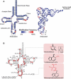
tRNAs are highly post-transcriptionally modified. (A) Heat map showing modification frequency for each position in the tRNA on the 2D (left) and 3D (right) tRNA structures. Modification data for 715 unique tRNA sequences from archaea, bacteria and eukaryotes accessed from the MODOMICS database [http://genesilico.pl/modomics/; [282]]. Circled bases are not modified in any of the 715 unique tRNA sequences in the database. (B) tRNA modifications found across all S. cerevisiae tRNAs [pseudouridine (Ψ); 2ʹ-O-methylcytidine (Cm); 2ʹ-O-methyladenosine (Am); 1-methyl-guanosine (m1G); N2-methylguanosine (m2G); N4-acetylcytidine (ac4C); dihydrouridine (D); 2ʹ-O-methylguanosine (Gm); N2,N2-dimethylguanosine (m2,2G); 3-methylcytidine (m3C); inosine (I); 5-methylcytidine (m5C); 5-methoxycarbonylmethyluridine (mcm5U); 5-methoxycarbonylmethyl-2-thiouridine (mcm5s2U); 5-carbamoylmethyluridine (ncm5U); 5-carbamoylmethyl-2ʹ-O-methyluridine (ncm5Um); 1-methylinosine (m1I); N6-isopentenyladenosine (i6A); wybutosine (yW); N6-threonylcarbamoyladenosine (t6A); 2ʹ-O-methyluridine (Um); 7-methylguanosine (m7G); ribothymidine (rT); 1-methyladenosine (m1A); 2ʹ-O-ribosyladenosine phosphate (Ar(p))]. The nucleobases of select-modified nucleotides are shown on the right
Figure 5.
Figure 5.
Aminoacyl-tRNA at the ribosome. Aminoacyl-tRNAs are recognized by GTP-bound EF-Tu in bacteria or EF1A in eukaryotes and recruited to the elongating ribosome in an mRNA-independent manner. During initial selection (i), codon–anticodon interactions are monitored by the ribosome and non-cognate tRNAs are rejected. A cognate codon–anticodon interaction triggers GTP hydrolysis by EF-Tu/EF1A and a conformational change that puts the aminoacyl branch of the tRNA into the peptidyl transferase centre. Proofreading (ii) occurs as a second check to ensure proper codon–anticodon pairing. Here, near-cognate matches that may have made it past initial selection are rejected. A cognate codon–anticodon interaction triggers peptide bond formation (iii) and the amino acid is added to the growing polypeptide chain. Translocation (iv) moves the tRNA containing the polypeptide chain into the P site and the cycle repeats
Figure 6.
Figure 6.
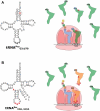
tRNA variants can increase levels of mistranslation. (A) A S. cerevisiae proline tRNA variant with a C70U mutation that creates a G3:U70 base pair in the acceptor stem is aminoacylated with alanine instead of proline, leading to alanine misincorporation [188]. The toxicity of mistranslation is buffered by other wild-type copies of tRNAPro that compete for decoding of CCA codons with the mistranslating tRNA variant. (B) A serine tRNA variant with a UGG anticodon is aminoacylated with serine, but decodes proline codons, leading to serine misincorporation. A secondary mutation of G26A dampens tRNA function allowing non-lethal levels of mistranslation [377]
Figure 7.
Figure 7.
tRNA-derived small RNAs regulate diverse cellular processes. Mature tRNAs can be cleaved into 5ʹ (yellow) and 3ʹ tRNA (purple) halves by Rny1 in yeast or angiogenin in mammals. Cleavage can also occur in the D- or T-loops by Dicer in mammals creating 5ʹ and 3ʹ CCA tRNA fragments (tRFs). Selected examples of the roles of tRNA fragments in regulating cellular processes are shown in the boxes. *For some tRFs, other nucleases have been implicated

References

    1. Hoagland MB, Stephenson ML, Scott JF, et al. A soluble ribonucleic acid intermediate in protein synthesis. J Biol Chem. 1958;231:241–257. - PubMed
    1. Crick FH. On protein synthesis. Symp Soc Exp Biol. 1958;12:138–163. - PubMed
    1. Crick FHC, Barnett L, Brenner S, et al. General nature of the genetic code for proteins. Nature. 1961;192:1227–1232. - PubMed
    1. Dounce AL. Duplicating mechanism for peptide chain and nucleic acid synthesis. Enzymologia. 1952;15:251–258. - PubMed
    1. Nirenberg M, Leder P, Bernfield M, et al. RNA codewords and protein synthesis, VII. On the general nature of the RNA code. Proc Natl Acad Sci. 1965;53:1161–1168. - PMC - PubMed

Publication types