Review
. 2020 Nov;30(11):904-915.
doi: 10.1016/j.tcb.2020.08.005. Epub 2020 Sep 6.

Photoreceptor Discs: Built Like Ectosomes

Affiliations
Review

Photoreceptor Discs: Built Like Ectosomes

William J Spencer et al. Trends Cell Biol. 2020 Nov.

Abstract

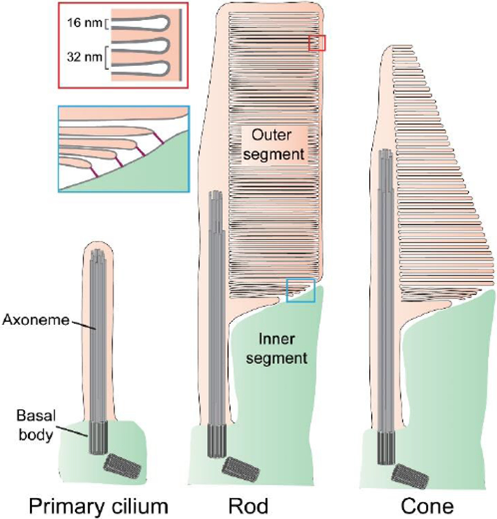
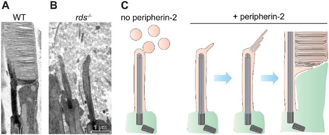
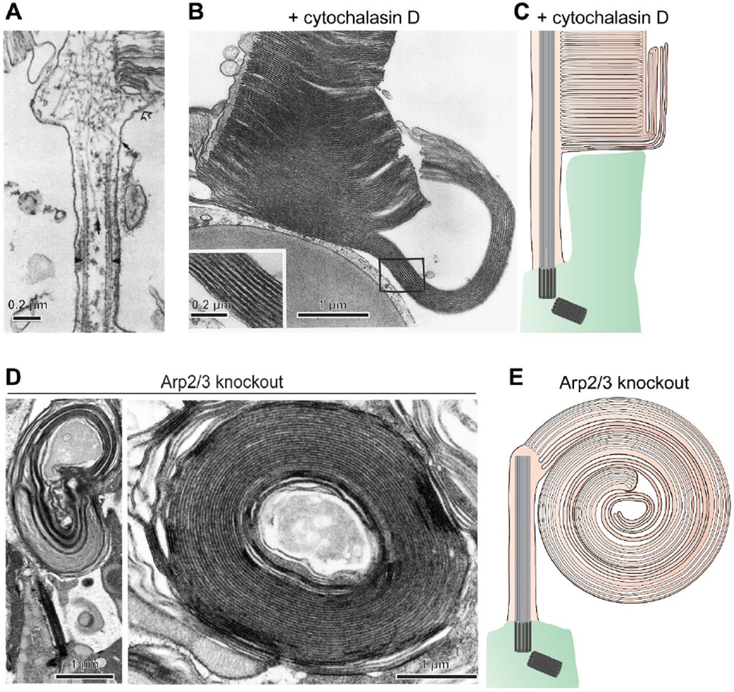
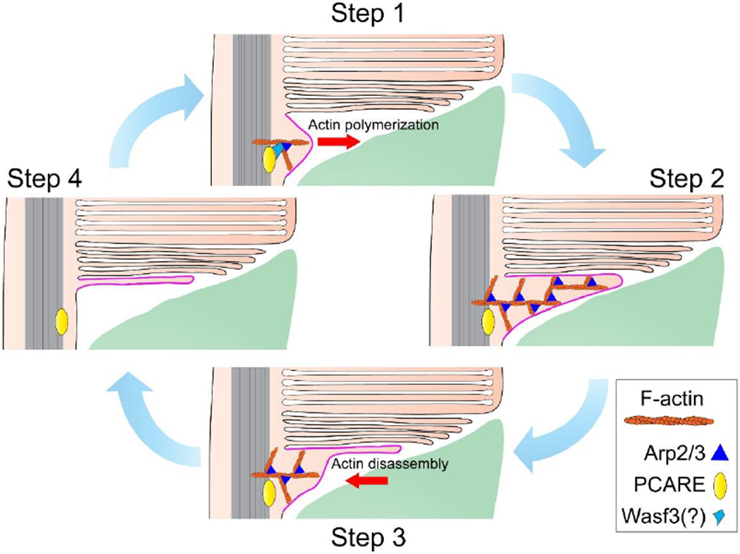
The light-sensitive outer segment organelle of the vertebrate photoreceptor cell is a modified cilium filled with hundreds of flattened 'disc' membranes that provide vast light-absorbing surfaces. The outer segment is constantly renewed with new discs added at its base every day. This continuous process is essential for photoreceptor viability. In this review, we describe recent breakthroughs in the understanding of disc morphogenesis, with a focus on the molecular mechanisms responsible for initiating disc formation from the ciliary membrane. We highlight the discoveries that this mechanism evolved from an innate ciliary process of releasing small extracellular vesicles, or ectosomes, and that both disc formation and ectosome release rely on the actin cytoskeleton.

Keywords: actin cytoskeleton; cilia; extracellular vesicle; outer segment; photoreceptor; vision.


Figures

Figure 1.
Figure 1.. Photoreceptor outer segments are highly modified primary cilia.
A prototypic primary cilium and outer segments of rod and cone photoreceptor cells are illustrated to highlight their similarity. Each structure is stabilized by a microtubular axoneme extending from a basal body. Photoreceptor discs form at the base of the outer segment as evaginations of the ciliary plasma membrane. In rods, discs eventually enclose inside the outer segment. In cones of lower vertebrates (as illustrated), discs remain as lamellar folds of the plasma membrane. In mammalian cones, discs undergo partial enclosure. The red inset highlights the periodicity of disc spacing. The blue inset illustrates that newly forming discs are connected to the inner segment through linking structures.
Figure 2.
Figure 2.. The outer segment evolved through the retention of ciliary ectosomes.
(A) An electron micrograph of an inner-outer segment juncture from a WT mouse rod. The retinas were contrasted with tannic acid to discern between discs exposed to the extracellular space (darkly stained membranes) and those enclosed inside the outer segment (lightly stained membranes). (B) An electron micrograph of a comparable region from an rds−/− mouse lacking peripherin-2 (adapted from [16]). The photoreceptor cilium fails to elaborate an outer segment and resembles a prototypic primary cilium surrounded by massive amounts of ectosomes. (C) A hypothesis on the evolutionary origin of the outer segment. The outer segment evolved through introduction of the photoreceptor-specific protein, peripherin-2, which enabled prototypic photoreceptors to retain ectosomes at the cilium. Subsequent adaptations allowed flattening, elongation and enclosure of retained membrane material.
Figure 3.
Figure 3.. Branched actin polymerization initiates disc formation.
(A) An electron micrograph showing myosin-labeled actin filaments at the site of disc morphogenesis in a rat rod (adapted from [40]). (B) An electron micrograph of a cone from a Xenopus laevis retina acutely treated with the actin polymerization inhibitor cytochalasin D reveals overgrowth of newly forming discs (adapted from [43]). (C) A cartoon illustrating the effect of cytochalasin D treatment. The drug halted the initiation of new disc formation without preventing delivery of new membrane material to the outer segment. This material incorporates into the newly forming discs which were undergoing expansion at the time of treatment, causing their uncontrolled overgrowth. (D) Electron micrographs of overgrown membranes in rods of Arp2/3 knockout mice which have elaborated into large whorls. (E) A cartoon illustrating a massive membrane whorl in the Arp2/3 knockout rod. A permanent halting of the initiation of new disc formation by this knockout causes much more extended membrane outgrowth than an acute cytochalasin D treatment. Images in (C-E) are adapted from [44].
Figure 4.
Figure 4.. A cycle of actin dynamics during disc formation.
A cycle of actin polymerization/disassembly begins in Step 1 when PCARE recruits an actin nucleation promoting factor, likely Wasf3, to the site of disc morphogenesis. This actin nucleation promoting factor associates with the Arp2/3 complex to nucleate branched actin filaments (F-actin). In Step 2, continued actin polymerization pushes the ciliary membrane outwards. The Arp2/3 complex remains as a structural component of these filaments. In Step 3, while the membrane remains protruded, actin filaments are disassembled and retracted allowing the evaginated membrane to flatten. In Step 4, actin has fully retracted and the initiation step of disc formation is complete. Meanwhile, the newly forming disc continues to elongate through ongoing addition of membrane material. The cartoon is updated from [44]. See also Movie S1 to view the entire process of disc formation.
Figure 5.
Figure 5.. PRCD is required for the efficient flattening of newly forming discs.
(A) An electron micrograph of PRCD knockout rods reveals the presence of ectosomes accumulated at the outer segment base. (B) An image of a 1 nm z-section from a 3D electron tomogram taken at the base of a PRCD knockout rod outer segment showing that newly forming discs fail to efficiently flatten, forming bulges filled with cytoplasm (marked by arrowheads). These bulges shed as ectosomes before disc enclosure, allowing mature discs to be flat inside the outer segment. (C) An illustration of the PRCD knockout phenotype. Images are adapted from [61].
Figure 6.
Figure 6.. Peripherin-2 fortifies the disc rim.
(A) Immunogold labeling shows that, in newly forming discs, peripherin-2 localizes only to the disc rims adjacent to the axoneme and is absent from the elongating disc edges. In mature discs, peripherin-2 localizes to the entire disc rim, including incisures. Shown is an uncropped version of an image from [16]. (B) Illustration of peripherin-2 localization in a disc. Long, self-assembling chains of peripherin-2 oligomers extend circumferentially throughout the disc rim to form and maintain its hairpin-like shape. Peripherin-2 oligomers similarly regulate the formation of disc incisures and fortify their rims. The image is modified from [71]. The rectangular inset illustrates the organization of the peripherin-2 molecule. The tetraspanin body (purple) is critical for supporting disc rim structure, while the C-terminus (yellow) is essential for preventing the release of ciliary ectosomes. (C) The C150S mutation of peripherin-2, which prevents its high-order oligomerization, causes major outer segment abnormalities in knockin mice. Tannic acid staining reveals the presence of both darkly stained (open) and lightly stained (enclosed) membrane structures, suggesting that high-order peripherin-2 oligomerization is not essential for disc enclosure.

Similar articles

Cited by

References

    1. Sjostrand FS (1953) The ultrastructure of the outer segments of rods and cones of the eye as revealed by the electron microscope. J Cell Physiol 42 (1), 15–44. - PubMed
    1. Goldberg AF et al. (2016) Molecular basis for photoreceptor outer segment architecture. Prog Retin Eye Res 55, 52–81. - PMC - PubMed
    1. Pearring JN et al. (2013) Protein sorting, targeting and trafficking in photoreceptor cells. Prog Retin Eye Res 36, 24–51. - PMC - PubMed
    1. Wensel TG et al. (2016) Structural and molecular bases of rod photoreceptor morphogenesis and disease. Prog Retin Eye Res 55, 32–51. - PMC - PubMed
    1. Nickell S et al. (2007) Three-dimensional architecture of murine rod outer segments determined by cryoelectron tomography. J Cell Biol 177 (5), 917–25. - PMC - PubMed

Publication types