. 2020 Dec 1;26(23):6321-6334.
doi: 10.1158/1078-0432.CCR-20-0357. Epub 2020 Sep 8.

CSPG4-Specific CAR.CIK Lymphocytes as a Novel Therapy for the Treatment of Multiple Soft-Tissue Sarcoma Histotypes

Affiliations

CSPG4-Specific CAR.CIK Lymphocytes as a Novel Therapy for the Treatment of Multiple Soft-Tissue Sarcoma Histotypes

Valeria Leuci et al. Clin Cancer Res. .

Abstract

Purpose: No effective therapy is available for unresectable soft-tissue sarcomas (STS). This unmet clinical need prompted us to test whether chondroitin sulfate proteoglycan 4 (CSPG4)-specific chimeric antigen receptor (CAR)-redirected cytokine-induced killer lymphocytes (CAR.CIK) are effective in eliminating tumor cells derived from multiple STS histotypes in vitro and in immunodeficient mice.

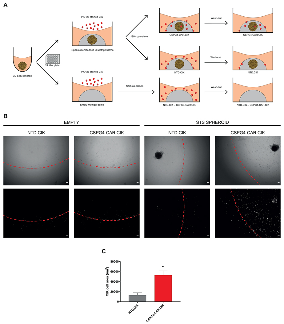
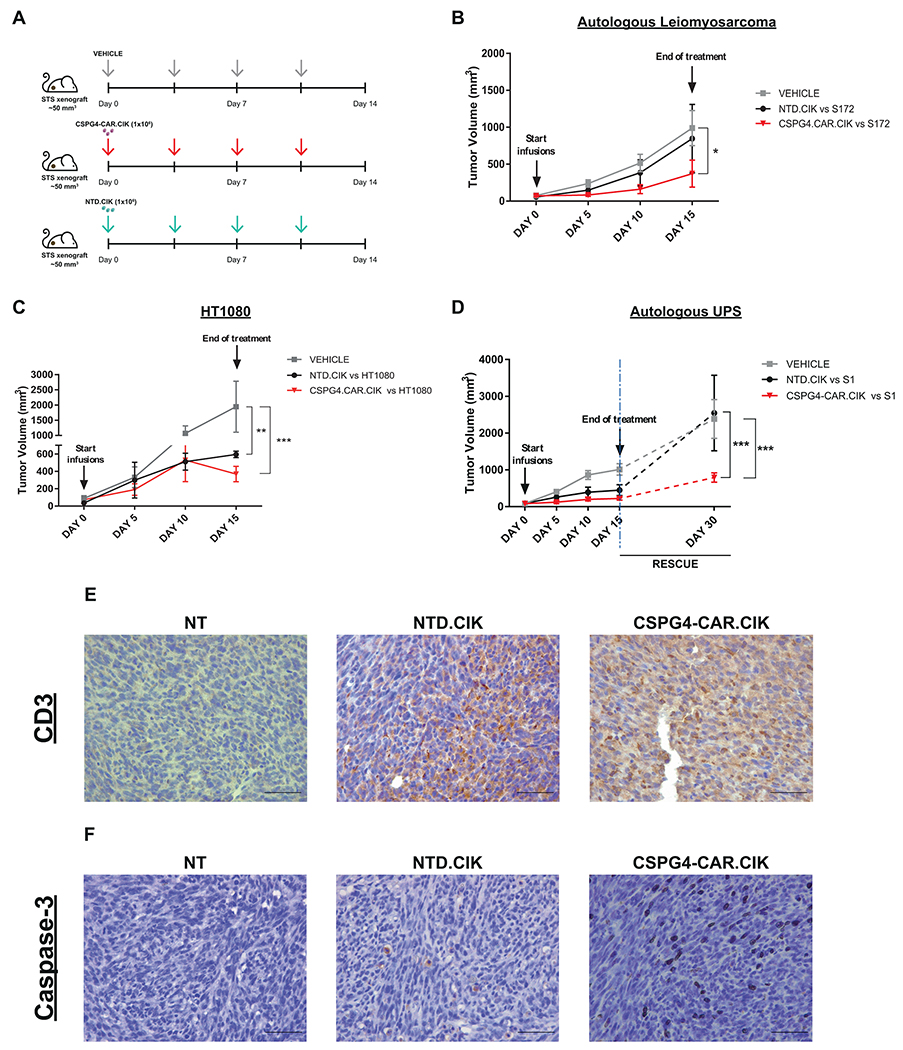
Experimental design: The experimental platform included patient-derived CAR.CIK and cell lines established from multiple STS histotypes. CAR.CIK were transduced with a retroviral vector encoding second-generation CSPG4-specific CAR (CSPG4-CAR) with 4-1BB costimulation. The functional activity of CSPG4-CAR.CIK was explored in vitro, in two- and three-dimensional STS cultures, and in three in vivo STS xenograft models.

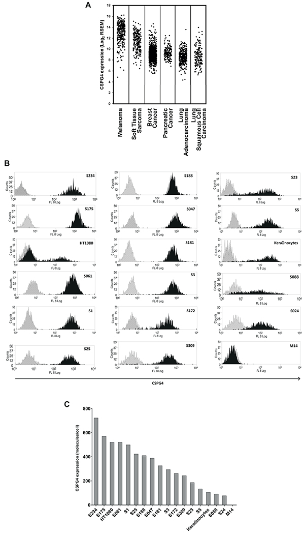
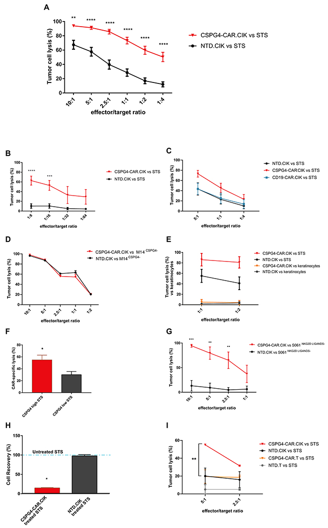
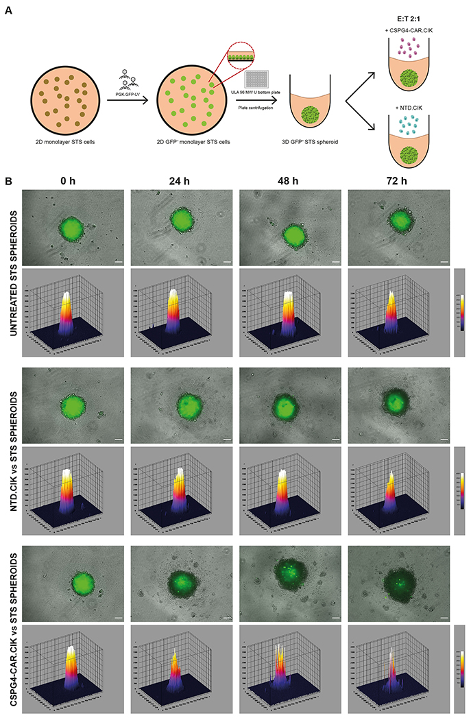
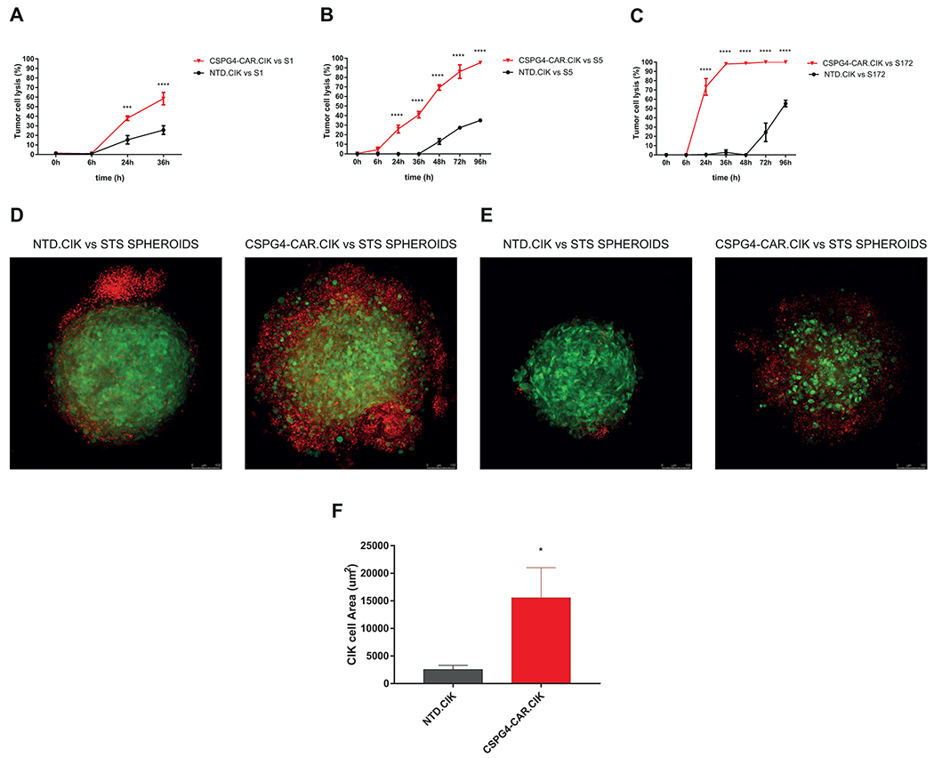
Results: CSPG4-CAR.CIK were efficiently generated from patients with STS. CSPG4 was highly expressed in multiple STS histotypes by in silico analysis and on all 16 STS cell lines tested by flow cytometry. CSPG4-CAR.CIK displayed superior in vitro cytolytic activity against multiple STS histotypes as compared with paired unmodified control CIK. CSPG4-CAR.CIK also showed strong antitumor activity against STS spheroids; this effect was associated with tumor recruitment, infiltration, and matrix penetration. CSPG4-CAR.CIK significantly delayed or reversed tumor growth in vivo in three STS xenograft models (leiomyosarcoma, undifferentiated pleomorphic sarcoma, and fibrosarcoma). Tumor growth inhibition persisted for up to 2 weeks following the last administration of CSPG4-CAR.CIK.

Conclusions: This study has shown that CSPG4-CAR.CIK effectively targets multiple STS histotypes in vitro and in immunodeficient mice. These results provide a strong rationale to translate the novel strategy we have developed into a clinical setting.


Conflict of interest statement

Conflict of interest disclosure: G.D. and S.F. hold a patent on the CSPG4-CAR. G.D. has sponsor research agreements with Bluebird Bio, Cell Medica and Bellicum Pharmaceutical. G.D. serves in the scientific advisory board of MolMed S.p.A and Bellicum Pharmaceutical. The other authors declare no competing financial interests.

Figures

Figure 1.
Figure 1.. CSPG4 is highly expressed in multiple STS histotypes.
CSPG4 mRNA expression in multiple STS histotypes (Leiomyosarcoma; Dedifferentiated Liposarcoma; Undifferentiated pleomorphic sarcoma (UPS), Malignant Fibrous Histiocytoma, High-Grade Spindle Cell Sarcoma; Myxofibrosarcoma; Malignant Peripheral Nerve Sheath Tumor; Synovial Sarcoma). CSPG4 expression in STS was comparable to that observed in melanoma. RNA-sequencing expression data were selected and downloaded from the cBioPortal of the TCGA Pan-Cancer Collections. RSEM expression values were plotted after Log2 transformation with 0.5 jittering on the x-axis, using Microsoft Excel®. (A). CSPG4 expression was confirmed in patient-derived STS cell lines of various histologic types by flow cytometry. A representative flow-cytometry histogram is reported for each STS. The M14 melanoma cell line that lacks CSPG4 expression and normal keratinocytes were used for comparison. Isotype controls are shown in grey (B). Grey histograms show the number of CSPG4 molecules expressed on the cell surface of various patient-derived STS cell lines quantified as the CSPG4-specific mAb-binding capacity (sABC) on a per cell basis (C). Abbreviations: CSPG4, Chondroitin Sulfate proteoglycan 4; STS, Soft Tissue Sarcoma.
Figure 2.
Figure 2.. CSPG4-CAR.CIK effectively and specifically target STS cells in vitro.
Patient-derived CSPG4-CAR.CIK efficiently targeted STS cells in vitro when using either HLA mismatched (10 samples) or matched CIK (2 samples). Specific cytotoxicity of CSPG4-CAR.CIK was significantly higher than that obtained with unmodified NTD.CIK. Tumor cell-specific cytotoxicity values from 29 experiments are reported (mean ± SEM) (A). CSPG4-CAR.CIK retained their antitumor activity when challenged at very low E:T ratios (n=8) (B). In vitro cytotoxic activity of control CD19-CAR.CIK against STS was comparable to that of unmodified NTD.CIK (n=5) (C). CSPG4-CAR.CIK and paired unmodified NTD.CIK showed similar cytotoxic activity against the M14 control cell line that lacks CSPG4 expression (n=3) (D). Both CSPG4-CAR.CIK and paired unmodified NTD.CIK did not lyse normal keratinocytes (n=5) (E). CSPG4-CAR.CIK exhibited more intense killing of STS cells with high CSPG4 expression as compared with STS cells with low CPSG4 expression (n=7, * P ≤ 0.05 by t-test) (F). CSPG4-CAR.CIK, but not paired unmodified NTD.CIK, retained cytotoxic activity against a STS sample expressing CSPG4, but lacking NKG2D ligands (S061) (n=3; two-way ANOVA and Bonferroni’s post-test analysis) (G). STS cell growth assessed 48 hours following in vitro treatment was significantly delayed in STS exposed to CSPG4-CAR.CIK, as compared with unmodified NTD.CIK. The dashed line represents the control parallel growth of untreated STS cells (n=2, * P ≤ 0.05 by unpaired t-test) (H). CSPG4-CAR.CIK displayed significant and superior STS killing activity compared to paired “non-cytokine-induced”, but minimally activated with IL-2 (50U/ml) CAR.T expressing comparable membrane levels of CSPG4-specific CAR molecules (n=2, two-way ANOVA and Bonferroni’s post-test analysis) (I). All cytotoxicity assays were analyzed by two-way ANOVA and Bonferroni’s post-test analysis; statistical significance is reported as * P ≤ 0.05, ** P ≤ 0.01, *** P ≤ 0.001, and **** P ≤ 0.0001; “n” refers to the number of separate experiments. Abbreviations: CIK, Cytokine Induced Killer cells; CSPG4, Chondroitin Sulfate proteoglycan 4; CAR, Chimeric Antigen Receptor; NTD, Not Transduced; STS, Soft Tissue Sarcoma.
Figure 3.
Figure 3.. CSPG4-CAR.CIK effectively target STS cells in 3D spheroids.
Schematic showing the 3D assay to test the cytotoxic activity of CSPG4-CAR.CIK against GFP-expressing STS spheroids (green) (A). CSPG4-CAR.CIK showed superior tumor elimination, as compared to paired unmodified NTD.CIK. Shown are representative microscope fluorescence images and surface plot images of GFP-expressing STS spheroids (green) treated with CSPG4-CAR.CIK (up to 72 hours) and control CIK. Magnification: 10X; Scale bars: 100 μm (B). Abbreviations: CIK: Cytokine Induced Killer cells; CSPG4: Chondroitin Sulfate proteoglycan 4; CAR: Chimeric Antigen Receptor; NTD: Not Transduced; STS: Soft Tissue Sarcoma; GFP: Green Fluorescent Protein.
Figure 4.
Figure 4.. CSPG4-CAR.CIK infiltrate 3D STS spheroids.
CSPG4-CAR.CIK displayed cytolytic activity against 3D STS spheroids. Tumor cell elimination mediated by CSPG4-CAR.CIK was quantified by measuring GFP fluorescence loss overtime of S1 (A), S5 (B) and S172 (C) spheroids (pixel) by fluorescence microscopy. Values are reported as means (±SEM) from three independent wells (2:1 E:T ratio). Results were analyzed by two-way ANOVA and Bonferroni’s post-test analysis; statistical significance is reported as * P ≤ 0.05, ** P ≤ 0.01, *** P ≤ 0.001, and **** P ≤ 0.0001. Representative maximum intensity projections of confocal microscopy images for S5 (D) and S172 (E) spheroids (green) treated with unmodified PKH26-stained (red) NTD.CIK and CSPG4-CAR.CIK (2:1 E:T ratio). Confocal microscopy images taken 16 hours after co-incubating CIK and STS spheroids. 20X magnification and scale bars: 100 μm are shown. (F) CSPG4-CAR.CIK displayed superior infiltration within the STS spheroids (green) as compared with paired unmodified NTD.CIK (n=8, P≤0.05 by paired t-test). CSPG4-CAR.CIK density was determined as red fluorescent PKH26 area (μm2). Abbreviations: CIK: Cytokine Induced Killer cells; CSPG4: Chondroitin Sulfate Proteoglycan 4; CAR: Chimeric Antigen Receptor; NTD: Not Transduced; STS: Soft Tissue Sarcoma; GFP: Green Fluorescent Protein.
Figure 5.
Figure 5.. CSPG4-CAR.CIK penetrate Matrigel matrix toward the STS spheroids.
Schematic showing the Matrigel® based penetration assay. STS spheroids were embedded into Matrigel® domes and co-cultured with either PKH26-stained CSPG4-CAR.CIK or unmodified NTD.CIK at E:T ratios 10:1. Empty domes served as controls. Fluorescence microscope images were acquired at day 5 of co-culture (A). Representative imagines of CSPG4-CAR.CIK and unmodified NTD.CIK empty or STS spheroids embedded in Matrigel® domes are displayed. Dashed lines represent Matrigel® boundaries. Top panel includes bright field microscope images; bottom panel includes fluorescent images. Magnification: 4X; Scale bars: 100 μm (B). Summary showing that CSPG4-CAR.CIK displayed superior ability in penetrating Matrigel® domes as compared to paired NTD.CIK (n=5, P≤0.01 by paired t-test). CSPG4-CAR.CIK density was defined as the fluorescent PKH26 area (μm2) (C). Abbreviations: CIK: Cytokine Induced Killer cells; CSPG4: Chondroitin Sulfate Proteoglycan 4; CAR: Chimeric Antigen Receptor; NTD: Not Transduced; STS: Soft Tissue Sarcoma.
Figure 6.
Figure 6.. CSPG4-CAR.CIK are active against STS in xenograft models.
Schematic representation of the STS xenografts and treatment with CSPG4-CAR.CIK (red arrows) and NTD.CIK (blue arrows). Vehicle-treated mice were infused with PBS (grey arrows). CIK were infused intravenously (1x10⁶ cells/infusion) twice a week for 2 weeks (A). Autologous CSPG4-CAR.CIK caused a significant delay of the growth of the S172 leiomyosarcoma (CSPG4=72% and CSPG4 density=262 molecule/cell) as compared to unmodified NTD.CIK or vehicle-treated mice (n=6; p<0.05) (B). Autologous CSPG4-CAR.CIK caused a significant delay of the growth of the HT1080 fibrosarcoma (CSPG4=23% and CSPG4 density=521 molecule/cell) as compared to unmodified NTD.CIK or vehicle-treated mice (n=3, p<0.001). (C). Autologous CSPG4-CAR.CIK effectively delayed the growth of S1 UPS (CSPG4=95% and CSPG4 density=499 molecule/cell) as compared with controls (n=3, p<0.0001). All results were analyzed by two-way ANOVA and the Bonferroni post-test; statistical significance is reported as * P ≤ 0.05, ** P ≤ 0.01, *** P ≤ 0.001, and **** P ≤ 0.0001. (D). Tumor-homing of CSPG4-CAR.CIK and unmodified NTD.CIK was confirmed by IHC in explanted tumors using an anti-human CD3 antibody staining. Magnification: 40X; Scale bars: 50 μm (E). Apoptotic tumor cells were visualized by detecting cleaved caspase 3 by IHC in explanted tumors (F). Abbreviations: CIK: Cytokine Induced Killer cells; CSPG4: Chondroitin Sulfate Proteoglycan 4; CAR: Chimeric Antigen Receptor; NTD: Not Transduced; NOD/SCID: Non-obese diabetic/severe combined immunodeficiency mice; NSG: NOD/SCID gamma mice.

Similar articles

  • CSPG4 CAR-redirected Cytokine Induced Killer lymphocytes (CIK) as effective cellular immunotherapy for HLA class I defective melanoma.
    Giraudo L, Cattaneo G, Gammaitoni L, Iaia I, Donini C, Massa A, Centomo ML, Basiricò M, Vigna E, Pisacane A, Picciotto F, Berrino E, Marchiò C, Merlini A, Paruzzo L, Poletto S, Caravelli D, Biolato AM, Bortolot V, Landoni E, Ventin M, Ferrone CR, Aglietta M, Dotti G, Leuci V, Carnevale-Schianca F, Sangiolo D. Giraudo L, et al. J Exp Clin Cancer Res. 2023 Nov 22;42(1):310. doi: 10.1186/s13046-023-02884-x. J Exp Clin Cancer Res. 2023. PMID: 37993874 Free PMC article.
  • Cytokine-induced killer cells eradicate bone and soft-tissue sarcomas.
    Sangiolo D, Mesiano G, Gammaitoni L, Leuci V, Todorovic M, Giraudo L, Cammarata C, Dell'Aglio C, D'Ambrosio L, Pisacane A, Sarotto I, Miano S, Ferrero I, Carnevale-Schianca F, Pignochino Y, Sassi F, Bertotti A, Piacibello W, Fagioli F, Aglietta M, Grignani G. Sangiolo D, et al. Cancer Res. 2014 Jan 1;74(1):119-29. doi: 10.1158/0008-5472.CAN-13-1559. Epub 2013 Dec 19. Cancer Res. 2014. PMID: 24356422
  • CD44v6 as innovative sarcoma target for CAR-redirected CIK cells.
    Leuci V, Casucci GM, Grignani G, Rotolo R, Rossotti U, Vigna E, Gammaitoni L, Mesiano G, Fiorino E, Donini C, Pisacane A, Ambrosio LD, Pignochino Y, Aglietta M, Bondanza A, Sangiolo D. Leuci V, et al. Oncoimmunology. 2018 Feb 15;7(5):e1423167. doi: 10.1080/2162402X.2017.1423167. eCollection 2018. Oncoimmunology. 2018. PMID: 29721373 Free PMC article.
  • Adoptive immunotherapy against sarcomas.
    Mesiano G, Leuci V, Giraudo L, Gammaitoni L, Carnevale Schianca F, Cangemi M, Rotolo R, Capellero S, Pignochino Y, Grignani G, Aglietta M, Sangiolo D. Mesiano G, et al. Expert Opin Biol Ther. 2015 Apr;15(4):517-28. doi: 10.1517/14712598.2015.987121. Epub 2014 Dec 16. Expert Opin Biol Ther. 2015. PMID: 25516119 Review.
  • CSPG4 as Target for CAR-T-Cell Therapy of Various Tumor Entities-Merits and Challenges.
    Harrer DC, Dörrie J, Schaft N. Harrer DC, et al. Int J Mol Sci. 2019 Nov 26;20(23):5942. doi: 10.3390/ijms20235942. Int J Mol Sci. 2019. PMID: 31779130 Free PMC article. Review.

Cited by

References

    1. Siegel RL, Miller KD, Jemal A. Cancer statistics, 2019. CA Cancer J Clin 2019;69(1):7–34 doi 10.3322/caac.21551. - DOI - PubMed
    1. Pasquali S, Pizzamiglio S, Touati N, Litiere S, Marreaud S, Kasper B, et al. The impact of chemotherapy on survival of patients with extremity and trunk wall soft tissue sarcoma: revisiting the results of the EORTC-STBSG 62931 randomised trial. Eur J Cancer 2019;109:51–60 doi 10.1016/j.ejca.2018.12.009. - DOI - PubMed
    1. Pollack SM, Ingham M, Spraker MB, Schwartz GK. Emerging Targeted and Immune-Based Therapies in Sarcoma. J Clin Oncol 2018;36(2):125–35 doi 10.1200/JCO.2017.75.1610. - DOI - PubMed
    1. Wisdom AJ, Mowery YM, Riedel RF, Kirsch DG. Rationale and emerging strategies for immune checkpoint blockade in soft tissue sarcoma. Cancer 2018;124(19):3819–29 doi 10.1002/cncr.31517. - DOI - PMC - PubMed
    1. Alsaab HO, Sau S, Alzhrani R, Tatiparti K, Bhise K, Kashaw SK, et al. PD-1 and PD-L1 Checkpoint Signaling Inhibition for Cancer Immunotherapy: Mechanism, Combinations, and Clinical Outcome. Front Pharmacol 2017;8:561 doi 10.3389/fphar.2017.00561. - DOI - PMC - PubMed

Publication types

MeSH terms