. 2020 Sep 8;11(5):e02128-20.
doi: 10.1128/mBio.02128-20.

Network Rewiring: Physiological Consequences of Reciprocally Exchanging the Physical Locations and Growth-Phase-Dependent Expression Patterns of the Salmonella fis and dps Genes

Affiliations

Network Rewiring: Physiological Consequences of Reciprocally Exchanging the Physical Locations and Growth-Phase-Dependent Expression Patterns of the Salmonella fis and dps Genes

Marina M Bogue et al. mBio. .

Abstract

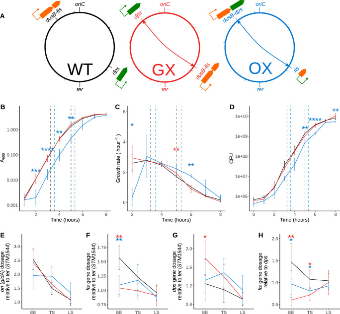
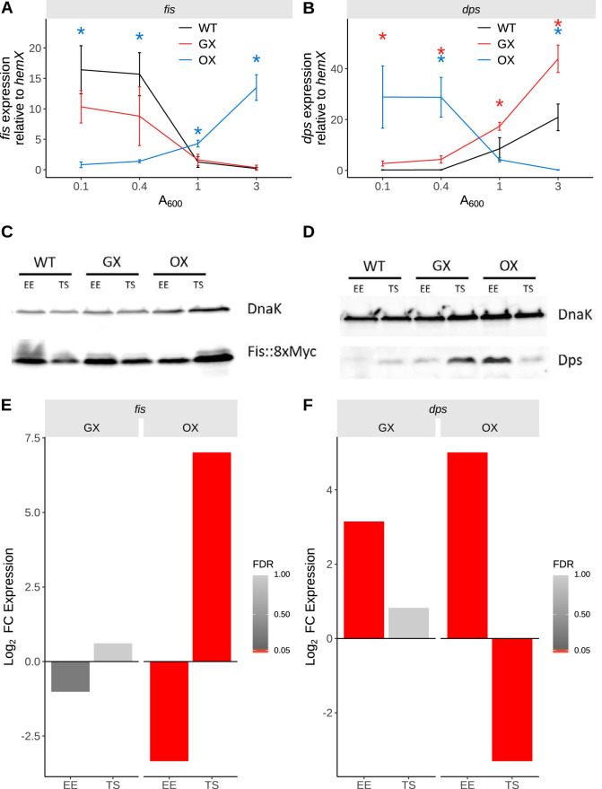
The Fis nucleoid-associated protein controls the expression of a large and diverse regulon of genes in Gram-negative bacteria. Fis production is normally maximal in bacteria during the early exponential phase of batch culture growth, becoming almost undetectable by the onset of stationary phase. We tested the effect on the Fis regulatory network in Salmonella of moving the complete fis gene from its usual location near the origin of chromosomal replication to the position normally occupied by the dps gene in the right macrodomain of the chromosome, and vice versa, creating the gene exchange (GX) strain. In a parallel experiment, we tested the effect of rewiring the Fis regulatory network by placing the fis open reading frame under the control of the stationary-phase-activated dps promoter at the dps genetic location within the right macrodomain, and vice versa, creating the open reading frame exchange (OX) strain. Chromatin immunoprecipitation sequencing (ChIP-seq) was used to measure global Fis protein binding levels and to determine gene expression patterns. Strain GX showed few changes compared with the wild type, although we did detect increased Fis binding at Ter, accompanied by reduced binding at Ori. Strain OX displayed a more pronounced version of this distorted Fis protein-binding pattern together with numerous alterations in the expression of genes in the Fis regulon. OX, but not GX, had a reduced ability to infect cultured mammalian cells. These findings illustrate the inherent robustness of the Fis regulatory network with respect to the effects of rewiring based on gene repositioning alone and emphasize the importance of fis expression signals in phenotypic determination.IMPORTANCE We assessed the impact on Salmonella physiology of reciprocally translocating the genes encoding the Fis and Dps nucleoid-associated proteins (NAPs) and of inverting their growth-phase production patterns such that Fis was produced in stationary phase (like Dps) and Dps was produced in exponential phase (like Fis). Changes to peak binding of Fis were detected by ChIP-seq on the chromosome, as were widespread impacts on the transcriptome, especially when Fis production mimicked Dps production. Virulence gene expression and the expression of a virulence phenotype were altered. Overall, these radical changes to NAP gene expression were well tolerated, revealing the robust and well-buffered nature of global gene regulation networks in the bacterium.

Keywords: ChIP-seq; Dps; Fis; RNA-seq; Salmonella enterica serovar Typhimurium; nucleoid-associated protein; transcriptome; virulence.


Figures

FIG 1
FIG 1
Genomic exchanges of fis and dps transcription units or ORFs. (A) Schematic depicting wild-type (WT) SL1344 strain with the original locations of dusB-fis and dps transcription units shown, the constructed gene exchange (GX) strain with the transcriptional units exchanged, and the ORF exchange (OX) strain with only the fis-dps ORFs exchanged. (B) Optical density-based growth curves showing the impact of these genetic changes on growth. Early exponential (EE), transition to stationary (TS), and late stationary (LS) time points selected for other analyses are shown. Significance was determined relative to the wild type using Welch's t test, and P values were adjusted using the Benjamini Hochberg (BH) method. Adjusted P values are indicated with asterisks (*, P < 0.05; **, P < 0.01; ***, P < 0.001; ****, P < 0.0001). (C) Growth rate curve derived from the OD-based growth curves. (D) CFU-based growth curves. (E) Origin-to-terminus gradients in these strains during the course of the growth curve analysis calculated by assaying the abundance of gidA (oriC proximal gene) DNA and STM1544 (ter proximal gene) DNA using qPCR. The late stationary (LS) phase is the 24-h time point in the growth curve not shown in the earlier plots. (F) Dosages of the fis gene relative to the terminus during the course of the growth curve analysis. (G) Dosages of the dps gene relative to the terminus. (H) Dosages of the fis gene relative to dps analyzed to understand the impact of the gene position swap.
FIG 2
FIG 2
Gene expression changes. (A) Expression of fis transcript during the course of the growth curve assayed using RT-qPCR. Significance was determined relative to the wild type using Welch's t test, and P values were adjusted using the BH method. Adjusted P values are indicated with asterisks (*, P < 0.05; **, P < 0.01; ***, P < 0.001; ****, P < 0.0001). Black, WT; red, GX; blue, OX. (B) Expression of dps transcript during the growth curve. Black, WT; red, GX; blue, OX. (C) Fis protein levels determined by immunoblotting with anti-Fis monoclonal antibody. (D) Dps protein levels determined by immunoblotting with anti-Dps polyclonal serum. (E and F) Expression changes in fis and dps genes determined by RNA-seq in the GX and OX strains at the EE and TS phases of growth. The color bar indicates FDR, and bars representing genes with FDR values of <0.05 are red.
FIG 3
FIG 3
Fis binding changes assayed by ChIP-seq during EE phase. (A) ChIP-seq was used to identify Fis peaks in WT, GX, and OX. The R package DiffBind was then used to determined Log2 fold changes in binding intensities of Fis in these peaks in the GX and OX strains relative to the wild type. Significant peak intensity changes (FDR < 0.05) are shown as red filled triangles. To determine if the medians of the distributions of Log2 fold changes in binding intensities differed significantly from 0 (no change in Fis binding relative to WT), the Wilcoxon rank sum test was performed with μ = 0; the P values are displayed over the individual distributions. The difference between the medians of the GX:WT distribution and the OX:WT distribution was not significant with the Wilcoxon rank sum test as shown above the square bracket corresponding to the data from the single-sample tests (using the less conservative Welch's t test here gives a P value of 0.0105). (B) Spearman’s correlation of peak intensity changes in the GX in comparison with the OX strain. Dashed line has a slope of 1 and indicates the position where the fold changes in Fis binding intensity in GX would be equal to those in OX. The blue solid line is the regression linear fit to the data. (C) Peak intensity changes along the chromosomal position shown in millions of base pairs. The blue curve shows the local regression peak intensity change calculated using the R locally estimated scatterplot smoothing (LOESS) function. In calculating these intensity changes, the appropriate mock data (WT mock for WT peaks, GX mock for GX peaks, OX mock for OX peaks) were used to control for ori-ter gradient changes (Fig. S5). (D) Fis ChIP-seq coverage of the WT, GX, and OX strains (two biological replicates and mock) of example regions near oriC and ter loci. The ranges of the data indicated on the y-axis scale were the same for all the coverage traces and have been omitted for brevity. For each strain, the two biological replicates are represented by the first two coverage traces in the darker color shading. The third, lighter color trace represents the mock coverage for that strain. Gray-shaded regions show peaks called in each sample relative to the mock. Genes (gray arrows) and Fis binding motifs (red blocks) are shown on the positive and negative reference strands. ChIP-seq data for plasmids can be found in Fig. S3.
FIG 4
FIG 4
Transcriptomic changes assayed by RNA-seq. (A) Counts of differentially expressed genes. (B) Box plot distribution representations of Log2 fold change in expression of genes in the GX and OX strains. The genes have been split into two groups based on whether or not they are predicted to be Fis targets. P values obtained using the two-sample Wilcoxon test for the comparison between Fis targets and nontargets are indicated over each pair of box plots. (C) Spearman’s correlation of Log2 fold changes in expression of genes in the GX with the OX strain. The dashed line has a slope of 1 and indicates the position where the fold changes are equal. (D) Log2 fold change in expression of genes along their position on the chromosome in millions of base pairs. The blue curve shows the local regression peak intensity change calculated using the R LOESS function. Significant gene expression changes (FDR < 0.05) are shown as red filled triangles.
FIG 5
FIG 5
Pathogenicity island expression changes assayed using chromosomal Pgfp+ reporters. (A) SPI-1 expression corresponding to the PprgH gfp+ chromosomal fusions and SPI-2 expression corresponding to the PssaG gfp+ chromosomal fusions are reported. Reporter expression is measured by determining the fluorescence (F) of gfp+ expression divided by A600. Means of results from biological replicates are shown as solid lines, and data from the individual biological replicates are shown in the background as lines with lighter shading. Significance was determined relative to the wild type using Welch's t test, and P values were adjusted using the BH method. Adjusted P values (WT versus GX and WT versus OX comparisons only) are indicated with asterisks (*, P < 0.05). (B) A slice of the data in panel A showing reporter expression at early time points (SPI-1, 5 h; SPI-2, 15 h) versus later time points (SPI-1, 15 h; SPI-2, 20 h). Bars represent median values, and the individual biological replicates are represented by the scatter. ns, not significant; *, P < 0.05; **, P < 0.01; ***, P < 0.001; ****, P < 0.0001. (C) Results of epithelial cell invasion assay carried out with subcultures grown to two different time points (early, 3.5 h; late, 6 h). Plotted are percent invasions of the strains relative to the WT (100% [not shown], depicted by the red dashed line) in HeLa cells determined 90 min postinfection. Median values of percent invasion are represented by the bars, and values from biological replicates are represented by the scatter. P values obtained from one-sample t tests (μ = 100) are shown in red above each strain designation. P values obtained from Welch’s two-sample t tests between GX and OX are indicated over these comparisons in black.

Similar articles

Cited by

References

    1. Browning DF, Butala M, Busby SJW. 2019. Bacterial transcription factors: regulation by Pick “N” Mix. J Mol Biol 431:4067–4077. doi:10.1016/j.jmb.2019.04.011. - DOI - PubMed
    1. Browning DF, Busby SJW. 2016. Local and global regulation of transcription initiation in bacteria. Nat Rev Microbiol 14:638–650. doi:10.1038/nrmicro.2016.103. - DOI - PubMed
    1. Li G-W, Burkhardt D, Gross C, Weissman JS. 2014. Quantifying absolute protein synthesis rates reveals principles underlying allocation of cellular resources. Cell 157:624–635. doi:10.1016/j.cell.2014.02.033. - DOI - PMC - PubMed
    1. Dillon SC, Dorman CJ. 2010. Bacterial nucleoid-associated proteins, nucleoid structure and gene expression. Nat Rev Microbiol 8:185–195. doi:10.1038/nrmicro2261. - DOI - PubMed
    1. Dorman CJ, Schumacher MA, Bush MJ, Brennan RG, Buttner MJ. 2020. When is a transcription factor a NAP? Curr Opin Microbiol 55:26–33. doi:10.1016/j.mib.2020.01.019. - DOI - PMC - PubMed

Publication types

MeSH terms

Substances