. 2020 Oct 27;94(22):e01683-20.
doi: 10.1128/JVI.01683-20. Print 2020 Oct 27.

Disruption of Adaptive Immunity Enhances Disease in SARS-CoV-2-Infected Syrian Hamsters

Affiliations

Disruption of Adaptive Immunity Enhances Disease in SARS-CoV-2-Infected Syrian Hamsters

Rebecca L Brocato et al. J Virol. .

Abstract

Animal models recapitulating human COVID-19 disease, especially severe disease, are urgently needed to understand pathogenesis and to evaluate candidate vaccines and therapeutics. Here, we develop novel severe-disease animal models for COVID-19 involving disruption of adaptive immunity in Syrian hamsters. Cyclophosphamide (CyP) immunosuppressed or RAG2 knockout (KO) hamsters were exposed to severe acute respiratory syndrome coronavirus 2 (SARS-CoV-2) by the respiratory route. Both the CyP-treated and RAG2 KO hamsters developed clinical signs of disease that were more severe than those in immunocompetent hamsters, notably weight loss, viral loads, and fatality (RAG2 KO only). Disease was prolonged in transiently immunosuppressed hamsters and was uniformly lethal in RAG2 KO hamsters. We evaluated the protective efficacy of a neutralizing monoclonal antibody and found that pretreatment, even in immunosuppressed animals, limited infection. Our results suggest that functional B and/or T cells are not only important for the clearance of SARS-CoV-2 but also play an early role in protection from acute disease.IMPORTANCE Syrian hamsters are in use as a model of disease caused by SARS-CoV-2. Pathology is pronounced in the upper and lower respiratory tract, and disease signs and endpoints include weight loss and viral RNA and/or infectious virus in swabs and organs (e.g., lungs). However, a high dose of virus is needed to produce disease, and the disease resolves rapidly. Here, we demonstrate that immunosuppressed hamsters are susceptible to low doses of virus and develop more severe and prolonged disease. We demonstrate the efficacy of a novel neutralizing monoclonal antibody using the cyclophosphamide transient suppression model. Furthermore, we demonstrate that RAG2 knockout hamsters develop severe/fatal disease when exposed to SARS-CoV-2. These immunosuppressed hamster models provide researchers with new tools for evaluating therapies and vaccines and understanding COVID-19 pathogenesis.

Keywords: COVID-19; SARS-CoV-2; Syrian hamster; animal models; cyclophosphamide; disease; monoclonal antibody.


Figures

FIG 1
FIG 1
CyP-treated, SARS-CoV-2-infected hamsters. Groups of 10 Syrian hamsters each were immunosuppressed with CyP. (A) Lymphocyte counts were determined from whole blood 3 days (closed symbols) or 4 days (open symbols) following the CyP loading dose. Hamsters were exposed to increasing doses of SARS-CoV-2 intranasally on day 0. (B) Weights were monitored for 35 days. (C) Viral RNA copies per pharyngeal swab were assayed at the indicated times postinfection. CyP administration is depicted by vertical dotted lines in panels B and C. Hamsters were monitored for (D) survival, and lung tissue collected either at the time of death or scheduled euthanasia 13 or 35 dpi was assessed for viral load by (E) Reverse transcription-PCR (RT-PCR) and (F) plaque assay. (G) Blood was collected from surviving hamsters at 35 dpi and assessed by plaque reduction or neutralization test. LU, lung.
FIG 2
FIG 2
SARS-CoV-2-infected RAG2 knockout (KO) hamsters. Either RAG2 KO (n = 7) or CyP-treated hamsters (n = 10, from Fig. 1) were exposed to 10,000 PFU SARS-CoV-2 on day 0. Vertical dotted lines (A, B) indicate CyP treatment for indicated animals. Hamsters were monitored for (A) weight and (C) survival. (B) Viral RNA copies per pharyngeal swab were assayed at the indicated times postinfection. (D) Organs collected at the time of death were homogenized and assayed for viral load. LU, lung; TR, trachea; HE, heart; LI, liver; SP, spleen; IN, intestine; BR, brain; KI, kidney.
FIG 3
FIG 3
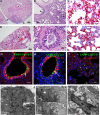
Pathology of SARS-CoV-2 in CyP-treated and RAG2 KO hamsters. Hematoxylin and eosin (H&E) sections (A, B) of lung tissue from CyP-treated hamsters euthanized at 13 dpi show extensive areas of consolidation with dense aggregates of inflammatory cells. (A) Bronchial lumina are lined by hyperplastic folds of respiratory epithelium (asterisk) and the pleural surface is multifocally thickened and expanded by fibrous connective tissue and inflammatory cells (arrows). (B) The bronchial lumina are lined by hyperplastic folds of respiratory epithelium (arrow). Areas of alveolar septa lined rows of type 2 pneumocytes (asterisks). (C) SARS-CoV-2 genomic RNA was frequently detected by in situ hybridization (ISH) in alveolar pneumocytes, alveolar infiltrates, and bronchiolar respiratory epithelial cells from CyP-treated hamsters. H&E sections (D, E) of lung tissue from RAG2 KO hamsters collected at the time of death. (D) Areas of hemorrhage (asterisk) and inflammation (arrowheads) expanding the interstitium and connective tissue surrounding the bronchi and arteries (arrows). (E) Necrotic bronchial epithelium (arrows) overlaid by hemorrhagic exudate. Peribronchial connective tissue is expanded by lymphocytes, heterophils (asterisks), and fewer macrophages that often contain hemosiderin (arrowheads). There is marked consolidation in surrounding alveoli with marked septal congestion and expansion by previously mentioned inflammatory cells. (F) SARS-CoV-2 genomic RNA was frequently detected by ISH in alveolar pneumocytes, alveolar infiltrates, and bronchiolar epithelial cells from RAG2 KO hamsters. (G to I) Immunofluorescence assays demonstrate that SARS-CoV-2 antigens (S or NP, green) were detected in bronchiolar epithelium-labeled anti-pan-cytokeratin antibody (red) (G) club (Clara) cells labeled by anti-CC10 antibody (red) (H) and alveolar epithelial cells labeled by anti-E-cadherin antibody (red) (I) in RAG2 KO hamsters. (J to L) Transmission electron microscopy of hamster lungs with increasing viral loads. (J) Lung section from hamster with 106 molecules of N2 per 100 ng RNA. Inset shows cytoplasmic vacuole with possible virus (black arrow). (K) Lung section from hamster with 107 molecules of N2 per 100 ng RNA. Potential mature viral particles (approximately 143 to 154 nm in diameter, arrowhead) are present at the cell periphery, and suspected immature virions are detected more internally in a cytoplasmic vacuole (approximately 62 to 97 nm in diameter, black arrow). (L) Lung section from hamster with 108 molecules of N2 per 100 ng RNA. Numerous cytoplasmic vacuoles of possible virus are evident (black arrows). Inset shows an example of swollen rough endoplasmic reticulum (rER) (asterisk) with virus forming within the swollen rER (black arrow). Bars are as follows: (A, D) 400 μm, (B) 100 μm, (E) 28 μm, (C, F) 100 μm, (G, H, I) 50 μm, and (J-L) 1 μm.
FIG 4
FIG 4
SARS-CoV-2 disrupts the tracheal epithelial layer. Tracheal sections were collected from SARS-CoV-2-infected, CyP-treated hamsters, sorted by lung viral load and analyzed by transmission electron microscopy. (A) The animal with a lung viral load of 106 molecules of N2 per 100 ng of RNA showed the most intact ciliated cells on the surface of the trachea (arrowheads). As viral load increases (from animals with lung viral loads of 107 [B] and 108 [C] molecules of N2 per 100 ng RNA, respectively), the presence of ciliated cells and epithelial cells lining the tracheal lumen decreases. (D) Cells from the low-viral-load animal show several cytoplasmic vacuoles with potential immature viral particles (arrows). (E) Release of cytoplasmic vacuole content (possible immature virions, arrows) into the luminal space of a cell that has detached from the epithelial layer. (F) From the animal with the highest viral load, very few ciliated cells were noted. Cytoplasmic vacuole with potential immature viral particles (arrow). Bars are as follows: (A to C) 1 μm and (D to F) 500 nm.
FIG 5
FIG 5
Rechallenge of previously infected SARS-CoV-2 hamsters. (A) Weight data from hamsters initially exposed to 10,000 PFU SARS-CoV-2 and rechallenged with 100,000 PFU SARS-CoV-2. (B) PRNT80 titers depicting the level of circulating neutralizing antibody prior to virus exposure on day 43. Disease progression was monitored by (A) weight and (C) pharyngeal swabs. Lung tissue collected on day 50 was assayed for (D) viral RNA and (E) infectious virus.
FIG 6
FIG 6
Passive transfer of anti-SARS-CoV-2 MAb Centi-F1 in immunosuppressed hamsters. Groups of 8 hamsters were immunosuppressed with CyP beginning at −3 dpi (indicated by the vertical lines in panels B and C), were passively transferred 30 mg/kg of Centi-F1 MAb, an equivalent volume of normal MAb, or PBS at −1 dpi, and were exposed to 1,000 PFU SARS-CoV-2 on day 0. (A) Levels of circulating neutralizing antibody from day 0 serum were assayed by PRNT. Disease progression was monitored by (B) weight and (C) pharyngeal swabs. Lung tissue collected at 13 dpi was assayed for (D) viral RNA and (E) infectious virus.

Similar articles

  • Syrian hamsters as a small animal model for SARS-CoV-2 infection and countermeasure development.
    Imai M, Iwatsuki-Horimoto K, Hatta M, Loeber S, Halfmann PJ, Nakajima N, Watanabe T, Ujie M, Takahashi K, Ito M, Yamada S, Fan S, Chiba S, Kuroda M, Guan L, Takada K, Armbrust T, Balogh A, Furusawa Y, Okuda M, Ueki H, Yasuhara A, Sakai-Tagawa Y, Lopes TJS, Kiso M, Yamayoshi S, Kinoshita N, Ohmagari N, Hattori SI, Takeda M, Mitsuya H, Krammer F, Suzuki T, Kawaoka Y. Imai M, et al. Proc Natl Acad Sci U S A. 2020 Jul 14;117(28):16587-16595. doi: 10.1073/pnas.2009799117. Epub 2020 Jun 22. Proc Natl Acad Sci U S A. 2020. PMID: 32571934 Free PMC article.
  • Pathogenesis and transmission of SARS-CoV-2 in golden hamsters.
    Sia SF, Yan LM, Chin AWH, Fung K, Choy KT, Wong AYL, Kaewpreedee P, Perera RAPM, Poon LLM, Nicholls JM, Peiris M, Yen HL. Sia SF, et al. Nature. 2020 Jul;583(7818):834-838. doi: 10.1038/s41586-020-2342-5. Epub 2020 May 14. Nature. 2020. PMID: 32408338 Free PMC article.
  • Sex Differences in Lung Imaging and SARS-CoV-2 Antibody Responses in a COVID-19 Golden Syrian Hamster Model.
    Dhakal S, Ruiz-Bedoya CA, Zhou R, Creisher PS, Villano JS, Littlefield K, Ruelas Castillo J, Marinho P, Jedlicka AE, Ordonez AA, Bahr M, Majewska N, Betenbaugh MJ, Flavahan K, Mueller ARL, Looney MM, Quijada D, Mota F, Beck SE, Brockhurst J, Braxton AM, Castell N, Stover M, D'Alessio FR, Metcalf Pate KA, Karakousis PC, Mankowski JL, Pekosz A, Jain SK, Klein SL; Johns Hopkins COVID-19 Hamster Study Group. Dhakal S, et al. mBio. 2021 Aug 31;12(4):e0097421. doi: 10.1128/mBio.00974-21. Epub 2021 Jul 13. mBio. 2021. PMID: 34253053 Free PMC article.
  • Hamsters as a Model of Severe Acute Respiratory Syndrome Coronavirus-2.
    Braxton AM, Creisher PS, Ruiz-Bedoya CA, Mulka KR, Dhakal S, Ordonez AA, Beck SE, Jain SK, Villano JS. Braxton AM, et al. Comp Med. 2021 Oct 1;71(5):398-410. doi: 10.30802/AALAS-CM-21-000036. Epub 2021 Sep 29. Comp Med. 2021. PMID: 34588095 Free PMC article. Review.
  • COVID-19: The Immune Responses and Clinical Therapy Candidates.
    Zhand S, Saghaeian Jazi M, Mohammadi S, Tarighati Rasekhi R, Rostamian G, Kalani MR, Rostamian A, George J, Douglas MW. Zhand S, et al. Int J Mol Sci. 2020 Aug 3;21(15):5559. doi: 10.3390/ijms21155559. Int J Mol Sci. 2020. PMID: 32756480 Free PMC article. Review.

Cited by

References

    1. Cohen J. 2020. From mice to monkeys, animals studied for coronavirus answers. Science 368:221–222. doi:10.1126/science.368.6488.221. - DOI - PubMed
    1. Chan JF, Zhang AJ, Yuan S, Poon VK, Chan CC, Lee AC, Chan WM, Fan Z, Tsoi HW, Wen L, Liang R, Cao J, Chen Y, Tang K, Luo C, Cai JP, Kok KH, Chu H, Chan KH, Sridhar S, Chen Z, Chen H, To KK, Yuen KY. 2020. Simulation of the clinical and pathological manifestations of coronavirus disease 2019 (COVID-19) in golden Syrian hamster model: implications for disease pathogenesis and transmissibility. Clin Infect Dis doi:10.1093/cid/ciaa325. - DOI - PMC - PubMed
    1. Rodgers TF, Zhao F, Huang D, Beutler N, Abbott RK, Callaghan S, Garcia E, He W, Hurtado J, Limbo O, Parren M, Peng L, Ricketts J, Ricciardi MK, Smith C, Song G, Woehl J, Yang L, Rawlings S, Smith DM, Nemazee D, Teijaro JR, Voss JE, Andrabi R, Briney B, Landais E, Sok D, Jardine JG, Burton DR. 2020. Rapid isolation of potent SARS-CoV-2 neutralizing antibodies and protection in a small animal model. bioRxiv 10.1101/2020.05.11.088674. - DOI - PMC - PubMed
    1. Schaecher SR, Stabenow J, Oberle C, Schriewer J, Buller RM, Sagartz JE, Pekosz A. 2008. An immunosuppressed Syrian golden hamster model for SARS-CoV infection. Virology 380:312–321. doi:10.1016/j.virol.2008.07.026. - DOI - PMC - PubMed
    1. Huang C, Wang Y, Li X, Ren L, Zhao J, Hu Y, Zhang L, Fan G, Xu J, Gu X, Cheng Z, Yu T, Xia J, Wei Y, Wu W, Xie X, Yin W, Li H, Liu M, Xiao Y, Gao H, Guo L, Xie J, Wang G, Jiang R, Gao Z, Jin Q, Wang J, Cao B. 2020. Clinical features of patients infected with 2019 novel coronavirus in Wuhan, China. Lancet 395:497–506. doi:10.1016/S0140-6736(20)30183-5. - DOI - PMC - PubMed