. 2020 Dec 11;295(50):16931-16948.
doi: 10.1074/jbc.RA120.014761. Epub 2020 Sep 8.

Cholesterol sensing by CD81 is important for hepatitis C virus entry

Affiliations

Cholesterol sensing by CD81 is important for hepatitis C virus entry

Machaela Palor et al. J Biol Chem. .

Abstract

CD81 plays a central role in a variety of physiological and pathological processes. Recent structural analysis of CD81 indicates that it contains an intramembrane cholesterol-binding pocket and that interaction with cholesterol may regulate a conformational switch in the large extracellular domain of CD81. Therefore, CD81 possesses a potential cholesterol-sensing mechanism; however, its relevance for protein function is thus far unknown. In this study we investigate CD81 cholesterol sensing in the context of its activity as a receptor for hepatitis C virus (HCV). Structure-led mutagenesis of the cholesterol-binding pocket reduced CD81-cholesterol association but had disparate effects on HCV entry, both reducing and enhancing CD81 receptor activity. We reasoned that this could be explained by alterations in the consequences of cholesterol binding. To investigate this further we performed molecular dynamic simulations of CD81 with and without cholesterol; this identified a potential allosteric mechanism by which cholesterol binding regulates the conformation of CD81. To test this, we designed further mutations to force CD81 into either the open (cholesterol-unbound) or closed (cholesterol-bound) conformation. The open mutant of CD81 exhibited reduced HCV receptor activity, whereas the closed mutant enhanced activity. These data are consistent with cholesterol sensing switching CD81 between a receptor active and inactive state. CD81 interactome analysis also suggests that conformational switching may modulate the assembly of CD81-partner protein networks. This work furthers our understanding of the molecular mechanism of CD81 cholesterol sensing, how this relates to HCV entry, and CD81's function as a molecular scaffold; these insights are relevant to CD81's varied roles in both health and disease.

Keywords: cholesterol-binding protein; hepatitis C virus (HCV); molecular dynamics; plasma membrane; tetraspanin; virus entry.


Conflict of interest statement

Conflict of interest—The authors declare that they have no conflicts of interest with the contents of this article.

Figures
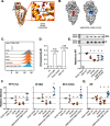
Figure 1.
Figure 1.
Mutations in the cholesterol-binding pocket of CD81 modulate HCV entry. A, cholesterol (red) is coordinated in the intramembrane cavity of CD81 by hydrogen bonds with inward-facing residues Asn18 and Glu219. We made various mutations at these sites to disrupt this interaction. B, the cholesterol molecule sits in the center of an intramembrane-binding pocket. In the V68W/M72W/A108W/V212W mutant, this space is occupied by tryptophan residues (blue). Molecular model images were created using Protein Data Bank code 5TCX (26). C, the cell-surface expression levels of each mutant CD81 was assessed by flow cytometry. D, Huh-7 CD81 KO cells were transduced with lentivector encoding WT CD81 or empty vector control. The cells were surface-labeled with anti-CD81 mAb and lysed in Brij-98 detergent buffer. CD81–mAb complexes were pulled down with protein G beads, and associated free cholesterol was measured. The negative control (−ve) contains no sample. The positive control contains 0.31 μm exogenous cholesterol and demonstrates the accuracy of the assay. The dashed line indicates the limit of detection. E, we assessed cholesterol association with WT and mutant CD81. The data are expressed relative to WT CD81, and an asterisk indicates statistical significance from WT (n = 4, one-way ANOVA, Prism). The Western blotting demonstrates equivalent levels of CD81 in the whole cell lysate (WCL) and pulldown (IP). F, Huh-7 CD81 KO cells were transduced with lentivectors encoding WT and mutant CD81, and cell-surface expression was confirmed by flow cytometry, as in C. HCV entry was assessed by challenge with a panel of HCVpp (including genotypes 1, 2, and 5). HCVpp infection, for three representative clones, is shown relative to cells expressing WT CD81 (n = 4). G, summary data displaying mean relative infection, as in E, for six HCVpp clones. An asterisk indicates statistical significance from WT (one-way ANOVA, Prism), and error bars indicate the standard deviation of the mean.
Figure 2.
Figure 2.
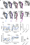
Conformational switching of CD81 in the absence of cholesterol. We performed five independent 500-ns MD simulations of WT CD81 with and without cholesterol. A, snapshots summarizing representative simulations from either condition. The Δ° measurement reflects the change in the angle between helix E of the EC2 and TMD4 (as annotated), by comparison with the CD81 crystal structure (Protein Data Bank code 5TCX). For each snapshot the region from which the measurement was taken is color-coded by time. Cholesterol is shown in red. Structures were orientated using TMD4 as a reference. Examples of the orientation of Asp196 and Lys201 are shown as insets. B, the angle between helix E and TMD4 was measured over time for each simulation, and 25° was chosen as a threshold to indicate conformational switching. C, the cumulative time spent in the open conformation was calculated across all simulations for either experimental condition. D, the distance between Asp196 and Lys201 was measured over time for each simulation, and the dashed line indicates the distance under which electrostatic interactions and hydrogen bonding occurs (10 Å). E, the average distance between Asp196 and Lys201 with and without cholesterol. The data points represent the mean value for each simulation, and the asterisk indicates statistical significance (n = 5 simulations, unpaired t test, Prism), and the error bars indicate standard deviation of the mean.
Figure 3.
Figure 3.
Conformational switch mutants modulate HCV entry. We mutated residues Asp196 and Lys201 to prevent stabilizing interactions across the EC2–TMD4 hinge. A, we performed five independent MD simulations of WT and D196A/K201A CD81 in the presence of cholesterol. Images provide overlaid snapshots from representative simulations. Helix E, TMD4, and cholesterol are color-coded by time. For clarity the remaining structure is shown in gray for the t = 0 ns snapshot only. Structures were orientated using TMD4 as a reference. B, the change in angle between helix E and TMD4, by comparison with the CD81 crystal structure, was measured over time for each D196A/K201A simulation (compare with Fig. 2B). The cumulative time spent in the open conformation across all simulations was 400 ns for WT and 1050 ns for D196A/K201A. C, the average distance between residues 196 and 201 for WT and D196A/K201A in the presence of cholesterol. The dashed line indicates the distance under which electrostatic interactions and hydrogen bonding can occur (10 Å). The data points represent the mean value for each simulation, and an asterisk indicates statistical significance (n = 5 simulations, unpaired t test, Prism). D, Huh-7 CD81 KO cells were transduced with lentivectors encoding WT CD81, N18A/E219A (cholesterol-binding mutant), D196A/K201A (open mutant), or K116A/D117A (closed mutant), equal cell-surface expression was confirmed by flow cytometry (representative data are provided in Fig. 4). HCV entry was assessed by challenge with a panel of HCVpp (including genotypes 1, 2, 4, and 5). HCVpp infection, from three representative clones, is shown relative to cells expressing WT CD81. An asterisk indicates statistical significance from WT (n = 4, one-way ANOVA, Prism). There was no significant difference between N18A/E219A and D196A/K201A. E, summary data displaying mean relative infection, as in D, for eight HCVpp clones. An asterisk indicates statistical significance from WT (n = 8, one-way ANOVA, Prism). In all plots error bars indicate standard deviation of the mean.
Figure 4.
Figure 4.
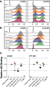
Cell-surface functionality of CD81 mutants. Huh-7 CD81 KO cells were co-transduced with lentivectors encoding human CD19 and CD81 or empty vector. A, representative flow cytometry histograms. All samples received CD19 lentivector plus the indicated CD81/control vector. Panel i demonstrates CD81 surface expression, and panel ii displays CD81-dependent trafficking of CD19 to the cell surface. B, CD81 expression on CHO cells confers binding on soluble HCV E2. Panel i demonstrates CD81 surface expression, and panel ii displays sE2 binding to transduced CHO cells. C, quantification of sE2 binding expressed relative to WT CD81. An asterisk indicates statistical significance from WT (n = 3, one-way ANOVA, Prism). Error bars indicate standard deviation of the mean.
Figure 5.
Figure 5.
Cholesterol sensing is important for authentic HCV infection. Huh-7 CD81 KO cells were transduced with lentivectors expressing the stated CD81 mutants and were then challenged with J6/JFH HCVcc. Equal cell-surface expression of WT and mutant CD81 was confirmed by flow cytometry (representative data are provided in Fig. 4). A, representative micrographs of HCVcc infection in transduced cells. The 4′,6-diamino-2-phenylindole nuclei shown in blue, and viral antigen NS5A is displayed in orange. Scale bar, 100 μm. B, quantification of infection. The data are expressed relative to infection in cells expressing WT CD81, and an asterisk indicates statistical significance from WT (n = 4, one-way ANOVA, Prism). C, Huh-7 Lunet N cells stably expressing the stated CD81 mutants were challenged with a panel of diverse HCVcc bearing the glycoproteins of genotypes 1, 2, 3, 4, and 5. Infection was quantified via a virally encoded luciferase reporter and is shown, relative to WT CD81, for three representative clones. An asterisks indicates statistical significance from WT (n = 3, one-way ANOVA, Prism). D, summary data displaying mean relative infection, as in C, for 12 HCVcc chimeras. An asterisk indicates statistical significance from WT (n = 12 one-way ANOVA, Prism). In all plots, error bars indicate standard deviation of the mean.
Figure 6.
Figure 6.
Conformational switch mutants exhibit altered protein interaction networks. A, volcano plot visualizing differences from co-IPs of Huh-7 Lunet N CD81 WT versus Lunet N control cells (n = 4 biological replicates for each cell line). LFQ intensity differences (log2) are plotted against the t test p value (−logP). Significant interactors were defined by a permutation-based FDR using S0 = 1 as described (93). Reference proteins (CD81, SCARB1, CLDN1, EGFR, TFRC, CAPN5 ITGB, and CD151) are highlighted and color-coded as in B. B, mean LFQ intensity differences (log2) of interactors in CD81 co-IP (Huh-7 Lunet N CD81 WT and mutants versus Lunet N control cells). Error bars indicate standard deviation of the mean (n = 4). C, Venn diagrams showing the overlap of significantly enriched proteins found in CD81 co-IPs from WT in gray, N18A/E219A (Chl) in orange, D196A/K201A (O) in purple, and K116A/D117A (C) in green. The values below each title indicate significant interactors for each CD81 variant, and the values in the center of each Venn diagram indicate overlapping interactors.

References

    1. Pileri, P., Uematsu, Y., Campagnoli, S., Galli, G., Falugi, F., Petracca, R., Weiner, A. J., Houghton, M., Rosa, D., Grandi, G., and Abrignani, S. (1998) Binding of hepatitis C virus to CD81. Science 282, 938–941 10.1126/science.282.5390.938 - DOI - PubMed
    1. Kinchen, V. J., Zahid, M. N., Flyak, A. I., Soliman, M. G., Learn, G. H., Wang, S., Davidson, E., Doranz, B. J., Ray, S. C., Cox, A. L., Crowe, J. E., Jr., Bjorkman, P. J., Shaw, G. M., and Bailey, J. R. (2018) Broadly neutralizing antibody mediated clearance of human hepatitis C virus infection. Cell Host Microbe 24, 717–730.e5 10.1016/j.chom.2018.10.012 - DOI - PMC - PubMed
    1. Flyak, A. I., Ruiz, S., Colbert, M. D., Luong, T., Crowe, J. E., Jr., Bailey, J. R., and Bjorkman, P. J. (2018) HCV broadly neutralizing antibodies use a CDRH3 disulfide motif to recognize an E2 glycoprotein site that can be targeted for vaccine design. Cell Host Microbe 24, 703–716.e3 10.1016/j.chom.2018.10.009 - DOI - PMC - PubMed
    1. Banse, P., Moeller, R., Bruening, J., Lasswitz, L., Kahl, S., Khan, A. G., Marcotrigiano, J., Pietschmann, T., and Gerold, G. (2018) CD81 receptor regions outside the large extracellular loop determine hepatitis C virus entry into hepatoma cells. Viruses 10, 207 10.3390/v10040207 - DOI - PMC - PubMed
    1. Charrin, S., Jouannet, S., Boucheix, C., and Rubinstein, E. (2014) Tetraspanins at a glance. J. Cell Sci. 127, 3641–3648 10.1242/jcs.154906 - DOI - PubMed

Publication types

Associated data

LinkOut - more resources