. 2020 Sep 8;10(1):14639.
doi: 10.1038/s41598-020-71633-3.

IRF2 maintains the stemness of colonic stem cells by limiting physiological stress from interferon

Affiliations

IRF2 maintains the stemness of colonic stem cells by limiting physiological stress from interferon

Kana Minamide et al. Sci Rep. .

Abstract

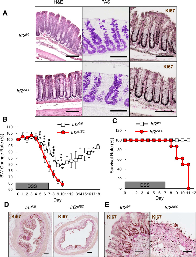
The physiological stresses that diminish tissue stem-cell characteristics remain largely unknown. We previously reported that type I interferon (IFN), which is essential for host antiviral responses, is a physiological stressor for hematopoietic stem cells (HSCs) and small intestinal stem cells (ISCs) and that interferon regulatory factor-2 (IRF2), which attenuates IFN signaling, maintains their stemness. Here, using a dextran sodium sulfate (DSS)-induced colitis model, we explore the role of IRF2 in maintaining colonic epithelial stem cells (CoSCs). In mice with a conditional Irf2 deletion in the intestinal epithelium (hereafter Irf2ΔIEC mice), both the number and the organoid-forming potential of CoSCs were markedly reduced. Consistent with this finding, the ability of Irf2ΔIEC mice to regenerate colon epithelium after inducing colitis was severely impaired, independently of microbial dysbiosis. Mechanistically, CoSCs differentiated prematurely into transit-amplifying (TA) cells in Irf2ΔIEC mice, which might explain their low CoSC counts. A similar phenotype was induced in wild-type mice by repeated injections of low doses of poly(I:C), which induces type I IFN. Collectively, we demonstrated that chronic IFN signaling physiologically stresses CoSCs. This study provides new insight into the development of colitis and molecular mechanisms that maintain functional CoSCs throughout life.


Conflict of interest statement

The authors declare no competing interests.

Figures

Figure 1
Figure 1
Defective regeneration of colonic epithelium in Irf2ΔIEC mice. (A) H&E (left) and PAS staining (middle) of distal colon tissue from Irf2fl/fl and Irf2ΔIEC mice (n = 3). Proliferating cells were detected by Ki67 staining (right). Scale bars: 100 μm. (B,C) Graphs show changes in body weight (BW) and mortality over time between Irf2fl/fl and Irf2ΔIEC mice that were given 2% DSS (w/v) in drinking water for 7 days followed by 11 days of plain water (n = 8 per group). (D) Low-magnification and (E) high-magnification images show histologic analysis of the distal colon in Irf2fl/fl and Irf2ΔIEC mice treated with 2% DSS in drinking water for 5 days followed by 6 days of plain water (n = 3). Regenerating crypts were detected by Ki67 staining. Scale bars: (D) 200 μm and (E) 100 μm. *P < 0.05, **P < 0.01, ***P < 0.001 by Student's t test.
Figure 2
Figure 2
IRF2 deficiency does not affect the composition of commensal bacteria or the mucus-layer structure. (A, B) 16S rRNA gene-sequencing analysis showing the relative abundance of bacteria identified at the phylum (A) and genus (B) level in fecal samples of co-housed Irf2fl/fl and Irf2ΔIEC mice (n = 10). At the genus level, the sequences from the samples represented 106 genera; the top 25 are listed in (B). Sequences that could not be classified into any known genus are shown as unclassified (Unc). (C) Rarefaction curves of bacterial alpha diversity using the Chao1 index. Significance was calculated by a non-parametric two-sample t test. (D) Principal coordinates analysis (PCoA) of beta diversity using Bray–Curtis distances. Significance was calculated by PERMANOVA. (E) Representative images of Alcian blue/PAS-stained distal colon sections of Irf2fl/fl and Irf2ΔIEC mice (n = 3). Arrowheads indicate the mucus layer. Asterisks indicate the area of mucus layer. Scale bars: 200 μm (left panels, low-magnification) and 100 μm (right panels, high-magnification).
Figure 3
Figure 3
IRF2 is essential for maintaining CoSCs. (A) Representative images show organoids formed from single colonic EpCAM+ cells from Irf2fl/fl and Irf2ΔIEC mice (n = 4). Scale bars: 100 μm. (B) Organoid-formation efficiency was determined by counting the viable organoids in each well on day 6; data represent the mean ± SD for five independent experiments. ***P < 0.001 by Student’s t test. (C) Representative FACS plots for Lgr5-GFPhi cells in colonic epithelial cells isolated from naïve Irf2fl/fl-Lgr5GFP and Irf2ΔIEC-Lgr5GFP mice (n = 3 each). (D) Average percentage of Lgr5-GFPhi cells among EpCAM+ cells; data represent mean ± SD for 3 mice. ***P < 0.001 by Student’s t test. (E) Confocal images of the base of crypts in the colon of control Irf2fl/fl-Lgr5GFP and Irf2ΔIEC-Lgr5GFP mice (n = 3 each), showing merged fluorescent and phase-contrast images (upper panels) and fluorescent images (lower panels). Scale bars: 200 μm. (F) Number of GFP-positive crypts per visual field. Five fields of view were counted per mouse. Data represent mean ± SD for 3 mice. *P < 0.05 by Student’s t test.
Figure 4
Figure 4
Loss of IRF2 promotes cell cycle and differentiation of CoSCs. (A) GSEA for colonic epithelial cells from Irf2fl/fl and Irf2ΔIEC mice (n = 3 for each group) using C5 MSigDB gene sets (C5: GO gene sets). The cell cycle process was significantly enriched in Irf2ΔIEC compared to Irf2fl/fl mice. NES, normalized enrichment score. (B) The top 20 most up-regulated genes in the gene set for cell-cycle processes in Irf2ΔIEC compared to Irf2fl/fl mice. Results are depicted as the log2 ratio determined by microarray. (C) Representative images of BrdU staining of the colon crypts of Irf2fl/fl and Irf2ΔIEC mice (n = 3). Mice were sacrificed 2 h after BrdU injection. Scale bars: 100 μm. (D) Number of BrdU+ cells per crypt. Approximately 100 crypts per mouse were analyzed. Data represent the means ± SD for 3 mice. *P < 0.05 by Student’s t test.
Figure 5
Figure 5
Chronic type I IFN signaling diminishes colonic epithelial stem-cell function. (A) Experimental protocol for poly(I:C) treatment. (B) H&E staining of the distal colon after 7 days of PBS or poly(I:C) treatment. Scale bars: 100 μm. (C, D) Representative images showing the organoid-forming efficiency of CoSCs from WT and (E,F) Ifnar1−/− mice treated with either poly (I:C) or PBS as in (A) (n = 3 for each group). Scale bars: 50 μm. Data represent mean ± SD of five independent experiments. ***P < 0.001 by Student’s t test. n.s., not significant. (G) Representative images of the 2-day BrdU-tracing experiment of the colon of WT mice treated with either PBS or poly(I:C). As shown in (A), BrdU was injected into mice on day 5 of poly(I:C) treatment. Mice were sacrificed on day 7. Scale bars: 100 μm. (H) Migration rate of BrdU-labeled cells 2 days after BrdU injection. The distance of the BrdU-labeled cells from the bottom of the crypt was divided by the total depth of the crypt. At least 30 crypts per mouse were analyzed. Data represent mean ± SD for 3 mice. *P<0.05 by Student's t test.

References

    1. Barker N, et al. Identification of stem cells in small intestine and colon by marker gene Lgr5. Nature. 2007;449:1003–1007. - PubMed
    1. Powell AE, et al. The pan-ErbB negative regulator Lrig1 is an intestinal stem cell marker that functions as a tumor suppressor. Cell. 2012;149:146–158. - PMC - PubMed
    1. Ramalingam S, Daughtridge GW, Johnston MJ, Gracz AD, Magness ST. Distinct levels of Sox9 expression mark colon epithelial stem cells that form colonoids in culture. AJP Gastrointest. Liver Physiol. 2012;302:G10–G20. - PMC - PubMed
    1. Jung P, et al. Isolation and in vitro expansion of human colonic stem cells. Nat. Med. 2011;17:1225–1227. - PubMed
    1. Asfaha S, et al. Krt19+/Lgr5- cells are radioresistant cancer-initiating stem cells in the colon and intestine. Cell Stem Cell. 2015;16:627–638. - PMC - PubMed

Publication types