. 2021 Jan 1;31(1):439-447.
doi: 10.1093/cercor/bhaa235.

Ultrahigh-resolution MRI Reveals Extensive Cortical Demyelination in a Nonhuman Primate Model of Multiple Sclerosis

Affiliations

Ultrahigh-resolution MRI Reveals Extensive Cortical Demyelination in a Nonhuman Primate Model of Multiple Sclerosis

Maxime Donadieu et al. Cereb Cortex. .

Abstract

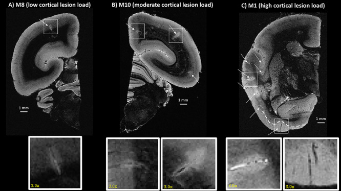
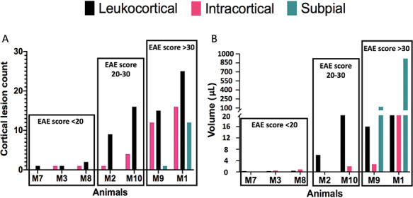
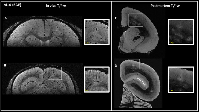
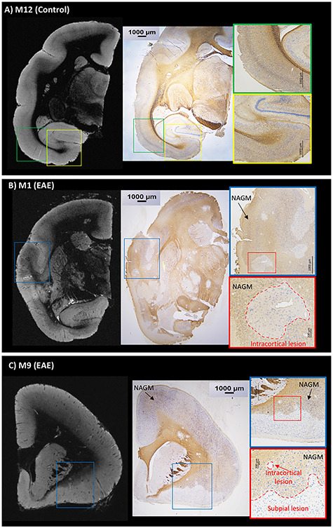
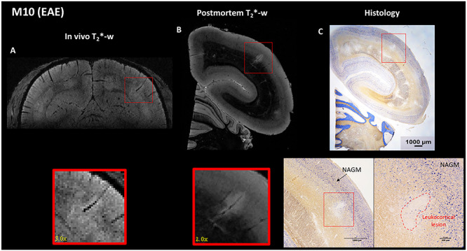
Cortical lesions are a primary driver of disability in multiple sclerosis (MS). However, noninvasive detection of cortical lesions with in vivo magnetic resonance imaging (MRI) remains challenging. Experimental autoimmune encephalomyelitis (EAE) in the common marmoset is a relevant animal model of MS for investigating the pathophysiological mechanisms leading to brain damage. This study aimed to characterize cortical lesions in marmosets with EAE using ultrahigh-field (7 T) MRI and histological analysis. Tissue preparation was optimized to enable the acquisition of high-spatial resolution (50-μm isotropic) T2*-weighted images. A total of 14 animals were scanned in this study, and 70% of the diseased animals presented at least one cortical lesion on postmortem imaging. Cortical lesions identified on MRI were verified with myelin proteolipid protein immunostaining. An optimized T2*-weighted sequence was developed for in vivo imaging and shown to capture 65% of cortical lesions detected postmortem. Immunostaining confirmed extensive demyelination with preserved neuronal somata in several cortical areas of EAE animals. Overall, this study demonstrates the relevance and feasibility of the marmoset EAE model to study cortical lesions, among the most important yet least understood features of MS.

Keywords: EAE; MRI; demyelination; marmoset; multiple sclerosis.


Figures

Figure 1
Figure 1
Postmortem 3D T2*-weighted gradient echo MRI showing different types of cortical lesions detected in three different animals (from low to high cortical lesion load). White arrows point to identified cortical lesions. (A) One leukocortical lesion (box) was identified in M8 in the occipital cortex. (B) Two leukocortical lesions were identified in M10 in the parietal cortex and the cingulate cortex (boxes). (C) M1 had many cortical lesions, including intracortical and subpial (boxes). The white dashed line shows the extent of the subpial lesion. All lesions are magnified by a factor of three in the inset.
Figure 2
Figure 2
Bar graphs presenting the count (A) and the volume (B) of each subtype of cortical lesions detected in EAE animals (from low to high cortical lesion load, dark green: subpial; black: leukocortical; pink: intracortical). Animals are subdivided into three groups, depending on their EAE scores: <20, 20–30, and >30.
Figure 3
Figure 3
Detection of the same cortical lesions in one EAE animal (M10) using in vivo (left) and postmortem (right) 3D T2*-weighted gradient-echo sequences. One intracortical (A) and one leukocortical (B) lesions were detected in vivo in two different areas: (A) right parietal cortex and (B) right occipital cortex. The same lesions were identified on postmortem MRI (C and D). In vivo lesions are magnified by a factor of three and postmortem lesions by a factor of two in the inset. Interestingly, cortical lesions appear larger in the in vivo images compared with postmortem images, likely due to water shifts.
Figure 4
Figure 4
Postmortem T2*-weighted gradient echo MRI and PLP/NeuN immunohistochemistry are well correlated. (A) Control animal (M12) with normal MRI contrast and normal PLP/NeuN staining. PLP (brown) staining is darker in the white matter but is still detectable in normal gray matter. NeuN staining (blue) demonstrates neuronal cell bodies in the gray matter. Interestingly, cortical myeloarchitecture is identifiable on T2*-weighted MRI. (B) EAE animal (M1) shows multiple cortical lesions on postmortem MRI corresponding to demyelinated areas on histology sections with sparing of neuronal somata. (C) EAE animal (M9) presents evident subpial and intracortical lesions on postmortem MRI corresponding to demyelinated areas on histology with preserved neuronal somata. Areas of normal-appearing gray matter (NAGM) are shown in the histology insets in the EAE animals (B and C).
Figure 5
Figure 5
EAE animal (M10). A leukocortical lesion in the right parietal cortex (red box) was identified on (A) in vivo T2*-weighted gradient echo MRI, (B) postmortem T2*-weighted gradient echo MRI, and (C) histology sections with PLP/NeuN staining. Interestingly, cortical lesions appear larger in the in vivo images compared with postmortem images, likely due to water shifts. The lesion detected on MRI corresponds to a demyelinated area on histology (red dotted line, with absence of PLP staining), with sparing of neuronal somata (persistence of NeuN staining). An area of normal-appearing gray matter (NAGM) is identified in the histology inset (C).

References

    1. Absinta M, Sati P, Reich DS. 2016. Advanced MRI and staging of multiple sclerosis lesions. Nat Rev Neurol. 12:358–368. - PMC - PubMed
    1. Ascherio A, Munger KL. 2016. Epidemiology of multiple sclerosis: from risk factors to prevention-an update. Semin Neurol. 36:103–114. - PubMed
    1. Beck ES, Sati P, Sethi V, Kober T, Dewey B, Bhargava P, Nair G, Cortese IC, Reich DS. 2018. Improved visualization of cortical lesions in multiple sclerosis using 7T MP2RAGE. Am J Neuroradiol. 39:459–466. - PMC - PubMed
    1. Bø L, Vedeler CA, Nyland HI, Trapp BD, Mørk SJ. 2003. Subpial demyelination in the cerebral cortex of multiple sclerosis patients. J Neuropathol Exp Neurol. 62:723–732. - PubMed
    1. Burrows DJ, McGown A, Jain SA, De Felice M, Ramesh TM, Sharrack B, Majid A. 2019. Animal models of multiple sclerosis: from rodents to zebrafish. Mult Scler. 25:306–324. - PubMed

Publication types