Review
. 2021 Jan;22(1):23-40.
doi: 10.3348/kjr.2019.0862. Epub 2020 Aug 28.

The Role of Imaging in Current Treatment Strategies for Pancreatic Adenocarcinoma

Affiliations
Review

The Role of Imaging in Current Treatment Strategies for Pancreatic Adenocarcinoma

Hyungjin Rhee et al. Korean J Radiol. 2021 Jan.

Abstract

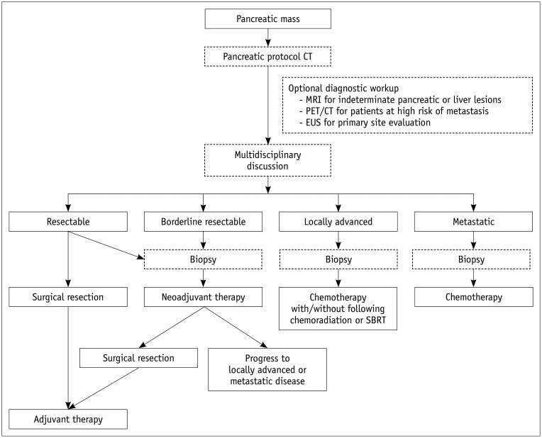
In pancreatic cancer, imaging plays an essential role in surveillance, diagnosis, resectability evaluation, and treatment response evaluation. Pancreatic cancer surveillance in high-risk individuals has been attempted using endoscopic ultrasound (EUS) or magnetic resonance imaging (MRI). Imaging diagnosis and resectability evaluation are the most important factors influencing treatment decisions, where computed tomography (CT) is the preferred modality. EUS, MRI, and positron emission tomography play a complementary role to CT. Treatment response evaluation is of increasing clinical importance, especially in patients undergoing neoadjuvant therapy. This review aimed to comprehensively review the role of imaging in relation to the current treatment strategy for pancreatic cancer, including surveillance, diagnosis, evaluation of resectability and treatment response, and prediction of prognosis.

Keywords: Diagnosis; Imaging; Pancreatic cancer; Prognosis; Resectability; Response evaluation.


Conflict of interest statement

The authors have no potential conflicts of interest to disclose.

Figures

Fig. 1
Fig. 1. Treatment strategy for pancreatic cancer.
CT = computed tomography, EUS = endoscopic ultrasound, MRI = magnetic resonance imaging, PET = positron emission tomography, SBRT = stereotactic body radiation therapy
Fig. 2
Fig. 2. Isoattenuating pancreatic cancer on CT.
A 55-year-old female patient was referred for a small pancreatic lesion that was detected in transabdominal ultrasound (image was not available). Both pancreatic (A) and venous phases (B) in CT showed no demonstrable lesion in the pancreas or significant pancreatic duct dilatation. MRI showed an approximately 1.5-cm focal pancreatic lesion (white arrows) with hypointensity in the T1-weighted image (C), hypoenhancement in the pancreatic phase (D), and moderate hyperintensity in the T2-weighted image (E). Magnetic resonance cholangiopancreatography (F) showed minimal pancreatic duct dilatation distal to the pancreatic mass, suggesting duct involvement (white arrowheads). The mass was seen as a hypoechoic mass in EUS (G).
Fig. 3
Fig. 3. Detection of small liver metastasis in MRI.
A 65-year-old male patient was admitted for chronic pancreatitis and pancreatic body cancer. Since there was no apparent vascular invasion in CT (A) and no demonstrable metastatic lesion in CT (B) and PET/CT (C), the pancreatic lesion was considered to be resectable. However, MRI showed multiple small hepatic lesions (white arrows) with peripheral enhancement in the pancreatic phase (D), hypointensity in the venous phase (E), decreased uptake in the hepatobiliary phase (F), hyperintensity in the T2-weighted image (G), and high signal intensity in the diffusion weighted image (b = 800) (H), suggesting liver metastases.
Fig. 4
Fig. 4. Resectability of pancreatic cancer determined by the National Comprehensive Cancer Network criteria.
A. Approximately 2-cm resectable pancreatic cancer (white arrow) confined to the pancreas showing no vascular involvement. B. Approximately 2.5-cm borderline resectable pancreatic cancer (white arrowhead) exhibiting superior mesenteric artery contact of less than 180°. C. Approximately 3.7-cm locally advanced pancreatic cancer (black arrow) showing encasement of the celiac artery and proximal common hepatic artery.
Fig. 5
Fig. 5. Pancreatic cancer showing partial response after a long period of chemotherapy.
A 55-year-old female was diagnosed with pancreatic cancer in the uncinate process. A, B. The pancreatic phase of initial CT showed an infiltrative hypoenhancing mass lesion (white arrows) involving the uncinate process and retroperitoneal margin, as well as encasing superior mesenteric artery (white arrowheads) and its jejunal branches, suggesting locally advanced tumor. C, D. After approximately 2 years of FOLFIRINOX chemotherapy, the lesion (black arrows) showed a reduction in size and extent of vascular involvement. It was apparent that the tumor became smaller with chemotherapy, but it was difficult to determine exactly how much of the viable tumor remained. The patient underwent pancreaticoduodenectomy, and margin negative (R0) resection was achieved without resection of vessels.

References

    1. Siegel RL, Miller KD, Jemal A. Cancer statistics, 2018. CA Cancer J Clin. 2018;68:7–30. - PubMed
    1. Malvezzi M, Carioli G, Bertuccio P, Boffetta P, Levi F, La Vecchia C, et al. European cancer mortality predictions for the year 2019 with focus on breast cancer. Ann Oncol. 2019;30:781–787. - PubMed
    1. Ferlay J, Colombet M, Soerjomataram I, Dyba T, Randi G, Bettio M, et al. Cancer incidence and mortality patterns in Eeurope: estimates for 40 countries and 25 major cancers in 2018. Eur J Cancer. 2018;103:356–387. - PubMed
    1. Jung KW, Won YJ, Kong HJ, Lee ES. Cancer statistics in Korea: incidence, mortality, survival, and prevalence in 2016. Cancer Res Treat. 2019;51:417–430. - PMC - PubMed
    1. Ducreux M, Cuhna AS, Caramella C, Hollebecque A, Burtin P, Goéré D, et al. Cancer of the pancreas: ESMO clinical practice guidelines for diagnosis, treatment and follow-up. Ann Oncol. 2015;26 Suppl 5:v56–v68. - PubMed