

Lack of effect of short-term DHEA supplementation on the perimenopausal ovary†
- PMID: 32901819
- PMCID: PMC7711893
- DOI: 10.1093/biolre/ioaa160
Lack of effect of short-term DHEA supplementation on the perimenopausal ovary†
Abstract
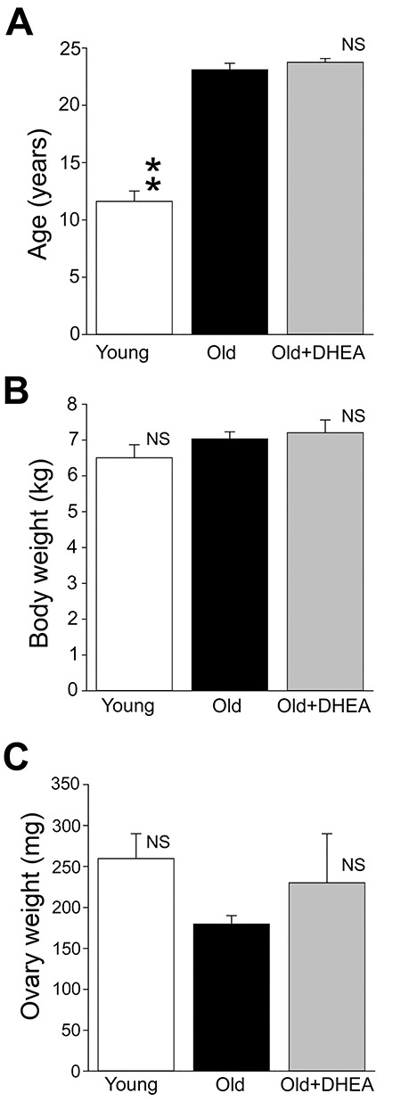
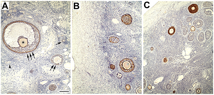
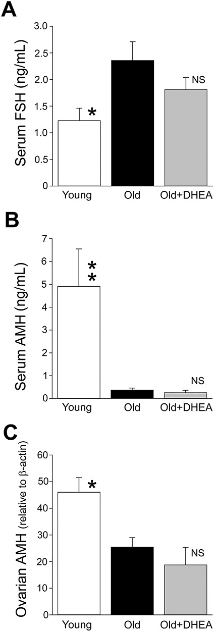
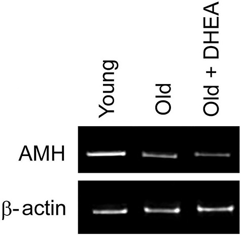
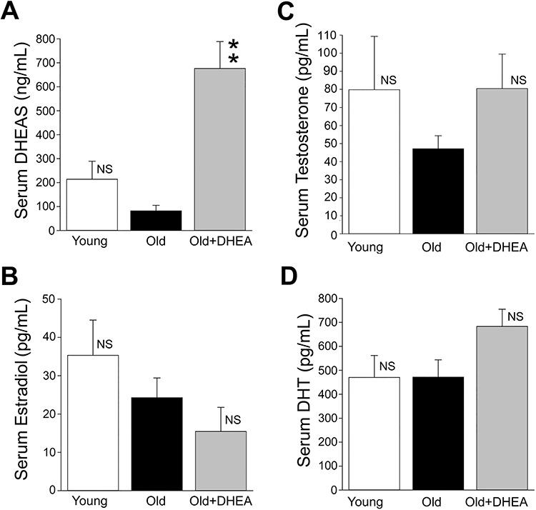
Dehydroepiandrosterone (DHEA) hormonal supplementation can improve oocyte quality in women with diminished ovarian function. However, it is unclear whether DHEA supplementation can also enhance ovarian function during the perimenopause (i.e., when the number of follicles in the ovary has undergone a marked reduction). To address this question, we examined the impact of 2.5-months of daily 5-mg oral DHEA supplementation on the number of ovarian follicles and the concentration of anti-Müllerian hormone (AMH) in perimenopausal rhesus macaques. Like women, these long-lived nonhuman primates have ~ 28-day menstrual cycles and eventually undergo menopause. They also show similar age-related neuroendocrine changes, including a marked decrease in circulating concentrations of DHEA and DHEA sulfate (DHEAS). Our experimental design involved the following three groups of animals (N = 6 per group): Young adult (mean age = 11.6 years), Old control (mean age = 23.1 years), and Old DHEA-treated (mean age = 23.5 years). Histological examination of the ovaries revealed a significant age-related decrease in the mean number of primordial follicles despite DHEA supplementation. Moreover, AMH concentrations within the ovaries and circulation, assessed by Western analysis and ELISA, respectively, showed significant age-related decreases that were not attenuated by DHEA supplementation. Taken together, these results fail to show a clear effect of short-term physiological DHEA supplementation on the perimenopausal ovary. However, they do not exclude the possibility that alternative DHEA supplementation paradigms (e.g., involving an earlier start date, longer duration and using pharmacological doses) may extend reproductive potential during aging.
Keywords: AMH; DHEA supplementation; FSH; menopause.
© The Author(s) 2020. Published by Oxford University Press on behalf of the American Physical Therapy Association. All rights reserved. For permissions, please email: journals.permissions@oup.com.
Figures

Similar articles
-
A randomized double-blinded placebo-controlled trial on the effect of dehydroepiandrosterone for 16 weeks on ovarian response markers in women with primary ovarian insufficiency.J Clin Endocrinol Metab. 2013 Jan;98(1):380-8. doi: 10.1210/jc.2012-3071. Epub 2012 Nov 8. J Clin Endocrinol Metab. 2013. PMID: 23144466 Clinical Trial.
-
Dehydroepiandrosterone supplementation improves predictive markers for diminished ovarian reserve: serum AMH, inhibin B and antral follicle count.Eur J Obstet Gynecol Reprod Biol. 2013 Jul;169(2):257-60. doi: 10.1016/j.ejogrb.2013.04.003. Epub 2013 May 9. Eur J Obstet Gynecol Reprod Biol. 2013. PMID: 23664458 Clinical Trial.
-
Effects of dehydroepiandrosterone on in vivo ovine follicular development.Hum Reprod. 2014 Jan;29(1):146-54. doi: 10.1093/humrep/det408. Epub 2013 Nov 20. Hum Reprod. 2014. PMID: 24256992
-
Causes and consequences of age-related steroid hormone changes: insights gained from nonhuman primates.J Neuroendocrinol. 2013 Nov;25(11):1062-9. doi: 10.1111/jne.12064. J Neuroendocrinol. 2013. PMID: 23796387 Free PMC article. Review.
-
DHEA and intracrinology at menopause, a positive choice for evolution of the human species.Climacteric. 2013 Apr;16(2):205-13. doi: 10.3109/13697137.2012.733983. Epub 2012 Nov 5. Climacteric. 2013. PMID: 23126249 Review.
Cited by
-
Effects of estradiol supplementation on the brain transcriptome of old rhesus macaques maintained on an obesogenic diet.Geroscience. 2022 Feb;44(1):229-252. doi: 10.1007/s11357-021-00453-8. Epub 2021 Oct 13. Geroscience. 2022. PMID: 34642852 Free PMC article.
-
Effect of hormone replacement therapy on amyloid beta (Aβ) plaque density in the rhesus macaque amygdala.Front Aging Neurosci. 2024 Jan 11;15:1326747. doi: 10.3389/fnagi.2023.1326747. eCollection 2023. Front Aging Neurosci. 2024. PMID: 38274989 Free PMC article.
-
hUC-MSC Combined with DHEA Alleviates Ovarian Senescence in Naturally Aging Mice through Enhancing Antioxidant Capacity and Inhibiting Inflammatory Response.Stem Cells Int. 2024 Jul 30;2024:3100942. doi: 10.1155/2024/3100942. eCollection 2024. Stem Cells Int. 2024. PMID: 39108701 Free PMC article.
-
Pathological Markers of Alzheimer's Disease and Related Dementia in the Rhesus Macaque Amygdala.J Alzheimers Dis Rep. 2024 Jan 9;8(1):25-32. doi: 10.3233/ADR-230184. eCollection 2024. J Alzheimers Dis Rep. 2024. PMID: 38229831 Free PMC article.
-
The aged female rhesus macaque as a translational model for human menopause and hormone therapy.Horm Behav. 2024 Nov;166:105658. doi: 10.1016/j.yhbeh.2024.105658. Epub 2024 Nov 11. Horm Behav. 2024. PMID: 39531811
References
-
- Labrie F, Luu-The V, Labrie C, Simard J. DHEA and its transformation into androgens and estrogens in peripheral target tissues: Intracrinology. Neuroendocrinology 2001; 22:185–212. - PubMed
-
- Gleicher N, Weghofer A, Barad DH. Improvement in diminished ovarian reserve after dehydroepiandrosterone supplementation. Reprod Biomed Online 2010; 21:360–365. - PubMed
-
- Yilmaz N, Uygur D, Inal H, Gorkem U, Cicek N, Mollamahmutoglu L. Dehydroepiandrosterone supplementation improves predictive markers for diminished ovarian reserve: Serum AMH, inhibin B and antral follicle count. Eur J Obstet Gynecol Reprod Biol 2013; 169:257–260. - PubMed
-
- Fusi FM, Ferrario M, Bosisio C, Arnoldi M, Zanga L. DHEA supplementation positively affects spontaneous pregnancies in women with diminished ovarian function. Gynecol Endocrinol 2013; 29:940–943. - PubMed
Publication types
MeSH terms
Substances
Grants and funding
LinkOut - more resources
Full Text Sources
Medical
