

Published Erratum
doi: 10.1042/CS-20200904_COR.
Correction: Unpuzzling COVID-19: tissue-related signaling pathways associated with SARS-CoV-2 infection and transmission
- PMID: 32901820
- PMCID: PMC7484393
- DOI: 10.1042/CS-20200904_COR
Item in Clipboard
Published Erratum
Correction: Unpuzzling COVID-19: tissue-related signaling pathways associated with SARS-CoV-2 infection and transmission
Clin Sci (Lond).
.
No abstract available
Keywords: COVID-19; Coronavirus; SARS-CoV-2; signaling pathway.
Figures
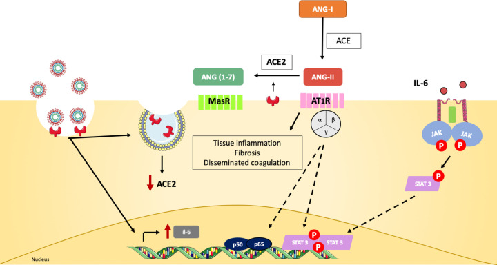
SARS-CoV-2 infection down-regulates ACE2 expression and leads to the production of pro-inflammatory mediators, such as IL-6 [1]. Angiotensin-I (Ang-I) is converted into Ang-II by the ACE in the extracellular space. ACE2 is able to further cleave Ang-II to Ang(1-7), which binds MasR receptors on the cell surface and promotes anti-inflammatory, vasodilation and anti-fibrotic effects [1]. Since ACE2 is down-regulated during viral infection, this event will lead to the accumulation of Ang-II and binding to AT1R receptors on cellular membrane. AT1R signals through JAK-STAT and induces fibrosis, pro-inflammatory gene expression and vasoconstriction [2,3]. Multiple organs express ACE2 and are target for SARS-CoV-2. As they lose ACE2-mediated protection, Ang-II signaling contributes to the pathological findings observed in COVID-19 patients, such as disseminated coagulopathy and acute tissue damage [4].
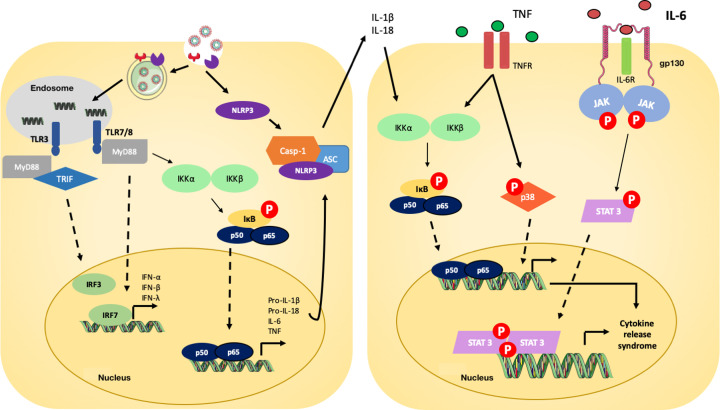
Toll-like receptors (TLRs) 3 and TLR 7/8 recognize SARS-CoV-2 RNA and initiate the inflammatory cascade via type I and type II IFN gene expression and NF-κB nuclear translocation [5,6]. Via NF-κB, the expression of multiple pro-inflammatory genes is stimulated, including pro-IL-1β, pro-IL-18, TNF and IL-6 [7–9]. The virus is also recognized by cytoplasmic NLRP3, which forms, together with ASC and caspase-1 (Casp-1), the inflammasome complex that will cleave and release mature forms of IL-1β and IL-18 [10]. The cytokines IL-1β, IL-18 and TNF bind to specific receptors and promote further NF-κB nuclear translocation and phosphorylation of p38 MAPK, which will lead to great expression of pro-inflammatory cytokines and chemokines [11,12]. IL-6, an important player in COVID-19, binds IL-6R and gp130 receptors to activate JAK/STAT-3 pathway and then contribute to the CRS observed in COVID-19 patients [13].
Erratum for
-
Unpuzzling COVID-19: tissue-related signaling pathways associated with SARS-CoV-2 infection and transmission.Clin Sci (Lond). 2020 Aug 28;134(16):2137-2160. doi: 10.1042/CS20200904. Clin Sci (Lond). 2020. PMID: 32820801 Free PMC article.
References
-
- Gheblawi M., Wang K., Viveiros A., Nguyen Q., Zhong J.-C., Turner A.J. et al. . (2020) Angiotensin-converting enzyme 2: SARS-CoV-2 receptor and regulator of the renin-angiotensin system: celebrating the 20th anniversary of the discovery of ACE2. Circ. Res. 126, 1456–1474 10.1161/CIRCRESAHA.120.317015 - DOI - PMC - PubMed
Publication types
LinkOut - more resources
Full Text Sources
Miscellaneous